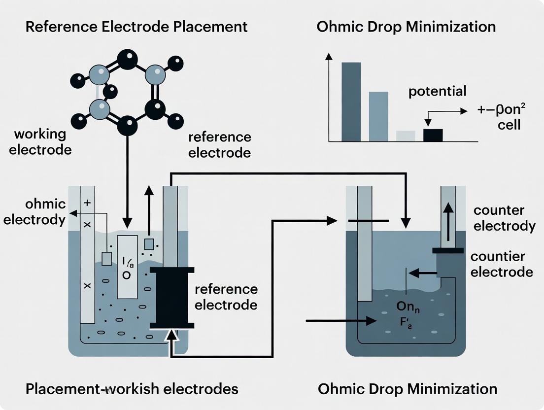
Minimizing Ohmic Drop in Electrochemical Measurements: A Strategic Guide to Reference Electrode Placement for Accurate Data
This article provides a comprehensive guide for researchers and development scientists on minimizing the detrimental effects of ohmic drop (iR drop) through strategic reference electrode placement.
Minimizing Ohmic Drop in Electrochemical Measurements: A Strategic Guide to Reference Electrode Placement for Accurate Data
Abstract
This article provides a comprehensive guide for researchers and development scientists on minimizing the detrimental effects of ohmic drop (iR drop) through strategic reference electrode placement. It covers the fundamental principles of iR drop, explores practical placement methodologies across various electrochemical cells (beaker, flow, multiplex), and addresses common troubleshooting scenarios. The guide also compares validation techniques, including Positive Feedback iR Compensation (IRFC) and Electrochemical Impedance Spectroscopy (EIS), to ensure measurement accuracy in critical applications like drug development, battery research, and corrosion studies.
Understanding Ohmic Drop: Why Reference Electrode Placement is Critical for Measurement Fidelity
In electrochemical measurements, Ohmic drop (iR drop) is an unwanted voltage loss that occurs due to the electrical resistance (R) of the electrolyte solution between the working and reference electrodes. When current (i) flows, this resistance causes a potential difference (iR) that is superimposed on the desired cell potential being measured. This artifact distorts voltammetric and potentiostatic data, leading to inaccurate interpretation of electrochemical kinetics, such as overstated overpotentials and miscalculated rate constants. This application note, framed within research on optimal reference electrode placement, details the nature, impact, and mitigation of iR drop.
Table 1: Effect of iR Drop on Common Electrochemical Techniques
| Technique | Primary Distortion | Typical Impact (Example System) | Mitigation Strategy |
|---|---|---|---|
| Cyclic Voltammetry (CV) | Peak potential separation (ΔEp) increases; peaks broaden and shift. | In a 0.1 M TBAPF6/MeCN solution with R=500 Ω and i=100 μA, iR drop = 50 mV, causing significant peak shift. | Positive Feedback iR Compensation, Ultramicroelectrodes. |
| Chronoamperometry (CA) | Applied step potential is effectively lower than set value, slowing current decay. | For a 1 V step with iR=100 mV, true driving force is 0.9 V, altering Cottrell slope analysis. | Current Interruption, Corrected potential application. |
| Electrochemical Impedance Spectroscopy (EIS) | High-frequency semicircle distortion; induces artificial inductance. | A 100 Ω uncompensated resistance distorts charge transfer resistance (Rct) estimation by same order. | Proper Reference Electrode Placement is critical. |
| Battery Cycling | Overestimates overpotential, reduces apparent energy efficiency. | In Li-ion cell with 10 mΩ internal R at 1A, iR drop is 10 mV per cycle. | Separated reference electrode (e.g., Li-metal wire). |
Table 2: Solution Resistance vs. Electrolyte Concentration (Theoretical, 25°C)
| Supporting Electrolyte Concentration (M) | Approx. Solution Resistivity (Ω·cm) | Estimated R (Ω) for 1 mm gap* | iR Drop (mV) at i=10 μA |
|---|---|---|---|
| 0.01 | ~1000 | 1000 | 10 |
| 0.1 | ~100 | 100 | 1 |
| 1.0 | ~10 | 10 | 0.1 |
*Assumes a simplified cell geometry with 1 cm² electrode area and 1mm electrode separation.
Experimental Protocols for iR Drop Analysis & Mitigation
Protocol 1: Measuring Uncompensated Resistance (Ru) via Current Interruption
Objective: Determine the uncompensated resistance in a three-electrode cell to assess iR drop magnitude. Materials: Potentiostat, standard electrochemical cell, working, counter, and reference electrodes, electrolyte solution. Procedure:
- Cell Setup: Configure a standard three-electrode cell. Ensure the reference electrode is in its initial test position (e.g., within the main cell compartment).
- Chronoamperometry Setup: Apply a small potential step (e.g., 5 mV) from open circuit potential for a short duration (e.g., 50 ms).
- Current Interruption: Utilize the potentiostat's current interruption function (or a fast sampling oscilloscope) to monitor the cell potential immediately (within µs) after the current is switched off.
- Data Analysis: The potential instantly drops by the value of the iR component. Calculate Ru: Ru = ΔV / i, where ΔV is the instantaneous potential change upon interruption, and i is the current just before interruption.
- Mapping: Repeat measurement for different reference electrode placements (e.g., variations in distance from WE, positions relative to Luggin capillary).
Protocol 2: Evaluating Reference Electrode Placement Using Ferrocene Redox Probe
Objective: Quantify the distortion in cyclic voltammetry caused by iR drop as a function of reference electrode distance. Materials: As in Protocol 1, plus 1 mM ferrocene in 0.1 M supporting electrolyte (e.g., TBAPF6 in acetonitrile). Luggin capillary. Procedure:
- Baseline CV: Obtain a CV of the Fc/Fc⁺ redox couple at 100 mV/s with the reference electrode positioned optimally using a Luggin capillary placed very close (<1 mm) to the working electrode surface. Record ΔEp and peak potentials.
- Variable Placement CV: Systematically increase the distance between the Luggin capillary tip and the working electrode (e.g., 1 mm, 3 mm, 5 mm, 10 mm). Acquire a CV at each position under identical conditions.
- Data Analysis: For each CV, measure the peak-to-peak separation (ΔEp) and the shift in formal potential (E1/2). Plot these parameters versus Luggin capillary distance.
- Resistance Calculation: Use the ΔEp increase from the theoretical value (59 mV for Fc) to estimate Ru from the equation: ΔEp = 59 mV + (ip * Ru) / α, where ip is peak current and α is transfer coefficient (~0.5).
Visualizing the Problem & Solutions
Diagram Title: Origin of iR Drop in a Three-Electrode Cell
Diagram Title: Impact of Reference Electrode Placement on Data Fidelity
The Scientist's Toolkit: Key Research Reagent Solutions
Table 3: Essential Materials for iR Drop Minimization Experiments
| Item | Function & Relevance |
|---|---|
| Luggin-Haber Capillary | A glass tube that brings the reference electrode tip into close, fixed proximity to the working electrode, dramatically reducing Ru by minimizing the current path in the solution. Critical for placement studies. |
| Non-aqueous Reference Electrode (e.g., Ag/Ag⁺) | Stable reference potential in organic electrolytes. Essential for battery or organic electrochemistry studies where calomel or Ag/AgCl are unsuitable. |
| High-Purity Supporting Electrolyte (e.g., TBAPF6, LiPF6) | Provides high ionic conductivity to minimize intrinsic solution resistance. Must be electrochemically inert in the potential window of interest. |
| Redox Probe (e.g., Ferrocene/Ferrocenium) | A well-characterized, reversible, single-electron couple used as an internal standard to quantify iR drop by measuring deviations in CV peak separation (ΔEp). |
| Ultramicroelectrode (UME, r < 10 μm) | Generates very low currents (nA scale), thereby minimizing the absolute iR product (iR drop). Useful for high-resistance media or as a diagnostic tool. |
| Potentiostat with iR Compensation | Offers electronic compensation (positive feedback) to subtract estimated iR drop in real-time. Caution: Over-compensation can cause instability. Manual Ru measurement is still required. |
| Separator (e.g., Glass Frit, Porous Cup) | Isolates reference electrode compartment to prevent contamination but can add series resistance. Placement must account for this additional R component. |
In electrochemical measurements for drug development and biosensor research, the accurate determination of electrode potential is critical. The measured potential (E_measured) at a working electrode is not the ideal interfacial potential (E_interfacial) due to the presence of an uncompensated solution resistance (R_u). This relationship is described by Ohm's Law:
Emeasured = Einterfacial ± I * R_u
Where I is the current flowing through the electrochemical cell. The sign depends on whether the working electrode is the anode or cathode. This ohmic drop (IR drop) introduces significant error, particularly in high-resistance solutions (e.g., low-ionic-strength buffers common in biological assays) or at high currents. Research on reference electrode placement is fundamentally aimed at minimizing R_u to obtain accurate E_interfacial values.
Table 1: Impact of Solution Resistance on Measured Potential at Various Currents
| Current (μA) | Solution Resistance, R_u (kΩ) | Ohmic Drop, I*R_u (mV) | Error in E_measured (%)* |
|---|---|---|---|
| 0.1 | 1 | 0.1 | Negligible |
| 10 | 1 | 10 | Significant for Nernstian systems |
| 100 | 1 | 100 | Severe distortion |
| 10 | 0.1 | 1 | Manageable with correction |
| 10 | 10 | 100 | Severe distortion |
| 1000 | 0.01 | 10 | Significant in plating/stripping |
Assumes a typical 100 mV redox event. Error is (IR_u / 100mV) * 100%.
Table 2: Common Electrolyte Conductivities and Approximate R_u*
| Electrolyte Solution | Ionic Strength | Approx. Conductivity (mS/cm) | Approx. R_u in a 1 cm cell (Ω) |
|---|---|---|---|
| 1 M KCl (Aqueous) | Very High | ~1100 | ~0.9 |
| 0.1 M PBS (Standard) | High | ~150 | ~6.7 |
| Pure Water | Extremely Low | ~0.055 | ~18,000 |
| Typical Cell Culture Media | Moderate | ~15 | ~67 |
| 0.1 M TBAP in Acetonitrile | Moderate (Organic) | ~10 | ~100 |
*R_u is geometry-dependent. Values are illustrative for a 1 cm² electrode spaced 1 cm apart.
Experimental Protocols
Protocol 1: Determination of Uncompensated Solution Resistance (R_u) via Current Interrupt
Objective: To experimentally determine R_u for IR compensation or assessment of reference electrode placement. Materials: Potentiostat, 3-electrode cell (WE, CE, RE), electrolyte solution. Procedure:
- Set up the electrochemical cell with the reference electrode in its standard position.
- Configure the potentiostat for a galvanostatic pulse experiment.
- Apply a small, short-duration current step (e.g., 10 μA for 50 μs). The current must be sufficiently small to avoid significant polarization.
- Record the potential transient at the working electrode versus the reference electrode with high temporal resolution.
- Analyze the potential-time trace. The instantaneous jump in potential (ΔE) at the moment the current is applied is primarily due to the ohmic drop across R_u.
- Calculate R_u using Ohm's Law: Ru = ΔE / Iapplied.
- Repeat with different reference electrode placements (e.g., positions within a Luggin capillary) to find the configuration that yields the lowest R_u.
Protocol 2: Assessing Reference Electrode Placement Efficacy via Cyclic Voltammetry of a Standard Redox Couple
Objective: To visualize the impact of R_u and validate placement strategies using a well-characterized electrochemical reaction. Materials: Potentiostat, 3-electrode cell, 1 mM Potassium Ferricyanide (K3Fe(CN)6) in 1 M KCl support electrolyte, Platinum working electrode. Procedure:
- Prepare the standard solution. For a high R_u comparison, also prepare a solution with 1 mM K3Fe(CN)6 in 0.01 M KCl.
- Place the reference electrode in the optimal position (e.g., at the tip of a Luggin capillary close to the WE).
- Record a cyclic voltammogram (CV) at a moderate scan rate (e.g., 50 mV/s) in the standard high-conductivity solution (1 M KCl). This CV serves as the ideal, low-IR-drop benchmark.
- Record a CV in the low-conductivity solution (0.01 M KCl) with the reference electrode in the same optimal position. Note the increased peak separation (ΔEp).
- Move the reference electrode to a position far from the working electrode (e.g., outside the primary current path).
- Record a CV in the low-conductivity solution with this suboptimal placement.
- Analysis: Compare ΔEp and peak shape. Increased ΔEp and distorted, drawn-out peaks indicate a larger uncompensated R_u. The optimal placement minimizes these distortions relative to the benchmark.
Visualizations
Diagram 1: Core Relationship Governing Measured Potential
Diagram 2: Experiment Workflow & IR Drop Mitigation
The Scientist's Toolkit: Research Reagent Solutions & Essential Materials
Table 3: Essential Materials for IR Drop Studies and Reference Electrode Placement Research
| Item | Function & Relevance to IR Drop |
|---|---|
| Potentiostat/Galvanostat | Essential instrument for applying controlled current/potential and measuring the electrochemical response. Must have current interrupt or positive feedback capability for R_u measurement/compensation. |
| Luggin Capillary | A glass capillary that allows the reference electrode to be positioned very close to the working electrode surface without shielding the current distribution. This is the primary physical tool for minimizing R_u in the measurement circuit. |
| Low-Resistance Reference Electrode (e.g., Ag/AgCl with 1-3 M KCl filling) | Provides a stable, low-impedance potential reference. A high-impedance reference electrode (e.g., some miniature electrodes) can exacerbate measurement error. |
| High-Purity Supporting Electrolyte (e.g., KCl, TBAPF6) | Provides ionic conductivity, minimizing bulk solution resistance (R_u). Inert over the potential window of interest. |
| Standard Redox Probe (e.g., Potassium Ferricyanide, Ferrocene) | A well-characterized, reversible redox couple used to diagnostically assess the impact of R_u via peak separation (ΔEp) in cyclic voltammetry. |
| Conductivity Meter | For pre-experiment measurement of bulk solution conductivity, which is inversely related to R_u for a given cell geometry. |
| Non-polarizable Counter Electrode (e.g., large Pt mesh/coil) | Minimizes polarization at the counter electrode, ensuring the applied current is primarily used to polarize the working electrode and that R_u is the main source of potential error. |
Within the broader thesis on optimizing reference electrode placement to minimize ohmic drop, this application note details the critical, often overlooked, consequences of uncompensated solution resistance (iR drop) in electrochemical kinetics. iR drop manifests as an error between the potential applied by the potentiostat (Eapp) and the actual potential at the working electrode surface (Esurface), defined as Esurface = Eapp - iRu, where i is the current and Ru is the uncompensated resistance. This error systematically distorts voltammetric data, leading to incorrect calculations of kinetic parameters (e.g., electron transfer rate constant, k⁰) and flawed mechanistic interpretations, particularly in drug development for analyzing redox-active metabolites or pharmaceutical compounds.
Quantitative Impact on Kinetic Parameters
The following tables summarize the effects of increasing R_u on key electrochemical parameters derived from simulated cyclic voltammograms (CVs) of a quasi-reversible one-electron transfer system (1 mM species, D=1e-5 cm²/s, A=0.1 cm², T=298 K, k⁰ true value = 0.01 cm/s).
Table 1: Effect of Uncompensated Resistance on Apparent Voltammetric Metrics
| R_u (Ω) | Peak Potential Separation, ΔE_p (mV) | Apparent k⁰ (cm/s) | Error in k⁰ (%) | Apparent Transfer Coefficient (α) |
|---|---|---|---|---|
| 0 | 70 | 0.0100 | 0% | 0.50 |
| 50 | 95 | 0.0058 | -42% | 0.52 |
| 100 | 125 | 0.0032 | -68% | 0.55 |
| 200 | 190 | 0.0014 | -86% | 0.59 |
Table 2: Impact on Tafel Analysis for Mechanism Elucidation
| Measurement | R_u = 0 Ω (Ideal) | R_u = 100 Ω (Distorted) | Consequence for Mechanism |
|---|---|---|---|
| Anodic Tafel Slope | 120 mV/dec | 140 mV/dec | Suggests incorrect rate-determining step or number of electrons. |
| Cathodic Tafel Slope | -120 mV/dec | -150 mV/dec | Masks true symmetry of energy barrier. |
| Apparent Exchange Current (i₀) | 1.00 µA | 0.65 µA | Underestimates intrinsic electrocatalytic activity by 35%. |
Experimental Protocols
Protocol 1: Determination of Uncompensated Resistance (Ru) via Current Interrupt Objective: Accurately measure Ru for subsequent compensation or data correction. Materials: Potentiostat, standard three-electrode cell, supporting electrolyte, known redox couple (e.g., 1-5 mM Ferrocenemethanol). Procedure: 1. Cell Setup: Place working, counter, and reference electrodes. Ensure reference is positioned in the optimal configuration under study (e.g., Luggin capillary). 2. Solution Preparation: Prepare a solution containing only supporting electrolyte (e.g., 0.1 M TBAPF6 in acetonitrile). Record an electrochemical impedance spectrum (EIS) from 100 kHz to 1 Hz at open circuit potential. Fit the high-frequency intercept on the real axis to obtain the initial Ru. 3. Current Interrupt Measurement: Add a reversible redox couple (Fc/Fc⁺). Apply a potential to generate a steady-state current (iss), typically in the range of 10-100 µA. 4. Interrupt: Use the potentiostat's current interrupt function. The instantaneous jump in potential (ΔE) upon interrupting the current is equal to iss * Ru. 5. Calculation: Ru = ΔE / iss. Perform at multiple current levels to verify linearity.
Protocol 2: Validating Kinetic Parameters Against iR Drop Objective: Obtain iR-corrected kinetic parameters to establish a baseline. Materials: As in Protocol 1, potentiostat with positive feedback iR compensation capability. Procedure: 1. Measure Ru: Determine Ru using Protocol 1 for your specific cell and electrode placement. 2. Acquire Baseline CV: Record a cyclic voltammogram of the redox couple at a slow scan rate (e.g., 10-50 mV/s) with full positive feedback iR compensation applied (set to 85-95% of measured Ru to avoid oscillation). 3. Analyze Data: Measure ΔEp. Calculate the true k⁰ using the Nicholson method for quasi-reversible systems. 4. Acquire Distorted Data: Disable iR compensation. Record CVs at identical scan rates. 5. Comparative Analysis: Fit the distorted CVs using software (e.g., DigiElch, GPES) without iR correction. Tabulate the apparent k⁰ and ΔE_p values as in Table 1.
The Scientist's Toolkit: Essential Research Reagent Solutions
| Item | Function & Rationale |
|---|---|
| Luggin Capillary | A glass tube that brings the reference electrode tip near the working electrode surface, physically minimizing the R_u component in the measured potential. Critical for accurate placement studies. |
| Non-Corrosive, Conductive Supporting Electrolyte (e.g., TBAPF6, KCl) | Provides ionic strength, minimizes migration current, and ensures a stable, known R_u. High purity is essential to avoid Faradaic contributions from impurities. |
| Well-Defined Redox Probes (e.g., Ferrocenemethanol, Potassium Ferricyanide) | Used for R_u calibration and benchmarking electrode kinetics. Their well-known, outer-sphere electron transfer provides a reliable reference point. |
| Potentiostat with Positive Feedback & Current Interrupt | Instrument must have active iR compensation circuits (positive feedback) and the ability to measure R_u in situ via interrupt or high-speed EIS. |
| Ultramicroelectrode (UME) | With radii ≤ 25 µm, UMEs generate very low currents, inherently minimizing iR drop. They serve as an excellent validation tool for mechanisms derived from macroelectrode data. |
Visualizations
Title: iR Drop Distortion Feedback Loop
Title: iR Drop Masks True Reaction Mechanism
Application Notes
Within the broader thesis research on the optimal placement of a reference electrode (RE) to minimize ohmic drop (iR drop) in electrochemical measurements, understanding and controlling solution resistance (Rs) is paramount. Uncompensated Rs leads to distorted voltammograms, inaccurate potential control at the working electrode (WE) surface, and erroneous data interpretation in key applications such as electrocatalysis, battery development, and pharmaceutical electroanalysis. This note details the three primary physical factors governing Rs and their practical implications for experimental design.
1. Electrolyte Conductivity: The ionic strength and mobility of the electrolyte directly determine its resistivity (ρ). Higher conductivity (lower ρ) minimizes Rs. In drug development, where studies often involve low-ionic-strength biological buffers or non-aqueous solvents, Rs can be significant. The use of supporting electrolytes (e.g., 0.1 M KCl, TBAPF6) is standard but must be evaluated for chemical inertness in the system under study.
2. Cell Geometry: The current distribution between the WE and counter electrode (CE) is geometry-dependent. Ideal geometries, such as concentric spheres or a small WE placed symmetrically within a large, distant CE, promote uniform current lines and simplify Rs calculation and compensation. Common lab cells (e.g., beaker-style) often have asymmetric geometries leading to non-uniform current distribution and spatially variable Rs.
3. Electrode Distance: The primary determinant of Rs in a given configuration is the distance between the WE and the Luggin capillary tip of the RE. Minimizing this distance is the most effective experimental intervention for reducing ohmic drop. However, the capillary must not be placed so close as to shield the WE surface or disturb diffusion layers. The optimal placement is typically 1.5-2 times the capillary outer diameter from the WE surface.
Critical Interaction with RE Placement: These factors are not independent. For instance, in a low-conductivity electrolyte, the negative impact of suboptimal cell geometry or increased WE-RE distance is greatly amplified. Therefore, the optimization protocol must be iterative: first maximize electrolyte conductivity within experimental constraints, then select an appropriate cell geometry, and finally, precisely position the RE Luggin capillary.
Experimental Protocols
Protocol 1: Determining Electrolyte Conductivity andRs
Objective: Quantify the conductivity of a prepared electrolyte solution and estimate the expected solution resistance for a given cell configuration. Materials: Conductivity meter and probe, electrochemical cell, supporting electrolyte, temperature-controlled bath. Procedure:
- Calibrate the conductivity meter using a standard solution (e.g., 0.01 M KCl).
- Immerse the conductivity probe in the test electrolyte (e.g., 0.1 M phosphate buffer + 0.1 M KCl). Ensure temperature is stabilized at 25.0 ± 0.1°C.
- Record the conductivity (κ) in Siemens per meter (S/m).
- Calculate resistivity: ρ = 1 / κ (Ω·m).
- Using a simplified cell geometry model (e.g., parallel plates), estimate Rs: Rs ≈ ρ * (d / A), where d is the WE-CE distance and A is the WE area.
Protocol 2: Optimizing Luggin Capillary Placement via Potentiostatic EIS
Objective: Empirically determine the optimal distance between the WE and the Luggin capillary tip to minimize Rs. Materials: Potentiostat, 3-electrode cell (WE, CE, RE with movable Luggin capillary), 1 mM K3Fe(CN)6 in 1.0 M KCl supporting electrolyte. Procedure:
- Assemble the cell with the Luggin capillary tip positioned approximately 5 mm from the WE surface.
- Perform an electrochemical impedance spectroscopy (EIS) measurement at the open-circuit potential. Apply a 10 mV RMS perturbation from 100 kHz to 1 Hz.
- Fit the high-frequency intercept of the Nyquist plot on the real (Z') axis to obtain the uncompensated solution resistance, Ru.
- Systematically move the Luggin capillary closer to the WE in 0.5 mm increments, repeating the EIS measurement at each position.
- Plot Ru vs. distance. Identify the point where further approach yields no significant decrease in Ru and before distortion of the low-frequency (diffusive) part of the EIS spectrum occurs, indicating shielding.
Protocol 3: Validating iR Compensation via Cyclic Voltammetry of a Standard Redox Couple
Objective: Assess the effectiveness of Rs compensation (positive feedback or post-experiment correction) on the shape of a reversible voltammogram. Materials: As in Protocol 2, with a known reversible redox couple (e.g., 1 mM ferrocenemethanol in 0.1 M KCl). Procedure:
- With the optimally placed RE (from Protocol 2), record a cyclic voltammogram (CV) at 100 mV/s without any iR compensation.
- Apply the potentiostat's positive feedback iR compensation, gradually increasing the compensation percentage until oscillation occurs, then back off by 5-10%.
- Record the compensated CV.
- Perform post-acquisition iR correction using the Ru value from EIS.
- Compare the peak separation (ΔEp) for the uncompensated, on-line compensated, and post-corrected CVs. A ΔEp* close to 59 mV confirms effective iR minimization.
Table 1: Impact of Electrolyte Conductivity on Solution Resistance
| Electrolyte Composition | Conductivity (S/m) at 25°C | Estimated Rs (Ω)* for d=1cm, A=0.1cm² |
|---|---|---|
| 0.1 M KCl (Aqueous) | 1.29 | 7.75 |
| 0.1 M Phosphate Buffer (pH 7.4) | ~0.75 | ~13.3 |
| 0.1 M TBAPF6 in Acetonitrile | ~0.10 | ~100 |
| Simulated Body Fluid (Ionic Strength ~0.15 M) | ~0.65 | ~15.4 |
*Estimated for simplified parallel plate geometry.
Table 2: Effect of Luggin Capillary Distance on Measured Ru
| Capillary Tip to WE Distance (mm) | Measured Ru (Ω) via EIS | Observed ΔEp in CV (mV) |
|---|---|---|
| 5.0 | 45.2 | 145 |
| 3.0 | 32.1 | 105 |
| 2.0 | 25.6 | 85 |
| 1.5 (Optimal) | 22.3 | 62 |
| 1.0 (Too Close) | 21.8 | 65* |
*Shielding/distortion of diffusion layer begins, causing slight peak broadening.
Diagrams
Title: Factors Governing Ohmic Drop in Electrochemical Cells
Title: RE Placement Optimization Workflow
The Scientist's Toolkit
Table 3: Essential Research Reagents and Materials for iR Drop Studies
| Item | Function & Rationale |
|---|---|
| Potassium Chloride (KCl), 0.1 M Aqueous | High-conductivity, inert supporting electrolyte for establishing baseline Rs and calibrating conductivity probes. |
| Potassium Ferricyanide (K3[Fe(CN)6]), 1-5 mM | Reversible, outer-sphere redox standard used in EIS and CV validation protocols due to its well-defined electrochemistry. |
| Tetrabutylammonium Hexafluorophosphate (TBAPF6) | Common supporting electrolyte for non-aqueous (organic solvent) electrochemistry, essential for studying low-dielectric systems. |
| Luggin Capillary | Salt bridge extension from the RE that allows close proximity to the WE without contamination, critical for controlling the WE-RE distance. |
| Platinum Counter Electrode | Inert, high-surface-area CE to prevent current limitation and ensure symmetric current distribution in ideal geometries. |
| Ag/AgCl (Sat'd KCl) Reference Electrode | Stable, low-impedance RE. The sat'd KCl electrolyte minimizes junction potential and provides stable potential for aqueous studies. |
| Conductivity Meter & Calibration Standards | For precise measurement of electrolyte conductivity (κ), enabling calculation of resistivity (ρ) and theoretical Rs. |
Practical Strategies for Optimal Reference Electrode Positioning in Diverse Experimental Setups
Within the critical research on Placement of reference electrode to minimize ohmic drop, the Luggin-Haber capillary is an indispensable tool. Its primary function is to isolate the reference electrode from the working electrode compartment, enabling accurate measurement of the working electrode's potential by minimizing the inclusion of solution resistance (iR drop or ohmic overpotential) in the measurement. This application note details its design, operational principles, and provides a standardized protocol for ideal positioning, which is central to validating experimental data in electrochemistry, corrosion science, and electrochemical drug development.
Design and Function
Core Design
The Luggin-Haber capillary is a slender, tapered glass or polymer tube filled with electrolyte. One end (the tip) is positioned close to the working electrode surface, while the other end connects to the chamber containing the reference electrode (e.g., Ag/AgCl, SCE). The electrolyte bridge maintains ionic conductivity while physically separating the test solution from the reference electrode compartment, preventing contamination.
Function: Minimizing Ohmic Drop
The uncompensated resistance (Ru) between the working electrode and the reference electrode tip leads to a measured potential error: ΔE = i * Ru. By bringing the capillary tip into close proximity to the working electrode, the path of current flow through the resistive solution is shortened, thereby reducing Ru and the associated error.
Table 1: Impact of Capillary Tip Distance on Ohmic Drop
| Tip-to-Working Electrode Distance (mm) | Approximate Relative Ru (%) | Typical Potential Error at 1 mA (mV) in 0.1 M KCl |
|---|---|---|
| 10 | 100 (Baseline) | ~15 |
| 2 | ~65 | ~10 |
| 1 (Recommended Max) | ~40 | ~6 |
| 0.5 (Optimal) | ~25 | ~4 |
| <<0.1 (Too close, risk of shielding) | <10 | <2 (but distorts current distribution) |
Ideal Positioning Protocol
Objective: To position the Luggin-Haber capillary to minimize ohmic drop without distorting the primary current distribution at the working electrode.
Materials & Reagent Solutions:
- Luggin-Haber Capillary: Filled with supporting electrolyte matching test solution or agar gel bridge.
- Reference Electrode: (e.g., Ag/AgCl in 3M KCl).
- Potentiostat/Galvanostat.
- 3-Axis Micromanipulator (for precise positioning).
- Electrochemical Cell with working, counter, and reference ports.
- Supporting Electrolyte Solution (e.g., 0.1 M Phosphate Buffer, 0.1 M KCl).
- Standard Redox Couple Solution (e.g., 1 mM Potassium Ferricyanide in 1 M KCl for validation).
Protocol Steps:
- Assembly & Filling: Fill the clean, dry Luggin-Haber capillary with the supporting electrolyte solution (or agar gel). Ensure no air bubbles are trapped in the tip. Connect it securely to the reference electrode compartment.
- Initial Placement: Mount the capillary on the micromanipulator. Submerge the tip in the electrolyte solution. Position the tip approximately two times the tip outer diameter (o.d.) away from the working electrode surface (~1-2 mm typically).
- Ohmic Drop Measurement (Positive Feedback Method):
- Using the potentiostat's current interrupt or automatic iR compensation function, measure the uncompensated resistance.
- Record this value as Ru, initial.
- Iterative Optimization:
- Using the micromanipulator, move the capillary tip closer to the working electrode in 0.1 mm increments.
- After each move, re-measure Ru. Continue until Ru stops decreasing significantly (plateaus).
- Validation & Shielding Check:
- Critical Step: Perform a cyclic voltammetry (CV) scan using a well-known, kinetically fast redox couple (e.g., 1 mM ferricyanide).
- Compare the peak-to-peak separation (ΔEp) with the theoretically expected value (~59 mV for a reversible, one-electron transfer).
- If ΔEp is near 59 mV: Positioning is optimal. Ohmic drop is minimized without significant current distortion.
- If ΔEp is > 59 mV but Ru is low: Residual ohmic drop may be present; check compensation settings.
- If ΔEp is < 59 mV or peaks are distorted: The tip is too close, causing "shielding" or blocking of diffusion. Retract the tip by 0.2-0.5 mm and re-run the CV until a symmetrical, well-defined voltammogram is obtained.
- Final Documentation: Record the final tip distance, measured Ru, and the ΔEp from the validation CV. This position should be used consistently for all subsequent experiments in that cell configuration.
The Scientist's Toolkit
Table 2: Essential Research Reagent Solutions & Materials
| Item | Function & Rationale |
|---|---|
| Ag/AgCl (3M KCl) Reference Electrode | Provides a stable, reproducible reference potential against which the working electrode potential is measured. |
| Luggin-Haber Capillary (Borosilicate Glass) | Creates a high-resistance bridge to isolate the reference electrode while allowing potential sensing near the working electrode. |
| Saturated KCl Agar Gel | A semi-solid electrolyte for filling capillaries; prevents convective mixing between cell and reference electrode solutions. |
| Potassium Ferricyanide/K Ferrocyanide (1 mM each in 1M KCl) | A reversible redox couple used as a standard to validate electrochemical cell setup and iR compensation. |
| High Purity Supporting Electrolyte (e.g., TBAPF6, KCl, PBS) | Provides ionic conductivity, minimizes migration current, and controls pH/ionic strength. Must be inert in the studied potential window. |
| 3-Axis Micromanipulator with Non-Conductive Tips | Enables precise, vibration-free positioning of the Luggin capillary to sub-millimeter accuracy. |
Visualization
Title: Protocol for Luggin Capillary Positioning
Title: Effect of Luggin Tip Position on Current Field
Accurate measurement of electrochemical potentials in in vitro systems is critical for research in electrophysiology, biosensor development, and drug discovery. A persistent challenge is the minimization of the uncompensated solution resistance, or ohmic drop (iR drop), which can distort measured potentials and lead to erroneous conclusions. This document provides application notes and detailed protocols for the placement of cells and associated reference electrodes across three common experimental platforms—beakers, flow cells, and multiplex electrode arrays (MEAs)—within the broader thesis research framework focused on optimizing reference electrode placement to minimize the ohmic drop. Proper geometric configuration is paramount for ensuring data fidelity.
The Scientist's Toolkit: Key Research Reagent Solutions
The following table lists essential materials and their functions for experiments across the described platforms.
| Item Name | Function/Brief Explanation |
|---|---|
| Ag/AgCl Pseudo-Reference Electrode | Provides a stable, low-polarizable reference potential for electrochemical measurements. |
| Luggin Capillary | A salt bridge extension that places the reference electrode sensing point close to the working electrode (cells) to reduce iR drop without contaminating the solution. |
| Cell Culture Media (e.g., DMEM, Neurobasal) | Provides the necessary nutrients and ionic environment to maintain cell viability during experiments. |
| Phosphate Buffered Saline (PBS) | Used for rinsing cells and as an electrolyte in simpler electrochemical setups. |
| Extracellular Matrix (e.g., Poly-L-Lysine, Matrigel) | Coats substrates to promote cell adhesion and spreading for stable recordings. |
| Electrode Impedance Reduction Gel | Applied to MEA electrodes to lower interface impedance, improving signal-to-noise ratio. |
| Flow Cell Gasket/Silicon Chamber | Defines the fluidic path and working volume in a flow cell, crucial for controlling fluid dynamics and electrode placement. |
| Faraday Cage | Encloses the experimental setup to shield from external electromagnetic interference. |
Protocol: Beaker-based Electrochemical Cell Setup
This protocol is for a classic three-electrode setup in a static beaker, common for initial characterization of cell monolayers or suspensions.
Materials
- Electrochemical workstation (potentiostat)
- Glass beaker (50-100 mL)
- Working Electrode (e.g., glassy carbon disk with adherent cells)
- Counter Electrode (Platinum wire coil)
- Ag/AgCl reference electrode with Luggin capillary
- Cell culture or PBS electrolyte solution
Detailed Methodology
- Cell Preparation: Seed and culture the chosen cell type (e.g., HEK293, cardiomyocytes) directly onto the conductive surface of the working electrode until the desired confluence is achieved.
- Electrode Assembly: Secure the working electrode in the beaker. Place the platinum coil counter electrode at the periphery of the beaker. Fill the beaker with pre-warmed, oxygenated electrolyte solution to immerse the electrodes.
- Critical Reference Placement: Fill the Luggin capillary with electrolyte. Position the tip of the Luggin capillary approximately 2 times its outer diameter away from the surface of the working electrode. This positioning is crucial—it brings the reference sensing point close to the cell layer to minimize iR drop while preventing physical disturbance or electrical shielding.
- Connection & Validation: Connect all electrodes to the potentiostat. Before introducing cells, perform an electrochemical impedance spectroscopy (EIS) scan (e.g., 100 kHz to 0.1 Hz) on the bare electrode in solution. The high-frequency real impedance intercept provides an estimate of the solution resistance (Rs).
- Measurement: After cell introduction, perform cyclic voltammetry or chronoamperometry. Use the potentiostat's positive feedback iR compensation function cautiously, if available, setting it to no more than 80% of the measured Rs to avoid circuit oscillation.
| Parameter | Typical Value/Range | Impact on Ohmic Drop |
|---|---|---|
| Luggin Tip to Working Electrode Distance | 1 - 2 mm | Primary control parameter. Halving distance can theoretically halve Rs. |
| Solution Conductivity (Culture Media) | ~1.5 S/m | Higher conductivity directly lowers Rs. |
| Estimated Rs (in PBS, typical geometry) | 50 - 200 Ω | Directly multiplies with current (i) to create iR drop error. |
| Recommended iR Compensation Level | ≤ 80% of measured Rs | Prevents oscillation; residual drop must be considered in analysis. |
Diagram Title: Beaker Setup with Luggin Capillary Placement
Protocol: Flow Cell System for Perfusion Experiments
Flow cells are used for controlled perfusion, mimicking vascular flow or enabling reagent exchange. Minimizing iR drop here is complicated by fluid dynamics.
Materials
- Laminar flow chamber with defined inlet/outlet
- Peristaltic or syringe pump
- Tubing and reservoir for perfusate
- Integrated or placed working electrode (e.g., gold film)
- Ag/AgCl reference electrode
- Counter electrode (may be integrated upstream)
Detailed Methodology
- Cell Seeding: Seed cells directly onto the conductive surface within the flow chamber. Allow for adhesion under static conditions.
- Flow Path Assembly: Connect the inlet to a reservoir containing pre-warmed, oxygenated media. Connect the outlet to waste. Ensure all bubbles are purged from the system.
- Reference Electrode Integration: Place the reference electrode in the flow stream DOWNSTREAM from the working electrode. Upstream placement causes potential shifts due to changes in upstream electrode overpotentials. The optimal location is in a dedicated, low-flow-rate side port or in the outlet reservoir, connected via a salt bridge.
- Flow Rate Calibration: Initiate flow at the desired rate (typically 0.1 - 2 mL/min). Use a dye or bubble to calibrate.
- iR Drop Assessment: Perform EIS under flowing conditions. Note that Rs may be slightly lower than in static conditions due to constant replenishment of ions, but is highly dependent on reference placement relative to flow and working electrode.
| Parameter | Typical Value/Range | Impact on Ohmic Drop |
|---|---|---|
| Reference Electrode Placement | Downstream of Working Electrode | Avoids contamination by upstream overpotentials. |
| Flow Rate | 0.5 - 2 mL/min | High flow can stabilize Rs but may affect cell layer. |
| Channel Height/Width | 0.1 - 1 mm | Smaller cross-section increases flow velocity but can increase Rs. |
| Estimated Rs in Microfluidic Flow Cell | 100 - 500 Ω | Highly geometry-dependent. |
Diagram Title: Flow Cell Reference Electrode Placement
Protocol: Microelectrode Array (MEA) Systems for Electrophysiology
MEAs record extracellular field potentials from electrically active cells (e.g., neurons, cardiac myocytes). The "reference" is often a dedicated electrode on the array.
Materials
- MEA chip (e.g., 60-electrode array)
- MEA amplifier and data acquisition system
- Cell culture insert for the MEA
- Incubator cape for long-term recording
- Electrolyte solution (culture media)
Detailed Methodology
- MEA Preparation & Coating: Sterilize the MEA. Coat the electrode region with an adhesion-promoting molecule (e.g., poly-L-lysine).
- Cell Plating: Plate primary neurons, stem-cell-derived neurons, or cardiomyocytes directly onto the active electrode area at high density to promote network formation.
- Reference Selection: Designate one or more electrodes far from active recording sites as the "reference" or "ground." In some systems, a large silver chloride pellet in the culture medium serves as the bath ground. The key is to ensure this reference has a stable, low-impedance connection to the bath.
- Placement for Ohmic Drop Minimization: In planar MEAs, the iR drop is contained within the thin electrolyte layer above the electrodes. Ensure the culture medium volume is consistent (typically 1-2 mm height). A dedicated, large-area reference electrode placed centrally and close to the array plane provides the most stable, low-impedance reference point, minimizing common-mode noise and localized iR effects.
- Recording & Validation: Begin recording after cells mature (e.g., 7-14 days for neuronal networks). Monitor spontaneous activity. Impedance for each electrode can be checked via the amplifier's test functions; typical values should be < 100 kΩ at 1 kHz for good signal quality.
| Parameter | Typical Value/Range | Impact on Signal & Ohmic Drop |
|---|---|---|
| MEA Electrode Diameter | 10 - 50 μm | Smaller electrodes have higher impedance, increasing thermal noise. |
| Electrode Impedance (at 1 kHz) | 20 - 100 kΩ | Lower impedance improves signal-to-noise ratio. |
| Culture Medium Height | 1 - 2 mm | Defines the volume resistor; consistent height stabilizes background. |
| Reference Electrode Area | Large Ag/AgCl pellet or dedicated MEA electrode | Larger area reduces reference impedance, stabilizing the reference potential for all recording sites. |
Diagram Title: MEA Reference Electrode Integration
Special Considerations for Low-Conductivity Media and Non-Aqueous Electrolytes
Within the broader thesis investigating reference electrode placement to minimize ohmic drop (iR drop), this application note addresses the significant challenges posed by low-conductivity aqueous media and non-aqueous electrolytes. Accurate electrochemical measurement in these systems is critical for research in organic synthesis, battery development, pharmaceutical analysis, and studies in non-polar solvents, where traditional three-electrode cell configurations fail due to high solution resistance.
Core Challenges and Quantitative Data
The primary impediment is excessive ohmic drop, which distorts voltammetric waveforms, shifts potentials, and reduces measurement accuracy. The table below summarizes the conductivity ranges and associated challenges.
Table 1: Conductivity Ranges and Measurement Challenges
| Medium Type | Typical Conductivity Range (S/m) | Primary Cause of Low Conductivity | Dominant Challenge for Electrochemistry |
|---|---|---|---|
| Ultra-Pure Water | ~5.5 × 10⁻⁶ | Low ion concentration | Extreme iR drop, unstable potentials |
| Organic Electrolytes (e.g., Li-ion battery) | 0.1 - 1.0 | Low dielectric constant, ion pairing | iR drop, limited electrochemical window |
| Pharmaceutical Solvents (MeOH, ACN, DMF) | 0.01 - 0.1 | Moderate dissociation | Significant iR drop, solvent purification effects |
| Supporting Electrolyte in Aprotic Solvents | 0.01 - 0.5 | Low ion mobility | iR drop, reference electrode compatibility |
Experimental Protocols
Protocol 1: Assessing iR Drop in Low-Conductivity Media
Objective: Quantify uncompensated resistance (Rᵤ) using electrochemical impedance spectroscopy (EIS). Materials: Potentiostat with EIS capability, 3-electrode cell (WE, CE, RE), low-conductivity test solution, supporting electrolyte. Procedure:
- Prepare the test solution with minimal added electrolyte to preserve low-conductivity state.
- Setup cell with conventional reference electrode (e.g., Ag/AgCl) positioned per standard geometry.
- Perform EIS at open circuit potential over a frequency range of 100 kHz to 1 Hz.
- Analyze Nyquist plot. The high-frequency real-axis intercept provides the solution resistance (Rₛ).
- Repeat measurement with alternative reference electrode placements (see Protocol 2). Analysis: Rₛ is a direct contributor to Rᵤ. Compare Rₛ values across placements to identify geometry minimizing iR.
Protocol 2: Optimized Reference Electrode Placement (Luggin-Haberl Capillary)
Objective: Minimize iR drop by precise positioning of a reference electrode via a capillary tip. Materials: Custom Luggin-Haberl capillary, reference electrode, micromanipulator. Procedure:
- Fabricate or obtain a fine-tipped glass capillary (tip diameter ~0.5-1x WE diameter).
- Fill capillary with electrolyte matching reference electrode (e.g., 0.1 M TBAPF₆ for non-aqueous).
- Insert reference electrode into capillary, ensuring electrolytic contact.
- Using a micromanipulator, position capillary tip at a distance of ~1.5-2 times the working electrode diameter from the WE surface.
- Critical: Ensure tip is aligned with the center of the WE and oriented perpendicular to the surface.
- Perform a test measurement (e.g., cyclic voltammetry of a known redox couple) and compare to theoretical shape. Note: In non-aqueous systems, ensure capillary electrolyte is compatible with solvent to prevent junction contamination.
Protocol 3: Determining Electrochemical Window in Non-Aqueous Electrolytes
Objective: Define the stable potential window for a non-aqueous system. Materials: High-purity anhydrous solvent (e.g., acetonitrile), supporting electrolyte (e.g., 0.1 M TBAPF₆), inert atmosphere glovebox, Pt working and counter electrodes, non-aqueous reference electrode (e.g., Ag/Ag⁺ in ACN). Procedure:
- Under inert atmosphere, prepare electrolyte solution with rigorous drying.
- Assemble cell with optimized Luggin capillary placement (Protocol 2).
- Record a cyclic voltammogram from 0 V to positive limits until current rapidly increases (solvent/electrolyte oxidation).
- Record a CV from 0 V to negative limits until current rapidly increases (reduction).
- The region between these rapid current increases is the electrochemical window. Safety: Be aware of potential solvent breakdown products.
The Scientist's Toolkit: Essential Research Reagents and Materials
Table 2: Key Reagent Solutions and Materials
| Item | Function | Key Consideration |
|---|---|---|
| Tetrabutylammonium Hexafluorophosphate (TBAPF₆) | Common supporting electrolyte for non-aqueous work. | Must be recrystallized and dried for low water content; provides wide potential window. |
| Luggin-Haberl Capillary | Enables precise placement of RE near WE to minimize iR drop. | Tip diameter and distance are critical; material must be inert to solvent. |
| Ag/Ag⁺ (in same solvent) Reference Electrode | Stable reference potential in non-aqueous systems. | Prepared with known concentration (e.g., 0.01 M AgNO₃ in 0.1 M electrolyte). |
| Ferrocene/Ferrocenium (Fc/Fc⁺) | Internal potential reference standard. | Added at end of experiment to reference potentials; redox potential is solvent-dependent. |
| Molecular Sieves (3Å or 4Å) | For drying organic solvents and electrolytes. | Activated under vacuum/heat prior to use; essential for reproducible results. |
| Potentiostat with Positive Feedback iR Compensation | Actively compensates for residual resistance. | Use with caution: overcompensation leads to oscillation; best used after physical minimization. |
Diagrams
Title: iR Drop Mitigation Workflow
Title: Optimal Luggin Capillary Placement
This application note, framed within a broader thesis on minimizing the uncompensated solution resistance (Ru, or ohmic drop) through reference electrode placement, details the experimental protocols and data for optimizing electrochemical cell configurations. Precise placement is critical for obtaining accurate kinetic parameters in hydrodynamic and micro-scale voltammetry, which are foundational to electrocatalysis and biosensing research in drug development.
The iR drop—the potential difference caused by current flowing through a solution's resistance—distorts voltammetric data, leading to inaccurate measurements of rate constants and overpotentials. While electronic compensation (positive feedback) is standard, it can cause instability. Physical optimization of the reference electrode (RE) position, especially in conjunction with a Luggin capillary, is a fundamental and necessary step to minimize Ru before compensation. This study quantifies the impact of RE placement in two critical setups: the Rotating Disk Electrode (RDE) for bulk electrocatalysis studies and microelectrodes for localized, high-resolution measurements.
The Impact of Reference Electrode Placement: Quantitative Data
Table 1: Measured Uncompensated Resistance (Ru) vs. Luggin Capillary Tip Position (RDE in 0.1 M KCl)
| Luggin Tip Distance from WE Surface (mm) | Ru (Ω) [Measured] | Estimated Potential Error (mV) at I=1 mA |
|---|---|---|
| 0.5 (optimal) | 85 ± 5 | 0.085 |
| 2.0 | 120 ± 8 | 0.120 |
| 5.0 | 210 ± 12 | 0.210 |
| 10.0 (with barrier) | 350 ± 20 | 0.350 |
Note: Working Electrode (WE): 5 mm diameter glassy carbon RDE. RE: Ag/AgCl (3M KCl). Counter Electrode (CE): Pt coil. Measurement via current-interrupt or high-frequency impedance.
Table 2: Effect of RE Placement on Apparent Electro kinetic Parameters for ORR (O2-saturated 0.1 M KOH)
| RE Configuration | Ru (Ω) | Apparent E1/2 (V vs. RHE) | Apparent JK (mA/cm²) | Corrected E1/2 (V vs. RHE) |
|---|---|---|---|---|
| Luggin ~0.5 mm from WE center | 90 | 0.801 | 5.10 | 0.801 |
| Luggin ~5 mm from WE, facing side | 250 | 0.778 | 4.65 | 0.803 |
| RE placed outside double-jacket, no Luggin | 550 | 0.745 | 4.05 | 0.805 |
Note: Catalyst: Polycrystalline Pt. Scan rate: 10 mV/s. Rotation: 1600 rpm. Correction applied post-measurement using measured Ru.
Experimental Protocols
Protocol 3.1: Optimizing Luggin Capillary Placement for RDE
Objective: To determine and set the minimal-distance, non-interfering position of the Luggin capillary for an RDE experiment. Materials: See "The Scientist's Toolkit" (Section 5). Procedure:
- Cell Setup: Fill the electrochemical cell with supporting electrolyte (e.g., 0.1 M KCl). Assemble the cell with the WE, CE, and RE with Luggin capillary. Ensure the RDE tip is perfectly vertical.
- Initial Positioning: Using a micromanipulator, bring the Luggin capillary tip close to the center of the stationary WE disk (~2 mm away). Visually align it just outside the path of the rotating shaft.
- Ru Measurement (AC Impedance): a. Apply the open circuit potential. b. Run a high-frequency impedance scan (e.g., 100 kHz to 10 kHz) with a small AC amplitude (10 mV). c. The high-frequency real-axis intercept in the Nyquist plot is the solution resistance (Rs), which is Ru.
- Distance Optimization: a. Systematically move the Luggin capillary closer to the WE in 0.2 mm steps using the micromanipulator. After each move, repeat Step 3 to measure Ru. b. Critical: Observe the electrolyte meniscus at the capillary tip. Stop advancing when the meniscus just touches the WE surface, then retract by 0.3-0.5 mm. This is the minimal non-contact distance.
- Hydrodynamic Verification: Start the RDE rotation at a typical speed (e.g., 900 rpm). Ensure no bubbles are drawn to the WE and that the measured Ru remains stable. The capillary must not disrupt laminar flow.
- Record Position: Document the final distance and corresponding stable Ru value for all subsequent experiments.
Protocol 3.2: RE Positioning for Microelectrode in Confined Environments
Objective: To place a quasi-reference electrode (QRE) optimally for measurements with a microelectrode in a low-volume or porous sample (e.g., tissue, hydrogel). Materials: See "The Scientist's Toolkit" (Section 5). Procedure:
- QRE Fabrication: Etch a Ag wire (250 µm diameter) in concentrated HNO3 for 30 s, rinse, and then chloridize it in 0.1 M HCl via galvanostatic oxidation (0.5 mA/cm² for 60 s) to create an Ag/AgCl QRE.
- Three-Electrode Probe Assembly: Align the microelectrode (e.g., 10 µm carbon fiber) and the Ag/AgCl QRE within a pulled glass dual-barrel capillary. The tip should be sealed with epoxy, with the two working ends exposed and separated by 50-200 µm.
- Calibration in Bulk Solution: a. Immerse the probe in a stirred standard solution (e.g., PBS). Perform cyclic voltammetry with the microelectrode using a known redox couple (e.g., 1 mM Ferrocenemethanol). b. Confirm the stability and potential of the QRE by checking the half-wave potential (E1/2) of the couple against its known value.
- Placement in Sample: a. Carefully insert the combined probe into the sample (e.g., a drug-loaded hydrogel or tissue slice). b. Using a micromanipulator, position the probe so that the QRE is as close as possible to the microelectrode's intended measurement site without physically damaging the sample microstructure. The goal is to keep the 50-200 µm separation.
- Stability Check: Monitor the open-circuit potential of the QRE for 60 seconds. A drift of < 2 mV/min is acceptable. A larger drift indicates a poorly conditioned or improperly placed QRE.
- iR Assessment: Perform fast-scan cyclic voltammetry (FSCV) at the microelectrode. The primary indicator of acceptable iR drop in microelectrode work is a symmetrical, non-tilted voltammogram for a known surface-bound redox reaction.
Visualizing Experimental Workflows and Principles
Diagram 1: Luggin Capillary Optimization Workflow
Diagram 2: RE Placement Principles for RDE vs. Microelectrode
The Scientist's Toolkit
Table 3: Essential Research Reagent Solutions & Materials
| Item & Specification | Function in Placement Optimization |
|---|---|
| Luggin Capillary (e.g., 1 mm OD, pulled tip) | Channels the reference electrode potential close to the working electrode, minimizing the measured solution resistance without physical interference. |
| Micromanipulator (3-axis, manual or motorized) | Allows for precise, sub-millimeter positioning of the Luggin capillary or microelectrode probes relative to the working electrode or sample. |
| Ag/AgCl Quasi-Reference Electrode (QRE) (Etched Ag wire, chloridized) | A miniaturized, low-profile reference for confined spaces. Its stability is crucial for microelectrode and localized measurements. |
| Dual-Barrel Glass Capillary (for probe assembly) | Houses and fixes the microelectrode and QRE in close, consistent proximity for measurements in tissues, gels, or small volumes. |
| 0.1 M KCl or KNO3 Solution (High-purity) | Standard, well-characterized electrolyte for initial cell setup and Ru measurement due to its known and stable conductivity. |
| Potentiostat with High-Frequency Impedance Capability | Required for accurate measurement of the uncompensated solution resistance (Ru) via AC impedance prior to DC experimentation. |
| Ferrocenemethanol (1-5 mM in electrolyte) | A stable, reversible outer-sphere redox couple used to benchmark and calibrate electrode performance and reference electrode stability. |
| Non-reactive Vacuum Grease or Electrolyte Jar | Used to create a physical barrier between RE and WE compartments if needed, to prevent contamination while assessing placement impacts. |
Diagnosing and Correcting Poor Placement: A Troubleshooting Guide for Common Pitfalls
Within the broader thesis on optimizing reference electrode (RE) placement to minimize uncompensated resistance (Ru) in electrochemical cells, this document details the identification of key experimental symptoms indicating suboptimal positioning. Unstable open-circuit potentials and distorted voltammograms are primary indicators, directly impacting data reliability in analytical assays and drug development research.
Core Symptoms & Quantitative Data
Manifestations of Poor Placement
Suboptimal RE placement leads to an increased ohmic drop (iRu), which manifests experimentally. The following table summarizes the key symptoms and their characteristics.
Table 1: Symptoms and Consequences of Suboptimal Reference Electrode Placement
| Symptom | Typical Observation | Direct Cause | Impact on Measurement |
|---|---|---|---|
| Unstable Open-Circuit Potential (OCP) | Drift > ±2 mV/min in stable electrolyte. | Fluctuating liquid junction potential; varying current line interception. | Erroneous baseline for all potential-controlled experiments. |
| Distorted Voltammetric Waveforms | Peak separation (ΔEp) > 59/n mV for reversible systems; asymmetric peaks. | Non-uniform iRu drop across potential scan. | Incorrect estimation of formal potential (E°') and kinetic parameters. |
| Increased Background Current & Noise | Non-steady, sloping baselines in cyclic voltammetry (CV). | Unstable electric field inducing capacitive fluctuations. | Reduced signal-to-noise ratio, obscured low-concentration analyte detection. |
| Scan Rate-Dependent Peak Shifting | Peak potentials (Ep) shift significantly with increasing scan rate (v) for reversible systems. | iRu distortion that scales with current (i), which scales with v^(1/2). | Invalidated diagnostic criteria for reaction mechanisms. |
Quantitative Benchmark Data
The following table presents acceptable vs. problematic metrics for common experimental setups using a 3 mm glassy carbon working electrode (WE) and a reversible redox couple (e.g., 1 mM Ferrocenemethanol).
Table 2: Benchmark Electrochemical Metrics for Optimal vs. Suboptimal RE Placement
| Metric | Optimal Placement (Proximal, aligned) | Suboptimal Placement (Distant, obstructed) | Experimental Conditions |
|---|---|---|---|
| Uncompensated Resistance (Ru) | 50 - 200 Ω | > 500 Ω | 0.1 M Bu₄NPF₆ in acetonitrile. |
| OCP Stability | < ±0.1 mV/min drift | > ±2 mV/min drift | Cell at rest, after equilibration. |
| Cyclic Voltammetry ΔEp | 59 - 70 mV (for n=1) | > 100 mV | 1 mM FcMeOH, 100 mV/s scan rate. |
| Peak Current Ratio (ipa/ipc) | 1.00 ± 0.05 | Deviates significantly from 1.0 | 1 mM FcMeOH, 100 mV/s scan rate. |
| Ru-derived Potential Error (iRu) | < 1 mV (at peak current) | Can exceed 10-50 mV | Calculated from Ru and peak current. |
Experimental Protocols
Protocol A: Diagnostic Test for RE Placement Quality
Objective: To systematically evaluate the stability and positioning of the reference electrode.
Materials:
- Electrochemical cell and potentiostat.
- Working, counter, and reference electrodes.
- Stable, reversible redox probe solution (e.g., 1.0 mM Potassium ferricyanide in 1.0 M KCl, or 1.0 mM Ferrocenemethanol in 0.1 M Bu₄NPF₆/ACN).
- Supporting electrolyte.
Procedure:
- Cell Setup: Fill the electrochemical cell with supporting electrolyte only. Position the RE in its intended experimental location.
- OCP Monitoring: Record the open-circuit potential for 300 seconds. Analyze the trace for linear drift (see Table 2).
- Ru Measurement: Using the potentiostat's built-in function (e.g., Current Interrupt, Positive Feedback, or Impedance at high frequency), measure the uncompensated resistance (Ru) of the cell. Note the value.
- Redox Probe Test: Add the reversible redox probe to the cell. Run a cyclic voltammogram at a slow scan rate (e.g., 50 mV/s) over an appropriate potential window.
- Data Analysis:
a. Calculate the peak-to-peak separation (ΔEp).
b. Determine the anodic-to-cathodic peak current ratio (ipa/ipc).
c. Optionally, use the
Rufrom step 3 and the peak current (ip) to calculate the approximateiRuerror:iRu = ip * Ru. - RE Repositioning: Move the RE to a new position—ideally closer to the WE, within the same plane, and ensuring no obstructing objects (e.g., cell walls, bubbles) are in the current path. Repeat steps 2-5.
- Comparison: Compare the metrics (OCP drift, Ru, ΔEp, ipa/ipc) before and after repositioning. Optimal placement yields values within the "Optimal" ranges of Table 2.
Protocol B: Mapping iRu Distortion as a Function of RE Position
Objective: To visualize the direct impact of RE geometry on voltammetric shape.
Procedure:
- Baseline Acquisition: With the RE in a "good" position (confirmed via Protocol A), acquire a CV of the reversible redox probe at 100 mV/s. Save this as the baseline "ideal" voltammogram.
- Controlled Repositioning: Systematically move the RE to predefined, suboptimal locations:
- Location 1: Far from the WE (e.g., opposite corner of the cell).
- Location 2: Behind a glass frit or obstructing cell component (if present).
- Location 3: At a significantly different height relative to the WE plane.
- Data Acquisition: At each location, repeat the CV measurement under identical parameters.
- Analysis: Overlay all CVs. Qualitatively note the increase in ΔEp, peak broadening, and asymmetry. Quantify the peak shift (in mV) for each location relative to the baseline.
Diagrams
Title: Diagnostic Path for Suboptimal RE Placement
Title: Protocol for Validating Reference Electrode Placement
The Scientist's Toolkit: Essential Research Reagents & Materials
Table 3: Key Reagents and Materials for RE Placement Diagnostics
| Item | Function & Rationale |
|---|---|
| Reversible Redox Probes (e.g., Potassium ferricyanide/KCl, Ferrocenemethanol) | Provides a known, stable electrochemical response. Deviation from ideal CV (ΔEp ~59/n mV, ipa/ipc ~1) signals iRu distortion. |
| High-Purity Supporting Electrolyte (e.g., TBAPF₆, KCl) | Minimizes background Faradaic processes and ensures known, high ionic conductivity to reduce intrinsic Ru. |
| Pseudoreference Electrode (e.g., Ag wire) | For rapid testing; can be positioned very close to the WE to estimate the "ideal" minimal Ru. Not used for formal reporting. |
| Luggin Capillary | A probe that allows the RE to be positioned extremely close to the WE without shielding, dramatically reducing Ru. Critical for precise kinetics studies. |
| Potentiostat with Ru Compensation (Positive Feedback, iR Compensation) | While compensation is not a substitute for good placement, it is a necessary tool. Its effectiveness is limited by stability; high required compensation % indicates poor placement. |
| Microreference Electrodes (e.g., miniaturized Ag/AgCl) | Enable placement in confined geometries or closer to micro-WEs, reducing cell geometry-related iRu. |
Avoiding Capillary Tip Blockage and Incorrect Angulation
Within the broader research thesis on "Placement of Reference Electrode to Minimize Ohmic Drop," achieving reliable and stable electrochemical measurements is paramount. A critical, yet often underestimated, practical challenge is the physical maintenance of the reference electrode's liquid junction, particularly when using capillary-based reference electrodes or pseudo-reference electrodes in confined electrochemical cells. Tip blockage of the capillary and incorrect angulation of the electrode assembly directly introduce measurement artifacts, increase solution resistance (Ru), and compromise data integrity. These practical failures contradict the theoretical goal of ohmic drop minimization. These Application Notes provide detailed protocols to prevent these issues, ensuring the experimental setup supports the core thesis objectives.
Quantitative Data on Blockage and Angulation Effects
Table 1: Impact of Capillary Condition on Electrochemical Parameters
| Parameter | Unblocked, Correct Angulation | Partially Blocked (50%) | Fully Blocked/ Severe Misangulation |
|---|---|---|---|
| Measured Ru (Ω) | 50 - 150 (Baseline) | 300 - 1000 | >2000 or unstable |
| Potentiostatic Stability | Stable (< ±1 mV drift/hr) | Unstable (±5-20 mV drift/hr) | Highly unstable or open circuit |
| CV Peak Separation (ΔEp) for 10 mM Ferrocene | 59 - 70 mV (near-Nernstian) | 80 - 150 mV | Non-detectable or distorted |
| Noise Level (pA RMS) | Low (1-5 pA) | High (20-100 pA) | Extreme (>200 pA) |
| Time to Stable OCP (s) | 30 - 60 | 120 - 600 | Never stabilizes |
Table 2: Recommended Angulation and Positioning Guidelines
| Cell/Setup Type | Optimal Capillary Angle (θ) to Working Electrode | Optimal Distance (d) | Rationale |
|---|---|---|---|
| Standard 3-Electrode (Beaker Cell) | 30° - 45° from horizontal | 1.5 - 2 x capillary tip diameter | Ensures electrolyte bridge, minimizes shielding. |
| Thin-Layer or Microfluidic Cell | As parallel as possible (θ ≈ 0° - 10°) | As close as cell design allows | Reduces path length for current, critical for low-volume. |
| RDE/RRDE Studies | Angled to avoid vortex interference | ≥ 2 mm from rotating surface | Prevents turbulence-induced blockage and noise. |
| Corrosion Studies (Large Cell) | 45° - 90° (vertical downward) | 2 - 3 mm | Ensures continuous electrolyte column, avoids gas bubble trapping. |
Experimental Protocols
Protocol 3.1: Fabrication and Maintenance of Unblocked Capillary Tips
Objective: To prepare and maintain a reference electrode capillary tip with consistent, unblocked flow of electrolyte. Materials: See "The Scientist's Toolkit" (Section 5.0). Procedure:
- Capillary Pulling: Using a micropipette puller, heat and pull a borosilicate glass capillary (1.2 mm OD, 0.69 mm ID) to achieve a tip inner diameter (ID) of 50 - 100 µm. Program the puller for a long, low-velocity final pull to create a gradual taper.
- Tip Inspection: Under a stereo microscope at 40x magnification, inspect the tip. It must be open, circular, and free of chips or sealed glass. Discard imperfect tips.
- Silanization (Optional, for Ag/AgCl): To prevent internal AgCl deposition and blockage, vapor-silanize the capillary by placing it in a desiccator with a few drops of dimethylchlorosilane for 1 hour. Bake at 120°C for 30 min.
- Filling: Back-fill the capillary using a fine-gauge needle and syringe with the chosen electrolyte (e.g., 3 M KCl saturated with AgCl for Ag/AgCl). Ensure no air bubbles are trapped in the shank.
- Storage: Store vertically (tip down) in a vial containing the same electrolyte. Never allow the tip to dry out.
Protocol 3.2: Validating Tip Patency and Electrode Placement
Objective: To experimentally verify a low-ohmic-drop connection and correct positioning. Materials: Potentiostat, dummy cell, calibration solution (1 mM Ferrocenemethanol in 0.1 M KCl), electrochemical cell. Procedure:
- Initial Ru Check: Using the potentiostat's built-in current-interrupt or positive-feedback iR compensation function, measure the uncompensated solution resistance (Ru) in the calibration solution with the working electrode. Record the value (Target: < 200 Ω for standard cells).
- OCP Stability Test: Measure the open-circuit potential (OCP) of the reference electrode versus a second, stable reference in the same solution. Record for 5 minutes. Acceptable drift is < 2 mV total.
- Angulation Setup: Mount the reference electrode capillary using a lab jack and adjustable holder. Position the tip at the recommended distance (d) and angle (θ) from the working electrode surface as per Table 2.
- Cyclic Voltammetry Validation: Perform a CV of the calibration solution from 0.0 V to 0.5 V at 100 mV/s. Measure the peak-to-peak separation (ΔEp). A value of 59-70 mV confirms minimal ohmic drop and proper setup.
- Flow Test (for fritless capillaries): Under the microscope, gently apply positive pressure via the back-fill syringe. A small, consistent droplet should form at the tip within 1-2 seconds.
Mandatory Visualizations
Diagram Title: Causes & Impacts of Capillary Blockage
Diagram Title: Protocol for Validating Electrode Placement
The Scientist's Toolkit: Research Reagent Solutions
Table 3: Essential Materials for Reference Electrode Placement Studies
| Item | Function in Context | Key Specification/Note |
|---|---|---|
| Borosilicate Glass Capillaries | Forms the physical liquid junction bridge. | OD: 1.2 mm, ID: 0.69 mm; ensures compatibility with pullers and holders. |
| Micropipette Puller | Creates a tapered, open tip of reproducible diameter. | Programmable heat and velocity for controlled taper (target ID: 50-100 µm). |
| Micro-positioning Holder | Allows precise control of angulation (θ) and distance (d). | XYZ translational and rotational stages with fine adjustment (< 0.1 mm). |
| Stereo Microscope | For visual inspection of tip patency and placement. | 20x-50x magnification with oblique illumination. |
| Potentiostat with iR Compensation | Measures and compensates for ohmic drop (Ru). | Must have current-interrupt or positive feedback iR compensation capability. |
| Ferrocenemethanol | Electrochemical calibration standard to validate minimal iR drop. | 1 mM in supporting electrolyte (e.g., 0.1 M KCl); provides known ΔEp. |
| Ag/AgCl Reference Electrode Filling Solution | Standard electrolyte for stable reference potential. | 3 M KCl, saturated with AgCl. Filtered (0.2 µm) to prevent particle blockage. |
| Dimethylchlorosilane | Hydrophobic silanizing agent for glass capillaries. | Prevents internal AgCl plating and salt creep, reducing blockage risk. |
| Micro-syringe & Fine-Gauge Needle | For back-filling capillary electrodes without bubbles. | Needle gauge ≤ 30G to fit capillary inner diameter. |
Mitigating Electrolyte Junction Potentials and Shielding Effects
Within the critical research on placement of reference electrodes to minimize ohmic drop, the control of junction potentials and electromagnetic interference represents a fundamental challenge. Unmitigated, these effects corrupt potential measurements, compromising data integrity in electrochemical experiments central to corrosion studies, battery development, and electrophysiological drug screening. This document provides application notes and protocols for identifying, quantifying, and mitigating these artifacts.
Key Concepts & Quantification
| Artifact Source | Typical Magnitude | Primary Impact | Common Experimental Context |
|---|---|---|---|
| Liquid Junction Potential (LJP) | 1 to 30 mV | Reference electrode stability, absolute potential accuracy | Ion-selective electrodes, bi-ionic conditions, patch-clamp. |
| Ohmic Drop (iR drop) | 5 mV to >1 V | Controlled potential accuracy, kinetic distortion | High-current experiments, low-conductivity electrolytes (organic solvents). |
| Electromagnetic Induction | µV to mV (AC noise) | Signal-to-noise ratio, low-current measurement | High-impedance measurements, fast potentiostats, urban lab environments. |
| Electrolyte Contamination | Drift (mV/min) | Reference electrode offset, drift | Leaking reference electrode frits, impurity introduction. |
Table 2: Comparison of Junction Potential Mitigation Strategies
| Strategy | Principle | Effectiveness (LJP Reduction) | Practical Complexity | Key Limitation |
|---|---|---|---|---|
| Salt Bridge (High [KCl]/AgCl) | Minimizes ionic activity gradient | High (70-95%) | Low | KCl contamination risk, clogging. |
| Intermediate Junction (e.g., LiAc) | Uses ion of similar mobility | Medium (50-80%) | Medium | Requires mobility data, extra preparation. |
| LJP Calculation & Software Correction (JPCalc) | Theoretical calculation & post-processing | High (for known systems) | Low-Medium | Requires precise solution composition. |
| Non-Fluid/Junctionless Reference | Solid-contact or ionic liquid | Eliminates LJP | High | Long-term stability, compatibility. |
Experimental Protocols
Protocol 1: Empirical Measurement of Liquid Junction Potential
Objective: Quantify the LJP between two specific electrolytes. Materials: Two matched reference electrodes (e.g., Ag/AgCl), high-impedance voltmeter (>10 GΩ), salt bridge solution (3 M KCl, agarose), experimental electrolytes A and B. Procedure:
- Fill two identical reference electrode assemblies with the standard filling solution (e.g., 3 M KCl). Confirm their potential difference is < ±0.2 mV.
- Immerse Reference 1 in Electrolyte A. Connect Reference 2 to a salt bridge (3 M KCl/agarose) terminating in a Vycor or ceramic frit.
- Immerse the salt bridge tip into Electrolyte B.
- Measure the potential difference between Reference 1 and Reference 2 using the high-impedance voltmeter. This measured potential (E_meas) approximates the LJP between A and B plus any cell potential. For symmetrical, non-reactive ions, it is essentially the LJP.
- Record the sign and magnitude. Repeat with reversed salt bridge connections to check for asymmetry.
Protocol 2: Implementation of a Guard Shield for Low-Current Measurements
Objective: Mitigate capacitive coupling and electromagnetic interference. Materials: Potentiostat, Faraday cage, coaxial cables, conductive mesh or foil, grounding strap, shielded working electrode lead. Procedure:
- Enclosure: Construct or utilize a grounded Faraday cage enclosing the electrochemical cell and working electrode lead.
- Guard Shield: For the working electrode connection, use a triaxial cable. The inner conductor carries the signal, the inner shield (guard) is connected to the potentiostat's guard output (which is driven at the same potential as the signal), and the outer shield is connected to earth ground.
- Cell Placement: Place the electrochemical cell on a grounded metal plate inside the cage. Ensure all shielding is electrically continuous.
- Connection: Keep the reference and counter electrode cables shielded and routed away from power lines. Connect the guard shield directly to the shielding of the working electrode compartment if possible.
- Validation: Run a low-current experiment (e.g., <1 nA) with and without the guard shield activated. Observe the reduction in 50/60 Hz noise and high-frequency baseline instability.
Protocol 3: Placement of Reference Electrode with Luggin Capillary to Minimize Ohmic Drop and Junction Effects
Objective: Position the reference sensor optimally to reduce iR drop while stabilizing the junction environment. Materials: Reference electrode, Luggin capillary (fine-tip glass or polymer), micromanipulator, electrolytic cell. Procedure:
- Capillary Preparation: Fill the Luggin capillary with the same electrolyte as the reference electrode filling solution (e.g., 3 M KCl). Attach it securely to the reference electrode body, ensuring no air bubbles.
- Initial Placement: Using a micromanipulator, position the capillary tip approximately 2 times its outer diameter away from the working electrode surface. This is a starting point.
- Probe for iR Compensation: If using a potentiostat with positive feedback iR compensation, engage the compensation and gradually increase it until oscillation occurs. Back off to 80-90% of this value. The optimal physical placement often coincides with the highest stable compensation value.
- Junction Stability Check: Monitor open-circuit potential or a low-current potential for 10-15 minutes after placement. Drift > 0.1 mV/min suggests junction instability, potentially due to clogging or excessive flow. A larger tip diameter may be needed, balanced against spatial resolution.
- Document Position: Precisely document the distance from the working electrode and angle relative to the working surface for experimental reproducibility.
Diagrams
Diagram Title: Artifact Diagnosis & Mitigation Workflow
Diagram Title: Optimal Reference & Shield Placement Schematic
The Scientist's Toolkit: Key Research Reagent Solutions
Table 3: Essential Materials for Junction & Shielding Control
| Item | Function & Rationale | Critical Specification / Note |
|---|---|---|
| High-Purity KCl (3M Solution) | Standard reference electrode filling solution. High concentration minimizes LJP contribution from the bridge itself. | 99.999% purity, AgCl-saturated for Ag/AgCl electrodes. |
| Agarose (Molecular Biology Grade) | Gelling agent for salt bridges. Prevents convective mixing while maintaining ionic conductivity. | Low-gelling temperature, purity to avoid organic contaminants. |
| Vycor or Ceramic Frit | Junction material for reference electrodes. Controls electrolyte flow, stabilizing the junction. | Pore size (e.g., 5-10 µm). Soak in filling solution before use. |
| Luggin-Habber Capillary | Glass or inert polymer capillary. Allows precise placement of reference sensing point near the working electrode. | Tip diameter ~0.5-1 mm. Distance to WE critical (2x dia). |
| Triaxial Cable & Guard Output | Cable with inner signal, driven guard, and outer ground shield. Actively shields the high-impedance WE signal from capacitance. | Must connect to potentiostat with active guard driver. |
| Faraday Cage | Enclosure of conductive mesh/foil. Attenuates external electromagnetic fields. | Must be securely grounded. All cell connections enter via shielded ports. |
| Non-Polarizable Reference Electrode (e.g., Hg/Hg₂SO₄) | Alternative reference system for non-aqueous or chloride-sensitive studies. Can have different junction properties. | Choose filling electrolyte compatible with your system (e.g., Na₂SO₄ for sulfate systems). |
| JPCalc or Equivalent Software | Calculates liquid junction potential using the Henderson or Planck equation. Allows for post-measurement correction. | Requires accurate knowledge of all ion identities, concentrations, and mobilities. |
This protocol is framed within a broader research thesis investigating the optimal placement of a reference electrode (RE) to minimize uncompensated ohmic drop (iRu) in electrochemical measurements. Accurate iRu compensation is critical for obtaining valid kinetic data in studies relevant to corrosion science, battery development, and electrochemical analysis in pharmaceutical research (e.g., drug redox properties). This checklist provides a step-by-step procedure to validate an electrochemical cell setup, ensuring that the measured potentials are not artifactually shifted by poor RE placement.
The Scientist's Toolkit: Essential Materials & Reagents
| Item | Function in iR_u Minimization Studies |
|---|---|
| Luggin-Haber Capillary | A glass tube extending from the RE salt bridge to near the working electrode (WE). Its fine tip proximity to the WE reduces the solution resistance in the measurement path. |
| Bipotentiostat/Galvanostat | Enables simultaneous control of WE and sense electrode, crucial for systems with integrated feedback for iR compensation (e.g., positive feedback, current interrupt). |
| Electrochemical Impedance Spectroscopy (EIS) Module | Used to measure solution resistance (Rs) between the WE and RE at high frequency. This Rs value is essential for accurate iR compensation. |
| Planar or Rotating Disk Electrode (RDE) | A well-defined, reproducible WE geometry simplifies the modeling of current distribution and ohmic drop effects. |
| High-Conductivity Supporting Electrolyte | (e.g., 0.1M – 1.0M KCl, TBAPF6). Minimizes the inherent solution resistance, though the optimal RE placement remains critical. |
| Ferrocene/Ferrocenium (Fc/Fc+) Redox Couple | A standard internal redox reference used to validate potential accuracy post-iR compensation. |
| 3-Electrode Cell with Precise Micrometer Stages | Allows for the controlled, reproducible 3D positioning of the RE capillary relative to the WE surface. |
| Digital Oscilloscope | Required for time-domain techniques like current interrupt to measure iR drop decay. |
Optimization Checklist & Validation Protocols
Checklist 1: Pre-Experiment Hardware & Geometry Setup
- Cell Configuration: Use a standard 3-electrode cell (WE, Counter Electrode (CE), RE with Luggin capillary) placed on a vibration-damped table.
- RE Capillary Positioning: Mount the RE/Luggin capillary on a 3-axis micromanipulator. The capillary tip orifice should face the WE.
- Initial Tip Distance: Set the capillary tip at a distance of ~2x the outer tip diameter from the WE surface (e.g., ~1-2 mm for a 0.5 mm tip). Record this distance (d).
- Alignment: Ensure the capillary tip is aligned with the center of a planar WE or the center axis of an RDE.
- CE Placement: Position the CE symmetrically and sufficiently far from the WE to ensure uniform current distribution.
Checklist 2: Quantitative Baseline Measurement of Solution Resistance (R_s)
Protocol 2.1: EIS Measurement for R_s
- Prepare a solution of known redox couple (e.g., 1mM K₃Fe(CN)₆ in 1.0M KCl).
- Apply the open circuit potential (OCP).
- Run a high-frequency EIS spectrum:
- Frequency Range: 100 kHz to 10 Hz
- AC Amplitude: 10 mV
- Fit the high-frequency intercept of the Nyquist plot on the real (Z') axis. This value is R_s.
- Repeat this measurement for at least three RE tip-to-WE distances (e.g., 1 mm, 2 mm, 5 mm).
Table 1: Measured Solution Resistance (R_s) vs. RE Capillary Distance
| RE Tip Distance from WE (mm) | Measured R_s (Ω) | Notes (e.g., capillary inner diameter) |
|---|---|---|
| 1.0 | 15.2 ± 0.3 | I.D. = 0.3 mm |
| 2.0 | 28.7 ± 0.5 | I.D. = 0.3 mm |
| 5.0 | 65.1 ± 1.1 | I.D. = 0.3 mm |
Checklist 3: Validation via Electrochemical Standards
Protocol 3.1: Cyclic Voltammetry (CV) of Fc/Fc+
- Switch to a non-aqueous system (e.g., 1 mM Ferrocene in 0.1 M TBAPF₆ / Acetonitrile).
- With RE at the closest stable position (from Checklist 1), run a CV at 100 mV/s.
- Activate the instrument's iR compensation (Positive Feedback or Automatic) using the R_s value from Protocol 2.1.
- Key Metric: The peak potential separation (ΔEp) should approach the theoretical value of 59 mV (for a reversible, diffusion-controlled, one-electron process at 25°C). A larger ΔEp indicates residual uncompensated resistance.
- Sweep Variable: Perform CVs at increasing scan rates (e.g., 50, 100, 200, 500 mV/s). At higher currents (higher scan rates), iR_u effects are magnified.
- Validate: The formal potential (E°) of Fc/Fc+ should be independent of scan rate with proper iR compensation and RE placement.
Table 2: Validation Metrics for Fc/Fc+ CV at Different RE Placements
| RE Tip Distance (mm) | R_s used for Comp (Ω) | ΔE_p at 100 mV/s (mV) | ΔE_p at 500 mV/s (mV) | E° vs. Ag/Ag+ (V) |
|---|---|---|---|---|
| 1.0 | 15.2 | 62 | 75 | 0.425 ± 0.005 |
| 2.0 | 28.7 | 68 | 110 | 0.415 ± 0.010 |
| 5.0 | 65.1 | 95 | 220 | 0.390 ± 0.020 |
Checklist 4: Final Functional Test with Target System
- Run Target Experiment: Perform the intended electrochemical measurement (e.g., CV of a drug candidate) with the optimized RE placement (shortest stable distance yielding stable R_s and valid standard metrics).
- Apply iR Compensation: Use the post-optimization R_s value.
- Verify Reproducibility: Conduct n≥3 replicate experiments. The key output parameters (peak potentials, limiting currents) must have a relative standard deviation (RSD) < 5%.
- Document Final Setup: Create a permanent diagram/sketch of the final cell geometry, including all distances.
Visualization of Protocols and Relationships
Diagram 1: Workflow for RE Placement Optimization and Setup Validation
Diagram 2: The Role of RE Placement and iR Compensation in the Measurement Circuit
Beyond Placement: Validating Low iR Drop with Compensation Techniques and Comparative Analysis
Within the broader research context of optimizing reference electrode placement to minimize ohmic drop, electronic iR compensation, specifically Positive Feedback iR Compensation (IRFC), remains a critical tool for accurate electrochemical measurements. Uncompensated resistance (Ru) distorts voltammetric signals, leading to inaccurate potentials and reduced peak currents. While physical optimization (e.g., Luggin capillary placement) is primary, electronic compensation is essential when physical minimization reaches its practical limits. These application notes detail the principles, appropriate use cases, and protocols for implementing positive feedback IRFC in modern electroanalytical research and drug development.
Principles and Decision Framework
Positive feedback IRFC operates by adding a fraction of the current flowing through the working electrode to the applied potential command. This added voltage aims to counteract the iR drop across the uncompensated solution resistance. Its application is not universal and requires careful consideration.
When to Use Positive Feedback IRFC:
- After maximizing physical minimization techniques (optimal reference electrode placement, increased electrolyte conductivity).
- In low-conductivity solvents common in pharmaceutical research (e.g., non-aqueous media for organic molecule studies).
- For fast-scan cyclic voltammetry (CV) or chronoamperometry, where transient iR drop is significant.
- In systems with low (< 1 mA) current magnitudes to avoid overcompensation.
- When studying the kinetics of fast electron transfer processes.
When to Avoid or Use Extreme Caution:
- In systems with very high currents (> several mA), as instability and oscillation are likely.
- As a substitute for poor physical cell geometry and electrode placement.
- For routine slow-scan CV in high-conductivity aqueous buffers where Ru is negligible.
Table 1: Comparison of iR Drop Mitigation Strategies
| Strategy | Mechanism | Advantages | Limitations | Best For |
|---|---|---|---|---|
| Optimal RE Placement | Physical reduction of Ru path | Inherently stable, no feedback risk | Geometric/practical limits, fixed cells | All experiments, primary strategy |
| Supporting Electrolyte | Increases solution conductivity | Simple, globally reduces Ru | May affect chemistry, solubility limits | Non-aqueous electrochemistry |
| Positive Feedback IRFC | Electronic negative resistance | Corrects residual Ru post-physical optimization | Risk of instability/oscillation | Fast scans, low-current kinetics |
| Current Interruption | Measures iR post-pulse | Potentially more stable | Not real-time, complex implementation | Specific instrument setups |
Experimental Protocols
Protocol 1: Determination of Uncompensated Resistance (Ru)
Objective: Accurately measure Ru prior to applying electronic compensation. Materials: Potentiostat with IRFC capability, electrochemical cell, working, counter, and properly placed reference electrodes, supporting electrolyte. Procedure:
- Set up cell with optimized Luggin capillary placement per primary thesis research.
- Prepare a solution containing a well-known, reversible redox couple (e.g., 1 mM Ferrocene in non-aqueous electrolyte or 1 mM K3Fe(CN)6 in 1 M KCl).
- Record a cyclic voltammogram at a moderate scan rate (e.g., 100 mV/s) with IRFC disabled.
- Measure the potential difference (ΔE) between the anodic and cathodic peak potentials (Epa - Epc).
- Calculate Ru using the formula: Ru = (ΔE - (59/n) mV) / (2 Ip), where Ip is the average peak current (in Amps) and n is the number of electrons transferred. For Fc/Fc⁺, n=1, ΔEideal ≈ 59 mV.
- Validate using potentiostat's current-interrupt or impedance-based auto-Ru measurement feature if available.
Protocol 2: Safe Implementation of Positive Feedback IRFC
Objective: Apply IRFC without inducing potentiostat instability. Materials: As in Protocol 1. Procedure:
- Determine Ru: Complete Protocol 1 to establish a baseline Ru value.
- Initialization: In the potentiostat software, enable the positive feedback IRFC function. Set the compensation level to 0%.
- Incremental Adjustment: Record a CV of the reversible couple. Gradually increase the compensation percentage (e.g., in 5% increments).
- Stability Monitoring: After each increment, observe the voltammogram for:
- Reduction of ΔE towards the theoretical value (59/n mV).
- Any signs of noise increase, baseline distortion, or oscillation in the current response (especially near E1/2).
- Termination Point: Stop increasing compensation when ΔE reaches the theoretical value or at the first sign of instability. Never exceed 100% of the measured Ru.
- Validation: Run CVs at multiple scan rates. A stable, scan-rate-independent ΔE indicates successful compensation.
Visualization
Title: Decision & Workflow for Applying Positive Feedback IRFC
The Scientist's Toolkit
Table 2: Key Research Reagent Solutions for iR Compensation Studies
| Item | Function & Relevance |
|---|---|
| Potentiostat/Galvanostat with IRFC | Instrument capable of applying positive feedback compensation; essential for protocol execution. |
| Luggin Capillary | Physical probe to position reference electrode close to working electrode, minimizing Ru primarily. |
| Non-Aqueous Electrolyte (e.g., 0.1 M TBAPF₆ in Acetonitrile) | Common low-conductivity medium for drug molecule electroanalysis; where IRFC is often needed. |
| Redox Potential Standard (e.g., Ferrocene/Ferrocenium) | Reversible couple to accurately determine Ru and validate compensation. |
| High-Purity Inert Salts (e.g., TBAPF₆, KCl) | To prepare supporting electrolytes of known and variable conductivity. |
| Precision Microelectrodes (e.g., Pt disk, Carbon fiber) | Working electrodes that generate low currents, reducing iR drop magnitude and easing stable compensation. |
Using Electrochemical Impedance Spectroscopy (EIS) to Measure Uncompensated Resistance (Ru)
Accurate measurement of uncompensated solution resistance (Ru) is a critical parameter in quantitative electrochemical analysis, particularly in kinetic studies and corrosion science. Ru causes an ohmic (iR) drop between the working and reference electrodes, distorting the applied potential and leading to significant errors in measured currents and derived parameters like rate constants. This Application Note details the use of Electrochemical Impedance Spectroscopy (EIS) for precise Ru determination. This work is framed within a broader thesis investigating the optimal placement of the reference electrode to minimize the ohmic drop. Precise Ru measurement via EIS provides the essential data to validate physical placement strategies and computational models aimed at iR compensation.
Key Principles of EIS forRuMeasurement
EIS measures the impedance of an electrochemical system over a wide frequency range. In a standard three-electrode cell, the high-frequency intercept of the impedance spectrum with the real (Z') axis in a Nyquist plot corresponds to the solution resistance between the working and reference electrodes, which is Ru. At sufficiently high frequencies, the contribution from the faradaic process (charge transfer) and double-layer capacitance is negligible, leaving only the ohmic resistance of the electrolyte.
Experimental Protocols
Protocol A: Basic EIS Measurement forRuDetermination
Objective: To obtain the uncompensated resistance (Ru) of an electrochemical cell using a high-frequency EIS measurement.
Materials & Setup:
- Potentiostat/Galvanostat with EIS capabilities.
- Standard three-electrode cell: Working Electrode (WE), Counter Electrode (CE), Reference Electrode (RE).
- Electrolyte solution of known, stable composition.
- Faraday cage (recommended for low-current measurements).
Procedure:
- Cell Assembly: Position the WE, CE, and RE according to the geometric configuration being studied (e.g., traditional Luggin capillary, coaxial placement, or symmetric cell). Ensure stable, bubble-free conditions.
- Open Circuit Potential (OCP) Measurement: Allow the system to reach a stable OCP (e.g., drift < 1 mV/min).
- EIS Parameters:
- DC Potential: Apply the OCP value.
- AC Amplitude: 5-10 mV RMS (ensure linear system response).
- Frequency Range: Typically from 100 kHz (or the instrument maximum) down to 1-10 Hz. The high-frequency limit is critical.
- Points per Decade: ≥ 10.
- Integration Time/Number of Cycles: Use auto or set to ensure good signal-to-noise at high frequencies.
- Run Measurement: Acquire the impedance spectrum.
- Data Analysis:
- Plot the data in a Nyquist representation ( -Z'' vs. Z' ).
- Identify the high-frequency intercept on the real axis. This value is Ru (in Ω).
- For a more robust fit, use a simple equivalent circuit model [Rs(RctCPE)] where Rs is fixed as the high-frequency intercept. The fitted Rs is Ru.
Protocol B:RuMapping via RE Position
Objective: To systematically quantify how Ru varies with the distance and geometry between the WE and RE, informing optimal placement.
Procedure:
- Establish a baseline using Protocol A with a standard RE placement (e.g., Luggin capillary tip ~2x diameter from WE surface).
- Variable Positioning: Using a micromanipulator, change the distance (d) between the RE tip and the WE surface in precise increments (e.g., 0.5 mm steps from 1 mm to 10 mm). For each position, perform Protocol A.
- Angular Variation (Optional): If studying asymmetric cells, vary the angle of the RE relative to the WE surface normal.
- Data Compilation: Tabulate Ru vs. distance (d) and/or angle. Plot Ru as a function of d. The slope relates to the solution conductivity (κ) and cell geometry (Ru ∝ d/κA for a uniform field).
Table 1: Measured Uncompensated Resistance vs. Reference Electrode Distance (Simulated data for 0.1 M KCl aqueous solution, planar 0.5 cm² Pt WE, Pt CE, Ag/AgCl RE)
| RE Distance from WE (mm) | Measured Ru (Ω) | Notes (Configuration) |
|---|---|---|
| 1.0 | 12.5 ± 0.3 | Luggin capillary, aligned normal |
| 2.0 | 18.2 ± 0.4 | Standard recommended position |
| 3.0 | 23.8 ± 0.5 | |
| 5.0 | 35.1 ± 0.7 | |
| 7.0 | 46.9 ± 1.0 | |
| 10.0 | 64.5 ± 1.5 | No Luggin, direct RE insertion |
Table 2: Impact of Ru on Potentiostatic Error Calculated iR drop at different currents for Ru = 20 Ω
| Applied Current (mA) | Ohmic Drop (iRu) (mV) | Consequence |
|---|---|---|
| 0.01 | 0.2 | Negligible for most applications |
| 0.1 | 2.0 | Significant for precise kinetics |
| 1.0 | 20.0 | Severe distortion of voltammogram |
| 10.0 | 200.0 | Makes controlled-potential experiment invalid |
Visualizations
Diagram: EIS Workflow for Ru Determination
Diagram: Thesis Context: Ru in iR Drop Minimization
The Scientist's Toolkit
Table 3: Essential Research Reagents & Materials for EIS-Based Ru Studies
| Item | Function & Relevance |
|---|---|
| Potentiostat with EIS Module | Provides the precise AC perturbation and measures the phase-sensitive current response. Required frequency range >100 kHz for accurate Ru. |
| Low-Impedance Reference Electrode | Minimizes its own impedance contribution. Ag/AgCl (3M KCl) or Hg/Hg₂SO₄ are common. Must have stable potential. |
| Luggin Capillary | A glass probe filled with electrolyte that allows the RE to be positioned close to the WE without shielding. Critical for minimizing and studying Ru. |
| Supporting Electrolyte | High-purity, inert salt (e.g., KCl, NaClO₄, TBAPF₆) at sufficient concentration (>0.1 M) to dominate solution conductivity. Must be electrochemically inert in the potential window. |
| Faraday Cage | A grounded metal enclosure that shields the electrochemical cell from external electromagnetic noise, crucial for stable high-frequency EIS measurements. |
| Micromanipulator | Allows precise, repeatable positioning of the RE relative to the WE. Essential for systematic studies of distance dependence. |
| Equivalent Circuit Fitting Software | Used to model the impedance spectrum and extract parameters (e.g., ZView, EC-Lab, pyimpspec). Confirms the high-frequency intercept is well-defined. |
| Standard Redox Couple Solution | (e.g., 5 mM K₃[Fe(CN)₆]/K₄[Fe(CN)₆] in 1 M KCl) A well-characterized system for validating the EIS measurement and Ru determination protocol. |
This application note is framed within a broader thesis investigating the placement of reference electrodes to minimize ohmic (iR) drop in electrochemical measurements, particularly for applications in electrophysiology and analytical drug development. Uncompensated iR drop distorts voltage control, slows electrode response, and reduces measurement accuracy. This analysis compares three strategies: optimal physical placement of the reference electrode (RE), suboptimal placement, and the use of full electronic iR compensation.
Core Concepts & Data Comparison
Table 1: Comparative Analysis of iR Mitigation Strategies
| Parameter | Optimal RE Placement | Suboptimal RE Placement | Full Electronic Compensation |
|---|---|---|---|
| Primary Principle | Minimize physical distance and solution resistance between RE and working electrode (WE). | RE placed at a significant, non-ideal distance from WE. | Uses positive feedback circuitry to actively nullify iR drop. |
| Typical iR Drop Remaining | < 1-5% (context-dependent) | 20-80% (often high and variable) | < 1% (theoretically near-zero) |
| Stability | High, passive. | High, passive. | Can be unstable; requires careful adjustment. |
| Implementation Complexity | Low (but requires careful cell design). | Low. | High (circuitry/algorithm needed). |
| Risk of Oscillation | None. | None. | Significant if over-compensated. |
| Impact on Time Response | Good, limited by physical geometry. | Poor, slowed by high series resistance. | Excellent when stable. |
| Best For | Routine, stable measurements; high-frequency work. | Demonstrating error magnitude (not recommended). | Precise in vitro kinetics studies where placement is constrained. |
| Key Artifact Introduced | Minimal. | Voltage error, distorted kinetics, reduced peak currents. | Inductive ringing or oscillations if misadjusted. |
Table 2: Quantitative Impact on Model Voltammetric Experiment (1mM analyte, 10Ω·cm resistivity)
| Condition | Theoretical Peak Current (mA) | Measured Peak Current (mA) | Observed Peak Potential Shift (mV) | Effective Time Constant |
|---|---|---|---|---|
| Optimal Placement (Luggin Capillary) | 1.00 | 0.99 | +2 | 1.1 * RC |
| Suboptimal (5mm away) | 1.00 | 0.85 | +35 | 5 * RC |
| Full Electronic Comp (100%) | 1.00 | 1.01 | 0 | 0.9 * RC |
Experimental Protocols
Protocol 1: Evaluating RE Placement for a Three-Electrode Cell
Objective: To quantify the iR drop for different RE placements and identify the optimal configuration. Materials: See "Scientist's Toolkit" below. Procedure:
- Cell Setup: Fill the electrochemical cell with a standardized electrolyte solution (e.g., 0.1 M KCl). Position the WE and CE.
- Baseline with Luggin Capillary: Position the RE within a Luggin capillary, with its tip approximately 2x its diameter from the WE surface. This is the "optimal" reference.
- Cyclic Voltammetry (CV) Test: Run a CV using a well-characterized redox couple (e.g., 1 mM Ferricyanide). Record peak current (Ip) and peak separation (ΔEp).
- Suboptimal Placement: Move the RE (or its capillary) to a position at least 5mm from the WE, near the wall of the cell. Ensure it remains immersed.
- Repeat CV: Run the identical CV scan. Note the decrease in Ip and increase in ΔEp.
- iR Drop Measurement: Using the current interrupt or positive feedback potentiostat function, measure the uncompensated resistance (Ru) for both placements.
- Analysis: Calculate iR drop (i * Ru) at a specific current. Correlate with observed peak broadening and shifting.
Protocol 2: Implementing and Tuning Full Electronic Compensation
Objective: To safely apply and optimize electronic iR compensation to correct for suboptimal placement. Procedure:
- Initial Suboptimal Setup: Set up the cell with the RE in a suboptimal position (as in Protocol 1, Step 4). Measure the initial Ru.
- Enable Compensation: Activate the potentiostat's iR compensation function (e.g., "Positive Feedback" or "Current Interrupt" mode).
- Gradual Adjustment: Begin with 0% compensation. Slowly increase the compensation percentage (e.g., 10% increments) while monitoring a steady-state current or a very slow scan rate CV.
- Stability Check: After each increase, observe the signal for noise or oscillation. Critical: Before proceeding to fast scans, ensure no oscillation at low overpotentials.
- Final Tuning at Fast Scan Rates: Once near 80-90% of the measured Ru, perform a faster CV scan. Fine-tune the compensation to achieve peak separation closest to the theoretical Nernstian value (59/n mV) without inducing baseline wobble or oscillation.
- Validation: Compare the compensated CV from the suboptimal placement to the optimal placement CV from Protocol 1. The curves should be nearly superimposable if compensation is correctly applied.
Diagrams
Diagram 1: Signal Pathway for iR Drop and Mitigation Strategies
Title: iR Drop Formation and Mitigation Pathways
Diagram 2: Experimental Workflow for Comparative Analysis
Title: Workflow for Comparative iR Drop Study
The Scientist's Toolkit: Research Reagent Solutions & Essential Materials
| Item | Function / Explanation |
|---|---|
| Potentiostat/Galvanostat | The core instrument for applying potential and measuring current in a controlled manner. Must have iR compensation functionality (positive feedback or current interrupt) for electronic compensation studies. |
| Faradaic Redox Couple (e.g., Potassium Ferricyanide) | A well-understood, reversible redox probe (Fe(CN)₆³⁻/⁴⁻) used to benchmark system performance and quantify iR-induced distortions via cyclic voltammetry. |
| Luggin Capillary | A glass capillary that houses the RE tip, allowing it to be positioned very close to the WE without shielding. Critical for achieving optimal placement and minimizing Ru. |
| Ag/AgCl Reference Electrode (with porous frit) | A stable, low-polarization reference electrode. The porous frit controls junction potential and limits chloride leakage. |
| Low-Resistivity Aqueous Electrolyte (e.g., 0.1M KCl, PBS) | Provides conductive medium. Lower resistivity directly reduces iR drop, making placement and compensation effects more or less critical. |
| Platinum Counter Electrode | An inert auxiliary electrode to complete the current path. Surface area should be larger than the WE to prevent it from being current-limiting. |
| Glassy Carbon Working Electrode | An inert, polished disk WE providing a reproducible surface for redox reactions. |
| Current Interrupt or EIS Module | Used to accurately measure the uncompensated solution resistance (Ru) prior to applying electronic compensation. EIS provides the most accurate Ru value. |
This application note details the critical protocols for minimizing ohmic (IR) drop in sensitive electrochemical measurements, a pivotal concern in pharmaceutical (e.g., drug redox potential studies) and energy research (e.g., battery electrolyte development). The accuracy of measurements such as potentiometric titrations, corrosion potential monitoring, and cyclic voltammetry is fundamentally compromised by uncompensated resistance between working and reference electrodes. This content is framed within a broader thesis investigating optimal reference electrode placement and system configuration to negate this error.
Experimental Protocols
Protocol 1: Luggin-Habber Capillary Placement for Three-Electrode Cells
Objective: To physically minimize the distance between the reference electrode and working electrode, thereby reducing the solution resistance in the measured potential. Materials: Standard electrochemical cell, working electrode (e.g., glassy carbon, Pt), counter electrode (Pt mesh), reference electrode (Ag/AgCl, SCE), Luggin-Habber capillary, electrolyte solution. Method:
- Fill the Luggin capillary with the same electrolyte as the main cell.
- Connect the capillary to the reference electrode via a salt bridge.
- Position the capillary tip approximately 2 times its outer diameter from the surface of the working electrode. Critical: Avoid positioning too close (<1x diameter) to avoid shielding the working electrode's diffusion profile.
- Secure the capillary in place using a micromanipulator or fixed holder.
- Perform electrochemical measurement (e.g., EIS, CV).
Protocol 2: Electronic iR Compensation via Positive Feedback
Objective: To electronically subtract the estimated ohmic drop from the applied potential in real-time. Materials: Potentiostat with built-in iR compensation functionality (e.g., current-interrupt or positive feedback), standard three-electrode cell. Method:
- Set up the electrochemical cell with standard configuration.
- Run a high-frequency electrochemical impedance spectroscopy (EIS) scan (e.g., 100 kHz) to determine the uncompensated resistance (Ru).
- In the potentiostat software, enable iR compensation and input the measured Ru value.
- Set the compensation level initially to 80-85% of the total Ru to avoid circuit oscillation.
- Run the desired experiment (e.g., CV at high scan rates). Gradually increase compensation, monitoring for stability.
Data Presentation
Table 1: Impact of iR Compensation on Measured Peak Potential Separation in Cyclic Voltammetry of 1 mM Ferrocenemethanol in 0.1 M KCl
| iR Compensation Level | ΔEp (mV) | Ohmic Drop (mV) at 100 μA | Notes |
|---|---|---|---|
| None | 125 | ~25 | Severe peak broadening and separation. |
| 85% (Positive Feedback) | 72 | ~3.75 | Near-ideal Nernstian behavior approached. |
| 100% (Theoretical) | 59 | 0 | Risk of potentiostat oscillation. |
| Luggin Capillary (Optimal) | 65 | ~5 | Physically minimized, stable. |
Table 2: Comparison of iR Mitigation Techniques for Different Applications
| Technique | Best Use Case | Advantages | Limitations |
|---|---|---|---|
| Luggin-Habber Capillary | Slow scan CV, steady-state corrosion monitoring. | No electronic artifact, stable. | Requires careful placement, can distort geometry. |
| Electronic Positive Feedback | Fast-scan CV, transient techniques. | Corrects in real-time, adjustable. | Can cause instability; overcompensation distorts data. |
| Current Interrupt | Battery impedance, systems with high DC current. | Direct measurement of Ru. | Not continuous; requires specialized hardware. |
| Geometric Cell Optimization | High-resistance organic electrolytes (energy research). | Fundamental solution. | May require custom cell design. |
Visualization
Title: Workflow for Minimizing Ohmic Drop in Sensitive Measurements
Title: Ohmic Drop (iR) and Luggin Capillary Role in Measurement
The Scientist's Toolkit: Essential Research Reagent Solutions
| Item | Function in iR Minimization Context |
|---|---|
| Luggin-Habber Capillary | Glass probe filled with electrolyte; bridges reference electrode close to WE to reduce solution path resistance. |
| Non-Aqueous Reference Electrode (e.g., Ag/Ag+) | Provides stable potential in organic electrolytes used in battery/photovoltaic research, reducing junction potentials. |
| Supporting Electrolyte (e.g., TBAPF6, KCl) | High-concentration inert salt to increase solution conductivity, thereby lowering bulk Ru. |
| Potentiostat with iR Comp | Instrument capable of real-time positive feedback or current-interrupt resistance compensation. |
| Electrochemical Impedance Spectrometer | Measures uncompensated resistance (Ru) accurately via high-frequency intercept. |
| Faraday Cage | Shields cell from external electromagnetic noise, crucial for stable iR compensation feedback loops. |
| Micropositioner | Allows precise, vibration-free placement of the Luggin capillary tip relative to the working electrode. |
Conclusion
Strategic reference electrode placement is a fundamental, cost-effective first line of defense against ohmic drop, essential for acquiring high-fidelity electrochemical data. While foundational understanding and meticulous positioning can minimize iR error significantly, validation with techniques like EIS and judicious use of electronic compensation provides a robust framework for accuracy. For researchers in drug development, battery science, and beyond, mastering these principles is not merely procedural but critical for deriving correct kinetic parameters, ensuring reproducible results, and making reliable conclusions from complex electrochemical experiments. Future directions include the development of smart cell designs with integrated, optimized reference probes and advanced real-time compensation algorithms for dynamic systems.