Mastering the Nernst Equation: A Comprehensive Guide to Concentration Cell Calculations for Biomedical Researchers
This article provides researchers, scientists, and drug development professionals with a detailed framework for understanding, applying, and validating the Nernst equation in concentration cell calculations.
Mastering the Nernst Equation: A Comprehensive Guide to Concentration Cell Calculations for Biomedical Researchers
Abstract
This article provides researchers, scientists, and drug development professionals with a detailed framework for understanding, applying, and validating the Nernst equation in concentration cell calculations. It moves from foundational principles to practical methodology, addressing common troubleshooting scenarios and offering comparative validation strategies. The content is designed to enhance the accuracy and reliability of electrochemical measurements in key applications such as ion-selective electrode calibration, membrane transport studies, and physiological ion gradient modeling, all crucial for modern biomedical and pharmaceutical research.
Understanding the Nernst Equation: The Electrochemical Foundation of Concentration Cells
A concentration cell is a specialized electrochemical cell where both electrodes are composed of the same material, and the electrolyte contains the same ions, but at different concentrations. The driving force for the cell's electrical potential is solely the difference in chemical potential (concentration gradient) between the two half-cells. This principle is a direct and elegant application of the Nernst equation. Within the broader thesis on Nernst equation applications, concentration cells serve as the purest experimental validation of the equation's predictive power for equilibrium potentials under non-standard conditions. In biomedical research, this concept underpins transmembrane potentials, ion-channel function, and electrochemical sensing platforms.
The Nernst Equation: The Governing Framework
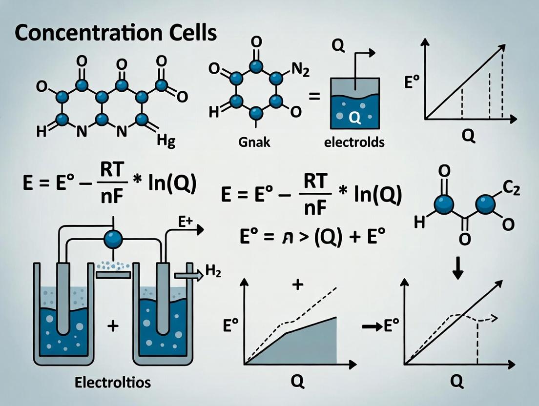
For a general reduction reaction: ( aA + ne^- \rightleftharpoons bB ), the Nernst Equation is: [ E = E^0 - \frac{RT}{nF} \ln Q ] Where (E) is the cell potential, (E^0) is the standard cell potential, (R) is the gas constant, (T) is temperature, (n) is the number of electrons transferred, (F) is Faraday's constant, and (Q) is the reaction quotient.
For a concentration cell with identical electrodes (e.g., Cu in Cu²⁺), (E^0 = 0). The equation simplifies to, for a cation cell: [ E{cell} = -\frac{RT}{nF} \ln \left( \frac{[M^{n+}]{dilute}}{[M^{n+}]{concentrated}} \right) = \frac{RT}{nF} \ln \left( \frac{[M^{n+}]{concentrated}}{[M^{n+}]_{dilute}} \right) ] Oxidation occurs in the dilute compartment (lower cation concentration), generating cations and electrons; reduction occurs in the concentrated compartment.
Table 1: Calculated Potentials for a Cu|Cu²⁺ Concentration Cell at 298.15 K
| [Cu²⁺] Concentrated (M) | [Cu²⁺] Dilute (M) | Concentration Ratio | Theoretical E_cell (mV) |
|---|---|---|---|
| 1.0 | 0.1 | 10 | +29.6 |
| 0.01 | 0.001 | 10 | +29.6 |
| 0.1 | 0.01 | 10 | +29.6 |
| 1.0 | 0.01 | 100 | +59.2 |
| 0.5 | 0.005 | 100 | +59.2 |
Note: Potential depends on the ratio, not absolute values. ( n=2 ) for Cu²⁺.
Experimental Protocol: Validating the Nernst Equation
This protocol demonstrates the direct relationship between concentration gradient and measured voltage.
A. Materials & Setup:
- Two identical copper wire electrodes.
- Two salt bridges (e.g., saturated KCl in agar).
- A high-impedance voltmeter/potentiometer.
- CuSO₄ solutions at prepared concentrations (e.g., 1.0 M, 0.1 M, 0.01 M, 0.001 M).
- Two beakers (half-cells).
B. Procedure:
- Clean the copper electrodes with dilute acid and rinse thoroughly.
- Fill one beaker with a known volume of a concentrated CuSO₄ solution (e.g., 0.1 M). Fill the second beaker with an equal volume of a more dilute solution (e.g., 0.01 M).
- Place a salt bridge between the two beakers to complete the circuit while minimizing liquid junction potential.
- Immerse one copper electrode in each beaker, ensuring no contact with the salt bridge.
- Connect the electrodes to the voltmeter. The electrode in the dilute solution will be the anode (negative terminal).
- Record the stable cell potential.
- Repeat steps 2-6 for different concentration pairs.
- Plot measured (E{cell}) vs. ( \ln([Cu^{2+}]{concd}/[Cu^{2+}]_{dil}) ). The slope should approximate (RT/nF) (0.0296 V at 298 K for n=2).
Biomedical Relevance and Applications
Transmembrane Potentials as Biological Concentration Cells: The resting membrane potential of a cell is fundamentally a concentration cell potential. The differential distribution of K⁺ (high intracellular, low extracellular) across a selectively permeable membrane generates the potential.
Ion-Selective Electrodes (ISEs): Modern biomedical sensors (e.g., for blood Na⁺, K⁺, Ca²⁺, pH) are advanced concentration cells. A membrane selective for the target ion separates the sample (unknown concentration) from a reference solution (fixed concentration). The measured potential is correlated to the sample's ion activity via the Nernst equation.
Corrosion and Implant Biocompatibility: Galvanic corrosion at implant sites can be modeled as a concentration cell, where electrolyte composition (e.g., O₂, Cl⁻) varies across the metal surface, creating anodic and cathodic regions.
The Scientist's Toolkit: Research Reagent Solutions
Table 2: Essential Materials for Concentration Cell Research & Development
| Item | Function in Experiment |
|---|---|
| Ion-Selective Membranes (e.g., Valinomycin for K⁺) | Provides selectivity for target ions in sensor construction, mimicking biological channels. |
| High-Impedance Potentiometer | Measures cell potential without drawing significant current, preventing polarization and ensuring accurate readings. |
| Salt Bridge (KCl-Agar) | Completes the electrical circuit between half-cells while minimizing liquid junction potential diffusion. |
| Standard Buffer Solutions (for pH ISEs) | Used to calibrate ion-selective electrodes by establishing a known concentration-potential relationship. |
| Reference Electrode (e.g., Ag/AgCl, Saturated Calomel) | Provides a stable, constant reference potential against which the indicator electrode's potential is measured. |
| Supporting Electrolyte (e.g., inert salt like NaNO₃) | Maintains constant ionic strength, ensuring activity coefficients are stable and simplifying Nernstian analysis. |
Visualizing Concepts and Workflows
Diagram 1: Logical flow of a concentration cell's operation.
Diagram 2: Ion-selective electrode calibration and use workflow.
Diagram 3: Transmembrane potential as a potassium concentration cell.
This analysis is framed within a broader thesis investigating the precision and limitations of the Nernst equation for calculating membrane potentials in biological concentration cells, a critical parameter in ion channel drug discovery and cellular electrophysiology research. While foundational, the equation's application to complex biological systems requires a rigorous, term-by-term deconstruction to understand its assumptions and guide experimental design.
Term-by-Term Deconstruction
The Nernst equation, for a single ion species, is given by:
E_ion = (RT / zF) * ln([X]_out / [X]_in)
Where E_ion is the equilibrium (reversal) potential.
Table 1: Quantitative Analysis of Nernst Equation Terms
| Term | Symbol | Physical Meaning | Typical Values & Units | Dependence & Notes |
|---|---|---|---|---|
| Gas Constant | R | Relates energy scale to temperature | 8.314462618 J·mol⁻¹·K⁻¹ | Fundamental constant. |
| Absolute Temperature | T | Absolute temperature of the system | 310.15 K (37°C) | Experimentally controlled. Directly proportional. |
| Ion Valence | z | Charge of the ion (with sign) | +1 (Na⁺, K⁺), +2 (Ca²⁺), -1 (Cl⁻) | Sign determines polarity of E_ion. |
| Faraday Constant | F | Charge per mole of electrons | 96485.33212 C·mol⁻¹ | Fundamental constant. |
| Outer Concentration | [X]_out | Ion concentration in extracellular space | Highly variable (see Table 2) | Logarithmic dependence. Critical for drug-induced changes. |
| Inner Concentration | [X]_in | Ion concentration in cytosol | Highly variable (see Table 2) | Logarithmic dependence. Often altered in disease models. |
| Nernst Potential | E_ion |
Theoretical equilibrium potential | Varies by ion (see Table 2) | Calculated output. Deviation indicates active transport or non-selectivity. |
Table 2: Physiological Ion Concentrations and Calculated Nernst Potentials (Mammalian Cell, ~37°C)
| Ion | Typical [Out] (mM) | Typical [In] (mM) | Ratio ([Out]/[In]) | Calculated E_ion (mV) |
|---|---|---|---|---|
| Na⁺ | 145 | 15 | 9.67 | +61.5 |
| K⁺ | 4 | 140 | 0.0286 | -96.9 |
| Ca²⁺ | 2.5 | 0.0001 | 25,000 | +129.2 |
| Cl⁻ | 110 | 10 | 11 | -64.2 |
Experimental Protocol: Validating the Nernst Potential for K⁺ in a Model Cell
This protocol outlines a method to empirically determine the reversal potential for K⁺ and compare it to the theoretical Nernst value.
Objective: To measure the reversal potential (E_rev) of a K⁺-selective current in the presence of a controlled K⁺ concentration gradient.
Key Reagents & Materials: Table 3: Research Reagent Solutions (Electrophysiology Toolkit)
| Item | Function & Explanation |
|---|---|
| Patch Pipette Puller | Creates glass micropipettes with sub-micron tips for electrical access to the cell. |
| Intracellular (Pipette) Solution | Mimics cytosol. For K⁺ validation: 140 mM KCl, 1 mM EGTA, 10 mM HEPES, pH 7.3. Sets [K⁺]_in. |
| Extracellular (Bath) Solution | Mimics interstitial fluid. Varied [KCl] (e.g., 4 mM, 20 mM, 40 mM) to set [K⁺]_out. |
| Ion Channel Expression System | HEK293 cells transiently transfected with cDNA for a selective K⁺ channel (e.g., Kir2.1). |
| Patch Clamp Amplifier | Measures tiny ionic currents (pA-nA) while applying controlled voltages (mV). |
| K⁺ Ionophore (Valinomycin) | Optional positive control. Creates a perfectly K⁺-selective membrane in artificial bilayers. |
Methodology:
- Cell Preparation: Culture and transfert HEK293 cells with a plasmid encoding a ligand-gated or constitutively active K⁺ channel.
- Solution Preparation: Prepare a standard intracellular solution with high [K⁺] (140 mM). Prepare three distinct extracellular solutions with [K⁺] at 4 mM, 20 mM, and 40 mM (replacing Na⁺ equimolarly).
- Whole-Cell Patch Clamp Setup: Establish the whole-cell configuration on a single cell using the standard intracellular solution and the 4 mM [K⁺]_out bath.
- Voltage Protocol: Apply a series of voltage steps (e.g., from -120 mV to +40 mV) from a holding potential.
- Current Recording: Record the resulting membrane currents. Identify the voltage step where the net K⁺ current is zero (
I_K = 0). This voltage is the observed reversal potential (E_rev). - Solution Perfusion: Gently perfuse the bath with the 20 mM [K⁺]out solution. Repeat steps 4-5. Repeat again for the 40 mM [K⁺]out solution.
- Data Analysis: For each [K⁺]out, plot the
E_revagainst the log of [K⁺]out. The data should follow a linear relationship. Fit the data to the Nernst equation. The slope should be close to RT/F * ln(10) ≈ 61.5 mV per decade change in [K⁺] at 37°C.
Conceptual and Experimental Workflow Diagrams
Diagram 1: Nernst Validation Research Cycle (92 chars)
Diagram 2: Patch Clamp Nernst Validation Workflow (98 chars)
Implications for Drug Development Research
Understanding each term's contribution is vital. For instance, drugs targeting NKCC1 cotransporters alter [K⁺]in and [Cl⁻]in, shifting their Nernst potentials and affecting neuronal excitability. Precision in T measurement is crucial for in vitro assays. The valence (z) dictates the sensitivity of E_ion to concentration changes, making divalent ions like Ca²⁺ potent signaling molecules. Discrepancies between measured membrane potential and E_ion highlight the activity of pumps or the simultaneous permeability to multiple ions, described by the Goldman-Hodgkin-Katz equation, which is the direct extension of this deconstruction for mixed ionic systems.
This whitepaper examines the fundamental physical chemistry principles that connect ionic activity to an experimentally measurable voltage, with a specific focus on the Nernst equation as it applies to electrochemical concentration cells. This discussion is framed within a broader research thesis aimed at refining the accuracy and applicability of Nernstian calculations for concentration cells, particularly under non-ideal conditions encountered in biological and pharmaceutical systems. For researchers in drug development, understanding this link is critical for applications ranging from ion-channel studies and membrane potential measurements to the characterization of ion-selective electrodes used in analyte sensing.
Theoretical Foundation: From Ion Activity to Electrode Potential
The measurable voltage (electromotive force, EMF) of an electrochemical cell arises from the thermodynamic drive to reduce free energy via charge transfer. For a reversible electrode responding to a specific ion i of charge z, the potential is governed by its electrochemical potential. The key link is the Nernst Equation:
E = E⁰ - (RT/zF) ln(a_i)
Where E is the measured potential, E⁰ is the standard electrode potential, R is the gas constant, T is temperature, F is Faraday's constant, and a_i is the ion activity. Activity (ai = γi * ci) incorporates both concentration (ci) and the non-ideal behavior captured by the activity coefficient (γ_i). In concentration cells, where identical electrode materials are immersed in solutions differing only in ion activity, E⁰ cancels, yielding:
Ecell = -(RT/zF) ln(ai(2)/a_i(1))
The central challenge in precise voltage calculation lies in accurately determining or controlling the single-ion activity, a thermodynamically immeasurable quantity that must be approximated via mean ionic activity coefficients in bulk solution or assumed in immobilized phases within ion-selective membranes.
Key Experimental Protocols for Validation
Protocol: Calibration of an Ion-Selective Electrode (ISE)
Objective: To empirically relate measured cell voltage to the activity of a target ion and verify Nernstian slope. Methodology:
- Setup: Construct a galvanic cell: Ag|AgCl|Reference Electrode || Salt Bridge || Test Solution | Ion-Selective Membrane | Internal Solution | Ag|AgCl.
- Solution Preparation: Prepare a series of standard solutions of the target ion with known concentrations spanning at least 3 orders of magnitude (e.g., 10⁻¹ M to 10⁻⁴ M). Maintain a constant, high background ionic strength using an inert electrolyte (e.g., NaNO₃) to stabilize the activity coefficient.
- Measurement: Immerse the ISE and a stable reference electrode (e.g., double-junction Ag/AgCl) in each standard solution under controlled temperature (25.0 ± 0.1°C). Allow potential to stabilize (1-3 mins).
- Data Analysis: Plot measured EMF vs. log10(ai), where ai is estimated using the Davies approximation for γ_i. Perform linear regression. A Nernstian response is indicated by a slope of ±(59.16/z) mV/decade at 25°C.
Protocol: Determination of Liquid Junction Potential (E_j)
Objective: To quantify and correct for the spurious potential arising from unequal ion mobilities at the salt bridge/solution interface. Methodology:
- Setup: Use a cell with a flowing junction: Hg|Hg₂Cl₂|KCl(satd) || Sample Solution | ISE.
- Procedure: Measure the cell potential with the sample solution. Replace the sample with a solution of known, unbiased composition (e.g., a matching ionic strength buffer) and measure again.
- Calculation: Estimate E_j using the Henderson approximation, integrating mobilities and concentrations of all ions at the junction. Modern practice uses the BaGGEL (Bodenschatz, Geisler, Gomm, Ehrlich, Lindner) empirical approach, measuring potentials with a symmetric cell setup and subtracting contributions.
- Correction: Apply the calculated E_j correction to the raw EMF to obtain the true membrane potential.
Data Presentation: Key Parameters & Experimental Outcomes
Table 1: Key Physical Constants for Nernst Equation Calculations
| Constant | Symbol | Value & Units | Relevance |
|---|---|---|---|
| Gas Constant | R | 8.314462618 J·mol⁻¹·K⁻¹ | Relates thermal energy to chemical potential |
| Faraday Constant | F | 96485.33212 C·mol⁻¹ | Converts molar charge to electrical charge |
| Nernst Slope (at 25°C) | (RT ln(10))/F | 59.157 mV/decade | Theoretical slope for a monovalent ion |
| Ideal Gas Constant (alternative) | R | 8.20574 × 10⁻² L·atm·mol⁻¹·K⁻¹ | For calculations involving pressure |
Table 2: Typical Nernstian Response Slopes for Common Ions at 25°C
| Ion | Charge (z) | Theoretical Slope (mV/decade) | Typical Experimental Slope (mV/decade)* | Common Application |
|---|---|---|---|---|
| H⁺ | +1 | +59.16 | 58.0 - 59.5 | pH electrodes |
| Na⁺ | +1 | +59.16 | 56.0 - 58.5 | Blood electrolyte analysis |
| K⁺ | +1 | +59.16 | 57.5 - 59.0 | Intracellular physiology |
| Ca²⁺ | +2 | +29.58 | 28.0 - 29.5 | Cell signaling studies |
| Cl⁻ | -1 | -59.16 | -57.5 to -59.0 | Reference electrode |
*Slopes can vary based on membrane composition and interference.
Table 3: Activity Coefficient (γ±) for HCl at 25°C (Davies Equation Estimate)
| Molality (mol/kg) | Mean Ionic Activity Coefficient (γ±) |
|---|---|
| 0.001 | 0.966 |
| 0.010 | 0.905 |
| 0.100 | 0.796 |
| 0.500 | 0.757 |
| 1.000 | 0.809 |
Visualizing the Pathways and Workflows
Title: From Ion Activity to Measured Voltage
Title: ISE Calibration Workflow
Title: Potential Contributions in a Measurement Cell
The Scientist's Toolkit: Key Research Reagent Solutions
Table 4: Essential Materials for Ion Activity-Potential Experiments
| Item | Function/Brief Explanation |
|---|---|
| Ion-Selective Membrane Cocktail | Contains ionophore (selective binder), ion exchanger, plasticizer, and polymer matrix (e.g., PVC). Forms the sensing element that generates the phase boundary potential. |
| High-Purity Ionic Salts (e.g., KCl, NaCl) | For preparing standard solutions and internal filling solutions. Purity is critical to avoid contamination that alters activity. |
| Ionic Strength Adjuster (ISA) | A concentrated, inert electrolyte (e.g., NH₄NO₃, ionic liquid) added to all standards and samples to fix the activity coefficient and minimize junction potentials. |
| Double-Junction Reference Electrode | Provides a stable, known reference potential. The outer filling solution is compatible with the sample to prevent contamination/clogging of the junction. |
| Symmetrical Cell Setup (H-Cell) | A two-chambered vessel with a removable salt bridge or frit. Essential for rigorous determination of membrane potential without significant liquid junction effects. |
| Activity Coefficient Calculator Software | Implements models (e.g., Debye-Hückel, Pitzer, SIT) to estimate single-ion activity from measurable mean ionic activities and composition. |
| Faraday Cage & Electrometer | Shields the experimental setup from external electrical noise. The electrometer provides high-impedance (>10¹² Ω) voltage measurement without current draw. |
| Thermostated Measurement Cell | Maintains constant temperature (±0.1°C), as the Nernst slope is temperature-dependent and thermal gradients induce spurious potentials. |
Concentration cells are electrochemical cells where the electromotive force (EMF) arises from a difference in the concentration of one or more electroactive species between the two half-cells. This discussion is framed within a broader thesis on applying the Nernst equation for the calculation and analysis of such cells. The fundamental Nernst equation for the EMF ((E)) of a concentration cell is: [ E = \frac{RT}{nF} \ln \frac{a2}{a1} ] where (R) is the gas constant, (T) is temperature, (n) is the number of electrons transferred, (F) is Faraday's constant, and (a1) and (a2) are the activities of the ionic species in the two half-cells.
The two primary categories are Electrode Concentration Cells and Electrolyte Concentration Cells, distinguished by the source of the concentration gradient.
Types and Core Mechanisms
Electrode Concentration Cells
In these cells, identical electrodes are immersed in an electrolyte of the same concentration. The EMF arises from a difference in the physical state or concentration of the electrode material itself.
- Mechanism: Common in amalgam cells (e.g., Zn(Hg) | Zn²⁺(aq) || Zn²⁺(aq) | Zn(Hg), with different Zn concentrations in the mercury amalgam). The potential difference originates from the differing activities of the metal in the two amalgams.
- Nernst Expression: For a cell M|Mⁿ⁺(C)|Mⁿ⁺(C)|M' (where M' is M in amalgam with different concentration), the EMF depends on the ratio of metal activities in the two electrodes.
Electrolyte Concentration Cells
These cells feature identical electrodes immersed in electrolytes containing the same ions but at different concentrations. The EMF is due solely to the tendency for ions to diffuse from a concentrated to a dilute solution.
- Sub-type 1: Cation-Transference Cells: The cation is the electroactive species (e.g., Ag|AgNO₃(C₁) || AgNO₃(C₂)|Ag, where C₁ ≠ C₂). The cell reaction involves the transfer of the cation from the higher to the lower concentration compartment.
- Sub-type 2: Anion-Transference Cells: The anion is the electroactive species (e.g., Pt, Cl₂(g)|HCl(C₁) || HCl(C₂)|Cl₂(g), Pt). The cell reaction involves the transfer of the anion.
Table 1: Core Comparison of Concentration Cell Types
| Feature | Electrode Concentration Cell | Electrolyte Concentration Cell |
|---|---|---|
| Electrodes | Different concentration/activity of same material | Identical |
| Electrolytes | Identical in composition and concentration | Same ions, different concentration (C₁, C₂) |
| Source of EMF | Difference in chemical potential of electrode material | Difference in chemical potential of electrolyte ions |
| Typical Example | Zn(Hg)(c₁) | ZnSO₄(aq) | Zn(Hg)(c₂) | Ag | Ag⁺(aq, c₁) | Ag⁺(aq, c₂) | Ag |
| Primary Research Use | Study of alloy thermodynamics, metal activity coefficients | Determination of transport numbers, solubility products, ion activity coefficients |
Research Applications and Experimental Protocols
Determination of Transport Numbers (Ionic Mobilities)
Electrolyte concentration cells are pivotal for measuring transport numbers (the fraction of current carried by a given ion).
Detailed Experimental Protocol: Hittorf Method using a Concentration Cell Setup
- Apparatus: A Hittorf cell or a multi-compartment electrolytic cell with electrodes (often Ag/AgCl) and reversible to the anion or cation under study.
- Procedure: a. Fill the cell with electrolyte (e.g., HCl) at a known, uniform concentration. b. Pass a precise quantity of electricity (Q = I·t, measured with a coulometer) through the cell. c. After electrolysis, carefully separate the anodic, cathodic, and middle compartments. d. Titrate the electrolyte from the anolyte and catholyte to determine the change in the amount of the ionic species.
- Calculation: The transport number of the cation (t₊) is calculated from the change in concentration in the cathode compartment relative to the total moles of electrons passed. The concentration cell EMF data before and after can be used to cross-verify concentration changes via the Nernst equation.
Solubility Product Constant (Ksp) Determination
A concentration cell can be constructed to measure the extremely low concentration of an ion from a sparingly soluble salt.
Detailed Experimental Protocol for Ksp of AgCl
- Cell Construction: Create a cell with two silver electrodes:
- Half-cell A: Ag(s) | Ag⁺(sat'd AgCl, known low [Cl⁻], e.g., 0.0100 M KCl).
- Half-cell B: Ag(s) | Ag⁺(known high concentration, e.g., 0.0100 M AgNO₃).
- Measurement: Measure the EMF (E_cell) of this cell accurately using a high-impedance voltmeter at 25°C.
- Calculation: a. The [Ag⁺] in Half-cell A is unknown and related to Ksp: [Ag⁺] = Ksp / [Cl⁻]. b. Apply the Nernst equation: Ecell = 0.05916 V * log([Ag⁺]B / [Ag⁺]A). c. Solve the equation for [Ag⁺]A, then calculate Ksp = [Ag⁺]_A * [Cl⁻].
Biochemical and Pharmaceutical Sensing
Concentration cells form the basis of ion-selective electrodes (ISEs) used in drug development for monitoring key ions (K⁺, Na⁺, Ca²⁺, H⁺) in biological fluids.
Table 2: Quantitative Data from Representative Applications
| Application | Measured Parameter | Typical Concentration Range | Achievable Precision (EMF) | Key Reference (Example) |
|---|---|---|---|---|
| Transport Number | t₊ (for H⁺ in HCl) | 0.01 - 1.0 M | ±0.001 in t value | Hittorf, Ann. Phys., 1853 |
| Solubility Product | Ksp (AgCl) | ~1.8 × 10⁻¹⁰ M² | ±0.5% in Ksp | MacInnes, JACS, 1919 |
| Biochemical Sensing | pH, pCa in serum | pH 6-8; pCa 2-5 | ±0.01 pH unit | Buck, RP, Anal. Chem., 1976 |
| Stability Constant | log β (Metal-Ligand) | 10² - 10¹⁰ M⁻¹ | ±0.05 log unit | Rossotti, The Determination of Stability Constants, 1961 |
The Scientist's Toolkit: Key Research Reagents & Materials
Table 3: Essential Materials for Concentration Cell Research
| Item | Function/Description | Example in Protocol |
|---|---|---|
| Reversible Electrodes | Electrodes reversible to the ion of interest, providing stable, reproducible potential. | Ag/AgCl electrode for Cl⁻ studies; Zn amalgam for electrode cells. |
| Salt Bridge | High-concentration electrolyte in gel (e.g., KCl-agar) to minimize liquid junction potential between half-cells. | Used in all electrolyte concentration cells with different solutions. |
| Coulometer | Device to accurately measure the total charge (Q) passed during electrolysis. | Essential for transport number determination experiments. |
| High-Impedance Voltmeter | Measures cell EMF without drawing significant current, which would alter concentrations. | Digital pH/mV meter with >10¹² Ω input impedance. |
| Ionophore-doped Membranes | For ISEs; selective organic ligands that bind target ions, creating the concentration gradient. | Valinomycin for K⁺-selective electrodes used in drug R&D. |
| Standard Reference Solutions | Solutions of known, precise activity for calibrating concentration cell responses. | NIST-traceable pH buffers, standard AgNO₃ solutions. |
Visualized Workflows and Pathways
Diagram Title: Electrolyte Concentration Cell Experimental Workflow
Diagram Title: Nernst Equation Application Logic Pathway
The Nernst equation (E = (RT/zF) ln([Cout]/[Cin])) is the foundational thermodynamic model for predicting membrane potentials and ion fluxes in concentration cells. In biological research and drug development, it serves as the essential starting point for understanding electrochemical gradients. However, its derivation assumes standard conditions—dilute solutions, ideal behavior, and a single permeable ion—that starkly contrast with the crowded, regulated, and multi-ionic reality of living cells. This whitepaper, framed within broader thesis research on refining concentration cell calculations, examines the critical divergences between the Nernstian ideal and biological systems, presenting current experimental data and methodologies for bridging this gap.
The Nernstian Ideal: Core Assumptions and Limitations
The Nernst equation provides the equilibrium potential for a single ion species across a membrane. Its standard assumptions are systematically violated in biology.
| Nernst Equation Assumption | Biological Reality | Consequence for Prediction |
|---|---|---|
| Ideal, Dilute Solution | Crowded, non-ideal cytosol & extracellular matrix. | Activity coefficients (γ) deviate from 1; effective concentration ≠ bulk concentration. |
| Single Permeable Ion | Multiple ions (K⁺, Na⁺, Cl⁻, Ca²⁺) with variable permeabilities. | Membrane potential is a weighted average (Goldman-Hodgkin-Katz equation). |
| Perfect Selectivity | Channels have finite selectivity and variable gating states. | Potential deviates from equilibrium potential of any single ion. |
| Passive, Equilibrium System | Active ion pumps (e.g., Na⁺/K⁺-ATPase) maintain steady-state. | System is not at equilibrium but at a dynamic steady-state. |
| Uniform Compartmentalization | Subcellular microdomains and organelles create gradients. | Local potentials and concentrations differ from whole-cell averages. |
Quantitative Divergence: Experimental Data
Recent electrophysiological and fluorescence imaging studies quantify the discrepancies between Nernst predictions and measured values.
Table 1: Predicted vs. Measured Resting Membrane Potentials (Mammalian Neuron)
| Ion | Equilibrium Potential (E_ion) Nernst Prediction (mV) | Relative Permeability (P_ion) | GHK Prediction (mV) | Typically Measured (mV) |
|---|---|---|---|---|
| K⁺ | -102 | 1.0 | ||
| Na⁺ | +60 | ~0.05 | -72 mV | -65 to -70 mV |
| Cl⁻ | -45 | ~0.1 |
Assumptions: [K⁺]_out=5mM, [K⁺]_in=140mM, [Na⁺]_out=145mM, [Na⁺]_in=15mM, [Cl⁻]_out=110mM, [Cl⁻]_in=10mM, T=37°C. GHK = Goldman-Hodgkin-Katz voltage equation.
Table 2: Impact of Cytosolic Crowding on Ion Activity
| Ion | Bulk Concentration in Cytosol (mM) | Estimated Activity Coefficient (γ) | Effective Activity (mM) |
|---|---|---|---|
| K⁺ | 140 | 0.75 - 0.85 | 105 - 119 |
| Na⁺ | 15 | 0.75 - 0.85 | 11 - 13 |
| Ca²⁺ (resting) | 0.0001 | 0.2 - 0.3 | 0.00002 - 0.00003 |
Experimental Protocols: Moving Beyond the Nernst Starting Point
Protocol 1: Measuring the Goldman-Hodgkin-Katz (GHK) Voltage
Objective: To determine the resting membrane potential (V_m) accounting for multiple ion permeabilities. Methodology:
- Cell Preparation: Use patch-clamp electrophysiology on a cultured neuron in whole-cell configuration.
- Ionic Control: Utilize a perfusion system to alter extracellular ionic solutions (e.g., high K⁺, low Na⁺).
- Voltage Measurement: In current-clamp (I=0) mode, record the resting V_m.
- Permeability Ratio Determination:
- Replace extracellular Na⁺ with an impermeant ion (e.g., NMDG⁺). The shift in V_m reflects sodium permeability.
- Apply specific channel blockers (e.g., TTX for NaV, TEA for KV) to isolate contributions.
- Data Analysis: Fit the recorded Vm changes under different solutions to the GHK voltage equation to solve for relative permeability ratios (PNa/PK, PCl/P_K).
Protocol 2: Fluorescence Imaging of Subcellular Ca²⁺ Microdomains
Objective: To visualize localized concentration gradients that violate the Nernst assumption of uniform compartments. Methodology:
- Dye Loading: Load cells with a ratiometric Ca²⁺ indicator (e.g., Fura-2 AM) and a organelle-specific dye (e.g., for ER).
- Calibration: Perform in situ calibration using ionophores (e.g., ionomycin) in Ca²⁺-free and saturating Ca²⁺ buffers.
- Stimulation: Use localized uncaging of IP3 or field stimulation to trigger Ca²⁺ release.
- Image Acquisition: Use high-speed, confocal, or TIRF microscopy to capture Ca²⁺ signals near channels (e.g., ryanodine receptors) versus bulk cytosol.
- Quantification: Generate time-course plots of Ca²⁺ concentration in microdomains vs. whole cell, demonstrating spatial heterogeneity.
Visualizing Key Concepts and Workflows
Title: From Nernst Assumptions to Biological Models
Title: GHK Permeability Measurement Protocol
The Scientist's Toolkit: Key Research Reagent Solutions
| Reagent/Material | Function in Context | Key Consideration |
|---|---|---|
| Patch Pipette Solution | Controls intracellular ionic composition during whole-cell recording. | Must include ATP, buffering agents (e.g., HEPES, EGTA), and mimic cytosolic ion concentrations. |
| Ion Channel Blockers (e.g., TTX, TEA, 4-AP) | Pharmacologically isolates specific ionic currents (Na⁺, K⁺). | Specificity and concentration are critical to avoid off-target effects. |
| Ionophores (e.g., Ionomycin, Gramicidin) | Creates defined ionic permeabilities for calibration (e.g., of fluorescent indicators). | Gramicidin used for perforated-patch to maintain intracellular signaling. |
| Ratiometric Fluorescent Dyes (e.g., Fura-2, Indo-1) | Measures intracellular ion concentration ([Ca²⁺], [H⁺], etc.) via emission/excitation ratio. | Ratiometric measurement corrects for dye concentration and path length. |
| Caged Compounds (e.g., caged IP₃, caged Ca²⁺) | Enables rapid, spatially localized release of signaling molecules via UV flash. | Allows precise initiation of signaling events to study microdomain dynamics. |
| Osmolytes & Crowding Agents (e.g., Ficoll, Dextran) | Mimics the crowded intracellular environment in in vitro experiments. | Used to measure effects on ion activity coefficients and reaction kinetics. |
The Nernst equation remains an indispensable starting point, providing the thermodynamic framework and null hypothesis for cellular electrochemistry. However, sophisticated drug development and mechanistic research require moving beyond its standard conditions. By integrating multi-ionic models like the GHK equation, employing advanced techniques like patch-clamp and fluorescence imaging, and accounting for cytoplasmic crowding and microdomains, researchers can develop quantitatively accurate models of biological concentration cells. This progression from ideal theory to biological reality is essential for predicting drug effects on excitability, signaling, and transport with high fidelity.
Step-by-Step Calculation Guide: Applying the Nernst Equation to Real-World Biomedical Problems
This technical guide details the complete workflow for calculating the cell potential of concentration cells using the Nernst equation. It is framed within a broader thesis research project that aims to refine and validate Nernstian predictions for non-standard biochemical conditions, particularly relevant to pharmaceutical electrolyte solutions and drug development. Accurate determination of membrane and liquid-junction potentials is critical in modeling drug transport and ion channel activity.
Foundational Theory: The Nernst Equation for Concentration Cells
For a concentration cell with identical electrodes but differing ion concentrations in the two half-cells, the cell potential ( E_{cell} ) is given by:
[ E{cell} = -\frac{RT}{nF} \ln \frac{a{red, anode}}{a{red, cathode}} = \frac{RT}{nF} \ln \frac{a{ox, cathode}}{a_{ox, anode}} ]
Where:
- ( R ) = Universal gas constant (8.314 J·mol⁻¹·K⁻¹)
- ( T ) = Temperature in Kelvin
- ( n ) = Number of electrons transferred per redox reaction
- ( F ) = Faraday constant (96485 C·mol⁻¹)
- ( a ) = Activity of the ionic species (often approximated by concentration [M] for dilute solutions)
At 298.15 K (25°C), and converting to base-10 logarithm, the equation simplifies to:
[ E{cell} = \frac{0.05916}{n} \log{10} \frac{C{cathode}}{C{anode}} \text{ V} ]
Table 1: Core Constants for Nernst Equation Calculations
| Constant | Symbol | Value | Units |
|---|---|---|---|
| Gas Constant | R | 8.314462618 | J·mol⁻¹·K⁻¹ |
| Faraday Constant | F | 96485.33212 | C·mol⁻¹ |
| Standard Temperature | T | 298.15 | K |
Experimental Data Acquisition Protocol
Materials & Setup for a Model Ag|Ag⁺ Concentration Cell
A common model system employs silver/silver ion electrodes.
Detailed Experimental Protocol
Title: Determination of Cell Potential for a Silver Concentration Cell
Principle: Two identical Ag electrodes are immersed in solutions of AgNO₃ at different concentrations. The potential difference arises solely from the difference in Ag⁺ ion activity.
Procedure:
- Electrode Preparation: Polish two silver wire electrodes with fine alumina slurry (0.05 µm). Rinse thoroughly with deionized water.
- Electrolyte Preparation: Prepare AgNO₃ solutions in deoxygenated, deionized water. Example concentrations: Anode Compartment: 0.001 M AgNO₃. Cathode Compartment: 0.1 M AgNO₃. Shield from light.
- Cell Assembly: Use a double-junction salt bridge (e.g., saturated KNO₃ in agar) to connect the two half-cells, minimizing liquid junction potential.
- Measurement: Connect electrodes to a high-impedance voltmeter (>10¹² Ω). Allow the system to stabilize for 300 seconds. Record the steady-state potential (( E_{obs} )) in triplicate.
- Temperature Control: Perform all measurements in a thermostated bath at 25.0 ± 0.1 °C.
- Data Recording: Record [Ag⁺] for each half-cell, temperature, and observed ( E_{obs} ).
Table 2: Sample Raw Experimental Data (Ag|Ag⁺ Cell)
| Trial | [Ag⁺]_anode (M) | [Ag⁺]_cathode (M) | T (K) | ( E_{obs} ) (V) |
|---|---|---|---|---|
| 1 | 0.00100 | 0.100 | 298.15 | +0.116 |
| 2 | 0.00100 | 0.100 | 298.15 | +0.118 |
| 3 | 0.00100 | 0.100 | 298.15 | +0.117 |
The Complete Calculation Workflow
Workflow Logic Diagram
Step-by-Step Calculation with Sample Data
Given Data from Trial 1: [Ag⁺]anode = 0.001 M, [Ag⁺]cathode = 0.100 M, T = 298.15 K, n = 1.
Step 1: Activity Coefficient Correction. For dilute solutions, use the Debye-Hückel limiting law or Davies approximation. For 1:1 electrolyte like AgNO₃: Ionic strength ( I = \frac{1}{2} \sum ci zi^2 \approx ) concentration for AgNO₃. Davies approximation: ( \log{10} \gamma{\pm} = -0.51 z^2 [ \frac{\sqrt{I}}{1 + \sqrt{I}} - 0.30 I ] ) at 298 K.
- For anode (I ≈ 0.001): ( \gamma{\pm} \approx 0.967 ). Activity ( a{anode} = 0.967 * 0.001 = 9.67 \times 10^{-4} )
- For cathode (I ≈ 0.100): ( \gamma{\pm} \approx 0.778 ). Activity ( a{cathode} = 0.778 * 0.100 = 0.0778 )
Step 2: Apply the Nernst Equation. [ E{calc} = \frac{0.05916}{1} \log{10} \frac{0.0778}{9.67 \times 10^{-4}} = 0.05916 \times \log_{10}(80.5) = 0.05916 \times 1.906 = 0.1128 \text{ V} ]
Step 3: Comparison with Observed Value. ( E{obs} = 0.116 V ); ( E{calc} = 0.113 V ). Discrepancy ( \Delta E = +0.003 V ) (3 mV). This residual may be due to residual liquid junction potential or slight electrode asymmetry.
Table 3: Complete Calculation Summary for Ag|Ag⁺ Cell
| Parameter | Anode Half-Cell | Cathode Half-Cell | Units |
|---|---|---|---|
| Concentration [Ag⁺] | 1.00 × 10⁻³ | 1.00 × 10⁻¹ | M |
| Ionic Strength (I) | 1.00 × 10⁻³ | 1.00 × 10⁻¹ | M |
| Activity Coeff. (γ±) | 0.967 | 0.778 | – |
| Activity (a) | 9.67 × 10⁻⁴ | 7.78 × 10⁻² | – |
| Theoretical Ecell (Ecalc) | 0.1128 | V | |
| Mean Observed Ecell (Eobs) | 0.117 | V | |
| Absolute Error (ΔE) | +0.004 | V |
The Scientist's Toolkit: Research Reagent Solutions
Table 4: Essential Materials for Concentration Cell Experiments
| Item | Function & Specification | Example Product/Catalog # |
|---|---|---|
| Ion-Selective or Pure Metal Electrodes | Senses specific ion activity; must be chemically identical for concentration cells. | Ag wire (99.99%), Sigma-Aldrich 327831 |
| High-Purity Electrolyte Salts | Provides the ionic species of interest; purity critical for accurate activity. | AgNO₃ (≥99.999% trace metals basis), Sigma-Aldrich 204390 |
| Double-Junction Salt Bridge Electrolyte | Minimizes liquid junction potential which can introduce error in E_cell. | KNO₃ for outer bridge, Thermo Fisher A22950 |
| Agarose (Molecular Biology Grade) | For gelling salt bridges to prevent convective mixing. | Invitrogen 16500100 |
| High-Impedance Voltmometer/Potentiostat | Measures potential without drawing significant current. | Keithley 6517B Electrometer |
| Thermostated Electrochemical Cell | Maintains constant temperature (e.g., 25.0°C) for stable, reproducible measurements. | Jacketed glass cell, e.g., Pine Research CEL-JAK-5 |
| Deoxygenation System | Removes dissolved O₂ to prevent redox interference (e.g., with Ag⁺). | N₂ or Ar gas sparging setup. |
Advanced Considerations & Error Analysis
Error Mitigation Protocols
- Liquid Junction Potential (LJP): Use a salt bridge with equitransferent ions (e.g., KCl, NH₄NO₃) or a double-junction bridge. Calculate LJP using the Henderson equation and apply correction if significant.
- Electrode Asymmetry: Pre-condition electrodes in the relevant ion solution. Use electrodes from the same batch and polishing protocol.
- Activity Coefficients: Move beyond the Davies equation for high concentrations (>0.1 M) or complex matrices (e.g., drug formulations) using Pitzer equations or experimental determination.
- Temperature Control: Use a calibrated thermometer and allow sufficient thermal equilibration time (>30 min) for the entire cell assembly.
This guide provides a rigorous, reproducible workflow for deriving cell potentials from experimental concentration data via the Nernst equation. The integration of proper activity corrections, detailed error analysis, and robust experimental protocol is paramount for thesis-level research. This validated methodology forms the foundation for applying Nernstian principles to complex, biologically relevant systems in pharmaceutical sciences, such as modeling transmembrane potentials of drug molecules or characterizing ion-selective sensors.
Within the broader research on the application of the Nernst equation to concentration cells, this guide provides a practical, experimental framework for quantifying potassium ion (K+) gradients across synthetic lipid bilayers. This model system is foundational for understanding cellular membrane potentials and is critical for researchers in biophysics and drug development, particularly those investigating ion channel function and electrogenic transporters.
Theoretical Foundation: The Nernst Equation
The Nernst equation calculates the equilibrium potential (E, in volts) for a specific ion across a membrane permeable to that ion. For K+, it is expressed as: EK = (RT/zF) * ln([K+]out / [K+]_in) Where:
- R = Universal gas constant (8.314 J·mol⁻¹·K⁻¹)
- T = Absolute temperature in Kelvin (e.g., 298.15 K for 25°C)
- z = Ion valence (+1 for K+)
- F = Faraday's constant (96485 C·mol⁻¹)
- [K+]out, [K+]in = Extracellular and intracellular K+ concentrations.
At 25°C (298.15 K), the equation simplifies to: EK ≈ (0.05916 V / z) * log₁₀([K+]out / [K+]_in)
Experimental Protocol: Measuring K+ Gradient Formation
This protocol details the formation of a model lipid bilayer and the establishment of a measurable K+ concentration gradient.
Materials & Preparation
- Lipid Solution: 1,2-diphytanoyl-sn-glycero-3-phosphocholine (DPhPC) dissolved in n-decane (10 mg/mL). DPhPC forms stable, solvent-containing bilayers.
- Aqueous Buffers:
- Compartment A (Cis): 10 mM KCl, 100 mM NaCl, 2 mM HEPES buffer, pH 7.4.
- Compartment B (Trans): 100 mM KCl, 10 mM NaCl, 2 mM HEPES buffer, pH 7.4. This creates a 10-fold K+ gradient ([K+]out / [K+]in = 10).
- Apparatus: A bilayer chamber with two compartments separated by a ~200 μm aperture, Ag/AgCl electrodes, a patch-clamp amplifier, and a data acquisition system.
Bilayer Formation (Painting Method)
- Clean the chamber and aperture thoroughly.
- Fill both compartments with their respective buffers.
- Using a small brush or pipette tip, apply a small amount of the DPhPC/decane solution across the aperture.
- Monitor membrane formation via capacitance measurements. A stable bilayer typically forms within minutes as the lipid solution thins, indicated by a sharp increase in measured capacitance to ~100-300 pF.
Electrophysiological Measurement
- Place an Ag/AgCl electrode in each compartment, connecting to the headstage of the amplifier.
- Set the amplifier to voltage-clamp mode. Hold the voltage at 0 mV.
- To confirm membrane integrity and ion selectivity, add a known K+ ionophore (e.g., valinomycin, 1-10 nM final concentration) to both sides from a stock solution in ethanol. Valinomycin selectively increases K+ permeability.
- Apply a voltage ramp (e.g., -100 mV to +100 mV over 2 seconds) to record the resulting current. The reversal potential (E_rev) of the current, where net current is zero, is determined.
Data Analysis
The measured reversal potential (Erev) is compared to the theoretical Nernst potential for K+ (EK). Under conditions where valinomycin makes the membrane highly selective for K+, Erev ≈ EK. The experimental gradient can be back-calculated using the measured E_rev.
Data Presentation
Table 1: Theoretical vs. Measured K+ Nernst Potentials at 25°C
| Gradient ([K+]out/[K+]in) | Theoretical E_K (mV) | Typical Measured E_rev (mV) * | Deviation (mV) |
|---|---|---|---|
| 0.1 | -59.2 | -57.5 ± 1.5 | +1.7 |
| 1 | 0.0 | 0.5 ± 0.5 | +0.5 |
| 10 | +59.2 | +58.0 ± 1.0 | -1.2 |
| 100 | +118.3 | +115.0 ± 2.0 | -3.3 |
*Data from representative experiments using the described protocol. Error represents standard deviation (n=5).
Table 2: Key Research Reagent Solutions
| Reagent/Material | Function in the Experiment |
|---|---|
| DPhPC in n-decane | Forms the model lipid bilayer (membrane matrix) across the aperture. |
| KCl/NaCl/HEPES Buffers | Establish controlled ionic strength, pH, and the defined K+ concentration gradient across the bilayer. |
| Valinomycin (Ethanol stock) | K+-specific ionophore used to induce selective K+ permeability, allowing measurement of the diffusion potential. |
| Ag/AgCl Electrodes | Reversible electrodes that facilitate stable electrical contact with the aqueous solutions without introducing junction potentials. |
| Bilayer Chamber with Aperture | Provides the physical support and partition for forming the separating lipid membrane. |
Visualizing the Experimental Workflow and Theory
Diagram 1: Bilayer Experiment Workflow & Nernst Comparison
Diagram 2: Ion Gradient & Potential Measurement Setup
Within the broader research on the application of the Nernst equation for concentration cell calculations, ion-selective electrodes (ISEs) serve as a quintessential real-world system. The chloride-selective electrode (CSE) provides a direct, potentiometric method for quantifying chloride ion activity, fundamentally governed by the Nernst equation: E = E° - (RT/zF)ln(a_Cl-). This guide details the practical calibration and application of a CSE in complex biological matrices like cell culture media, a critical step for researchers investigating chloride flux in cellular physiology, drug screening, and bioprocess monitoring.
Fundamentals of Chloride-Selective Electrode Operation
A CSE typically uses a membrane containing a silver chloride (AgCl) or liquid ion-exchanger selective for Cl- ions. The measured potential (EMF) relative to a reference electrode correlates to the logarithm of chloride ion activity. In concentrated, multi-ionic solutions like cell culture media, careful calibration is required to account for ionic strength, interfering ions (e.g., I-, Br-, SCN-), and matrix effects.
Experimental Protocols
Calibration Protocol in Simple Aqueous Solutions
Objective: Establish the electrode's slope, intercept, and detection limit prior to use in complex media.
Methodology:
- Standard Preparation: Prepare chloride standards (e.g., 10⁻¹ M to 10⁻⁴ M NaCl) in a background of constant, high ionic strength (e.g., 0.1 M KNO₃) using deionized water.
- System Setup: Connect the CSE and a double-junction reference electrode (with outer filling solution matching the sample ionic strength, e.g., 0.1 M KNO₃) to a high-impedance pH/mV meter.
- Measurement: Immerse electrodes in standards from lowest to highest concentration. Stir gently and record stable mV readings (typically after 1-2 minutes).
- Data Analysis: Plot mV vs. log10[Cl-]. Perform linear regression. A Nernstian slope at 25°C is -59.16 mV/decade.
Calibration Protocol in Cell Culture Media (Standard Addition Method)
Objective: To determine the chloride concentration in an unknown media sample while compensating for matrix effects.
Methodology:
- Sample Preparation: Aliquot a known volume (e.g., 50 mL) of cell culture media (pre-warmed to 37°C if simulating culture conditions). Measure background mV as E_sample.
- Standard Additions: Perform at least three sequential standard additions of small, known volumes of a concentrated NaCl standard (e.g., 1 M) to the sample.
- Measurement: After each addition, record the stable mV potential.
- Calculation: Use a standard addition plot (e.g., Gran's plot) or relevant software to back-calculate the original sample concentration, correcting for dilution.
Protocol for Continuous Monitoring in a Bioreactor
Objective: To track chloride concentration dynamically during cell culture.
Methodology:
- Sterilization & Installation: Autoclave or chemically sterilize (per manufacturer guidelines) a flow-through or immersible CSE assembly. Aseptically install it into a bioreactor port or sidestream cell.
- In-situ Calibration: Perform a two-point calibration in-situ using sterile chloride standards made in a matrix similar to the basal media.
- Monitoring: Continuously log the mV signal. Convert to concentration using the in-situ calibration curve, applying the Nernst equation.
Data Presentation
Table 1: Typical Calibration Data for a CSE in Aqueous 0.1 M KNO₃ Background
| [Cl⁻] (M) | log10[Cl⁻] | Mean EMF (mV) | Std. Dev. (mV, n=3) |
|---|---|---|---|
| 1.00E-01 | -1.00 | 45.2 | 0.3 |
| 1.00E-02 | -2.00 | 104.8 | 0.5 |
| 1.00E-03 | -3.00 | 163.5 | 0.7 |
| 1.00E-04 | -4.00 | 208.1 | 1.2 |
Linear Regression: Slope = -58.7 mV/decade, Intercept = -13.1 mV, R² = 0.999
Table 2: Standard Addition Data for DMEM Cell Culture Media
| Addition # | Total [Cl⁻] Added (mM) | Measured EMF (mV) | Calculated Original [Cl⁻] (mM) |
|---|---|---|---|
| 0 (Sample) | 0.0 | 122.4 | N/A |
| 1 | 1.5 | 118.9 | 102.1 |
| 2 | 3.0 | 115.8 | 101.8 |
| 3 | 4.5 | 113.0 | 101.5 |
Mean Original [Cl⁻] in DMEM: 101.8 ± 0.3 mM
Mandatory Visualizations
Title: CSE Calibration and Use Workflow
Title: Logic of Nernstian Measurement in Complex Media
The Scientist's Toolkit: Research Reagent Solutions & Essential Materials
Table 3: Essential Materials for CSE Experiments in Cell Culture
| Item | Function/Brief Explanation |
|---|---|
| Chloride-Selective Electrode | Sensor with membrane selective for Cl- ions. Requires proper conditioning in Cl- solution before use. |
| Double-Junction Reference Electrode | Provides stable reference potential. Outer fill solution (e.g., 0.1 M KNO₃) prevents contamination of sample with reference electrolyte (e.g., KCl) and clogging of junction. |
| High-Impedance pH/mV Meter | Measures the high-resistance potentiometric signal from the ISE without drawing current. |
| Ionic Strength Adjuster (ISA) | Concentrated salt solution (e.g., 5 M NaNO₃ or KNO₃) added to standards and samples to fix ionic strength, minimizing activity coefficient variation. |
| Chloride Standard Solutions | Certified NaCl solutions for calibration (e.g., 0.1 M, 0.01 M, 0.001 M). |
| Sterile, Chloride-Free Media Base | For preparing in-situ calibration standards that match the sample matrix without interfering Cl-. |
| Flow-Through or Immersible Electrode Housing | Enables sterile, continuous monitoring in bioreactor setups. |
| Electrode Storage/Conditioning Solution | Typically a dilute chloride solution (e.g., 0.01 M NaCl) to maintain membrane hydration and performance. |
1. Introduction
Within the broader research on refining concentration cell calculations, the need for precise, reproducible, and automated computation of electrochemical potentials is paramount. The Nernst equation, E = E⁰ - (RT/zF) ln(Q), is the cornerstone for determining ion concentrations or membrane potentials in contexts ranging from ion-channel studies to drug cytotoxicity assays. This technical guide provides an in-depth comparison of implementing the Nernst equation across three common platforms: the general-purpose languages Python and R, and specialized laboratory software (exemplified by GraphPad Prism). The objective is to equip researchers with standardized, error-minimizing protocols to enhance data integrity in experimental workflows.
2. Core Theoretical Framework & Quantitative Parameters
The Nernst equation for a concentration cell, where the standard electrode potential (E⁰) is zero, simplifies to: E = -(RT/zF) ln([C]₁/[C]₂) Where: E = Measured cell potential (Volts) R = Universal gas constant (8.314462618 J·mol⁻¹·K⁻¹) T = Temperature in Kelvin z = Charge number of the ionic species F = Faraday constant (96485.33212 C·mol⁻¹) [C]₁, [C]₂ = Ionic concentrations in the two half-cells
Table 1: Fundamental Constants and Typical Experimental Values
| Parameter | Symbol | Value & Units | Notes/Source |
|---|---|---|---|
| Gas Constant | R | 8.314462618 J·mol⁻¹·K⁻¹ | CODATA 2018 |
| Faraday Constant | F | 96485.33212 C·mol⁻¹ | CODATA 2018 |
| Physiological Temp. | T | 310.15 K | 37°C |
| Nernst Potential (K⁺, z=1) | E_K | ≈ -90 mV | For [K]ᵢ=150mM, [K]ₒ=4mM |
| Typical RT/F at 37°C | RT/F | 26.73 mV | Used in simplified form |
3. Implementation Protocols
3.1. Python Implementation
Python, with its numpy and scipy libraries, is ideal for batch processing and integration into larger data analysis pipelines.
3.2. R Implementation R is suited for statistical analysis and visualization of electrochemical data within a single environment.
3.3. Implementation in Laboratory Software (GraphPad Prism) Specialized software offers a GUI-based approach suitable for researchers less familiar with coding. Protocol:
- Create a new XY data table.
- Input concentration ratios (
[C]₁/[C]₂) into column A. - Into column B, input the corresponding measured potentials (mV) from your concentration cell experiment.
- Navigate to Analyze > Nonlinear regression.
- In the Equation selection, choose More Equations (or similar) and access the PFN (Prism File of Equations) library. You may need to import or define a custom model.
- Use a built-in equation for a straight line (
Y = B*X + A) and constrain the parameters to fit the Nernst equation:- Set
B(slope) equal to-(RT/zF)*1000. For a known ion valencezand temperatureT, calculate this constant and fix the slope. - Set
A(intercept) to 0, as E⁰ for a concentration cell is zero.
- Set
- Prism will fit the line, validating the Nernstian behavior of the system.
Table 2: Platform Comparison for Nernst Equation Implementation
| Feature | Python | R | Lab Software (e.g., Prism) |
|---|---|---|---|
| Primary Strength | High automation, integration with ML/AI libraries | Statistical modeling, integrated visualization | User-friendly GUI, rapid curve fitting |
| Reproducibility | High (script-based) | High (script-based) | Medium (manual steps in GUI) |
| Batch Processing | Excellent | Excellent | Limited |
| Customization | Very High | Very High | Moderate |
| Learning Curve | Steeper | Steeper | Gentle |
| Best For | High-throughput data, embedded systems | Statistical analysis of electrochemical data | Quick, one-off analyses & publication graphs |
4. Experimental Protocol: Validating Nernstian Response in a Model Concentration Cell
Objective: To experimentally determine the slope of a potassium chloride (KCl) concentration cell and validate it against the theoretical Nernst slope at 25°C.
Materials: See "The Scientist's Toolkit" below. Methodology:
- Prepare 0.1 M, 0.01 M, and 0.001 M KCl solutions using serial dilution from a certified 1.0 M stock. Use deionized water.
- Assemble two identical Ag/AgCl electrodes connected to a high-impedance voltmeter.
- Fill one half-cell with 0.1 M KCl (Reference). Immerse one electrode.
- Rinse the second electrode and the second half-cell thoroughly with the "Test" solution (begin with 0.01 M KCl).
- Fill the second half-cell with the Test solution, immerse the electrode, and connect the two half-cells via a salt bridge (agar-3M KCl).
- Allow the system to stabilize for 60 seconds. Record the potential difference (mV). Repeat for triplicate readings.
- Rinse the entire system and repeat steps 3-6 for all Test concentrations (0.01 M, 0.001 M) and a reverse gradient.
- Plot
log10([KCl]_test / [KCl]_ref)on the X-axis against measured potential (mV) on the Y-axis. - Perform linear regression. The slope should approximate the theoretical Nernstian slope of ~59.16 mV per decade change in activity for a monovalent ion at 25°C.
5. Visualization of the Computational Workflow
Title: Computational Workflow for Nernst Equation Automation
The Scientist's Toolkit
| Research Reagent / Material | Function in Experiment |
|---|---|
| Ag/AgCl Electrode | Provides a stable, reversible electrode potential for voltage measurement. |
| KCl Salt Bridge (3M in Agar) | Facilitates ionic current between half-cells while minimizing liquid junction potential. |
| Certified KCl Standards | Ensures accurate and known ion activities for calibrating the Nernstian response. |
| High-Impedance Voltmometer/pH Meter | Measures potential without drawing significant current, preventing polarization. |
| Thermostated Water Bath | Maintains constant temperature (e.g., 25°C or 37°C) for accurate theoretical slope. |
| NIST-Traceable Buffer Solutions | For calibrating pH meters used as voltmeters, ensuring measurement accuracy. |
6. Conclusion
Automating the Nernst equation across Python, R, and lab software platforms standardizes a critical calculation in electrochemical research. Each platform serves a distinct need: Python for scalable automation, R for statistical integration, and GUI-based software for accessibility. The provided protocols and validation method directly support the rigorous, reproducible data generation required for advancing thesis research on concentration cell phenomena and their applications in bioanalytical and pharmacological studies.
The rigorous prediction of a drug candidate’s absorption and distribution is foundational to pharmacokinetics (PK). This prediction is fundamentally rooted in physicochemical principles, most notably the Nernst equation for concentration cells. The Nernst potential describes the equilibrium potential for an ion across a membrane, a concept that extends to understanding passive diffusion of neutral and charged species. In drug development, the transmembrane concentration gradient of a compound, influenced by both passive permeability and active transporter interplay, dictates its bioavailability. This technical guide frames permeability assays and transporter studies within this quantitative electrochemical context, emphasizing how experimental data feed models predicting in vivo performance.
Permeability Assays: Quantifying Transcellular Movement
Permeability assays measure the rate of a compound's passage across a cellular or artificial membrane, a key determinant of intestinal absorption and blood-brain barrier (BBB) penetration.
Core Experimental Protocols
Protocol 1: Caco-2 Cell Monolayer Assay
- Objective: To predict human intestinal permeability.
- Methodology:
- Culture human colon adenocarcinoma (Caco-2) cells on porous filter supports for 21-28 days to form differentiated, polarized monolayers with tight junctions.
- Validate monolayer integrity by measuring Transepithelial Electrical Resistance (TEER) (>300 Ω·cm²) and low permeability of paracellular markers (e.g., Lucifer Yellow).
- Add test compound to the donor compartment (apical for A→B, basolateral for B→A).
- Sample from the acceptor compartment at regular intervals over ~2 hours.
- Quantify compound concentration using LC-MS/MS.
- Calculate Apparent Permeability: (P{app} = (dQ/dt) / (A \times C0)), where (dQ/dt) is the transport rate, (A) is the filter area, and (C_0) is the initial donor concentration.
Protocol 2: Parallel Artificial Membrane Permeability Assay (PAMPA)
- Objective: To assess passive transcellular permeability, devoid of transporter effects.
- Methodology:
- Prepare an artificial lipid membrane (e.g., phosphatidylcholine in dodecane) on a hydrophobic filter in a 96-well plate.
- Add test compound in buffer to the donor well.
- The acceptor well contains blank buffer.
- Incubate plate for 2-16 hours under agitation.
- Quantify compound in both compartments via UV spectroscopy or LC-MS.
- Calculate permeability using a similar equation as for Caco-2.
Table 1: Benchmark Permeability Values for Classification
| Assay Type | High Permeability (cm/s) | Low Permeability (cm/s) | Reference Compounds (High) | Reference Compounds (Low) |
|---|---|---|---|---|
| Caco-2 (A→B) | (P_{app} > 1 \times 10^{-5}) | (P_{app} < 1 \times 10^{-6}) | Propranolol, Metoprolol | Atenolol, Ranitidine |
| PAMPA | (P_e > 1.5 \times 10^{-5}) | (P_e < 1.0 \times 10^{-6}) | Testosterone, Verapamil | Furosemide, Mannitol |
| MDCK | (P_{app} > 2 \times 10^{-5}) | (P_{app} < 1 \times 10^{-6}) | — | — |
Diagram 1: Permeability Assay Decision Workflow
Transporter Inhibition Studies
Membrane transporters (e.g., P-gp, BCRP, OATPs) actively modulate drug distribution. Inhibition assays determine if a new compound will interfere with these transporters, risking drug-drug interactions (DDIs).
Core Experimental Protocol
Protocol: In Vitro Transporter Inhibition Assay for P-glycoprotein (P-gp)
- Objective: To determine the half-maximal inhibitory concentration (IC50) of a test compound against a key efflux transporter.
- Methodology:
- Use polarized cell lines overexpressing the human transporter (e.g., MDCKII-MDR1 or Caco-2).
- Pre-incubate cells with a range of test inhibitor concentrations (e.g., 0.1-100 µM) in both apical and basolateral buffers.
- Add a known fluorescent or radiolabeled probe substrate (e.g., Digoxin, N-methylquinidine for P-gp) to the donor compartment.
- Incubate for a predetermined time (e.g., 90-120 minutes).
- Measure the accumulated probe substrate in the acceptor compartment and inside cells.
- Calculate the net efflux ratio (ER) for each inhibitor concentration: (ER = P{app(B→A)} / P{app(A→B)}).
- Fit the data (ER vs. inhibitor concentration) to a sigmoidal model to derive the IC50 value.
Table 2: Regulatory Guidance for Transporter Inhibition Risk Assessment
| Transporter | Probe Substrate | Recommended [I1]/IC50 or [I2]/IC50 Threshold* for DDI Risk | Clinical Index Concentration [I1]/[I2] |
|---|---|---|---|
| P-gp | Digoxin | [I1]/IC50 ≥ 0.1 or [I2]/IC50 ≥ 10 | [I1]=Total Cmax; [I2]=Dose/250 mL |
| BCRP | Sulfasalazine | [I1]/IC50 ≥ 0.1 or [I2]/IC50 ≥ 50 | [I1]=Total Cmax; [I2]=Dose/250 mL |
| OATP1B1/3 | Rosuvastatin | R-value (1 + [I1]/IC50) ≥ 1.1 | [I1]=Total Cmax,unbound |
*[I1] = systemic inhibitor concentration; [I2] = intestinal inhibitor concentration.
Diagram 2: Drug-Transporter Interaction Pathways
The Scientist's Toolkit: Key Research Reagent Solutions
Table 3: Essential Materials for Permeability & Transporter Studies
| Item | Function/Brand Example | Application |
|---|---|---|
| Caco-2 Cells | Human colon adenocarcinoma cell line (ATCC HTB-37) | Gold-standard intestinal permeability model. |
| MDCKII-MDR1 Cells | Canine kidney cells overexpressing human P-gp | Specific transporter efflux and inhibition assays. |
| PAMPA Plate | Multiwell assembly with artificial membrane (e.g., Corning Gentest) | High-throughput passive permeability screening. |
| Transwell Inserts | Polycarbonate/cell culture-treated permeable supports (Corning) | Forming cell monolayers for bidirectional transport. |
| Probe Substrates | Digoxin (P-gp), Sulfasalazine (BCRP), Rosuvastatin (OATP1B1) | Marker compounds for specific transporter activity. |
| Reference Inhibitors | Verapamil (P-gp), Ko143 (BCRP), Rifampicin (OATP) | Positive controls for inhibition assays. |
| TEER Meter | Epithelial Voltohmmeter (EVOM) | Measures monolayer integrity and tight junction formation. |
| LC-MS/MS System | Triple quadrupole mass spectrometer (e.g., SCIEX, Agilent) | Sensitive and specific quantification of test compounds. |
Solving Common Pitfalls: Troubleshooting and Optimizing Nernstian Measurements in the Lab
This whitepaper addresses a critical phase in electrochemical research, specifically within a broader thesis investigating the Nernst equation for concentration cell calculations. The ideal Nernst potential (Ecell) for a concentration cell is given by Ecell = (RT/zF) ln(a2/a1), where a represents activity. In practice, measured potentials consistently deviate from this theoretical prediction due to non-ideal behavior. For researchers and drug development professionals, accurately diagnosing the source of these deviations is paramount, as it impacts the interpretation of ion channel assays, membrane permeability studies, and pH-dependent solubility measurements critical to pharmaceutical science.
Non-ideal behavior in electrochemical cells arises from systemic deviations from the core assumptions of the Nernst equation. The following table categorizes the primary sources, their quantitative impact, and diagnostic signatures.
Table 1: Sources of Non-Ideal Behavior in Electrochemical Concentration Cells
| Source of Deviation | Core Assumption Violated | Quantitative Impact on Potential (ΔE) | Key Diagnostic Signature |
|---|---|---|---|
| Activity Coefficients (γ ≠ 1) | Ideal dilute solution (activity ≈ concentration). | ΔE = (RT/zF) ln(γ2/γ1). Becomes significant at [ion] > ~10 mM. | Deviation increases non-linearly with concentration. Predictable via models (e.g., Debye-Hückel). |
| Liquid Junction Potential (E_LJP) | No potential difference between dissimilar electrolytes. | Typically 1-30 mV, can be >50 mV with large ion mobility differences. | Measured potential changes with choice of salt bridge/electrolyte. |
| Electrode Asymmetry & Drift | Identical, perfectly reversible electrodes. | Constant offset or drift over time (μV/h to mV/h). | Non-reproducible baseline between identical cells; time-dependent drift. |
| Solution Contamination | Purity of electrolytes, no interfering redox couples. | Unpredictable; can cause large offsets or instability. | Poor reproducibility, noisy signal, failure to respond to concentration changes. |
| Temperature Fluctuations | Constant, known temperature (T). | ΔE ≈ (E_cell / T) * ΔT. ~0.2 mV/°C for a 50 mV cell. | Correlation between measured potential and ambient temperature. |
| Non-Selective Electrode Interference | (For ISEs) Perfect ion selectivity. | Described by Nikolsky-Eisenman equation. | Measured response to primary ion is attenuated by presence of interfering ion. |
Experimental Protocols for Systematic Diagnosis
Protocol 1: Assessing Activity Coefficient Effects
Objective: To decouple concentration from ionic strength effects. Methodology:
- Prepare a primary concentration series (e.g., 1, 10, 100 mM KCl) using a single stock.
- Prepare a matched ionic strength series using an inert electrolyte (e.g., 1, 10, 100 mM KCl, each adjusted to I=100 mM with KNO₃).
- Measure cell potential (E_meas) for each solution pair using a high-impedance voltmeter and identical Ag/AgCl electrodes.
- Plot Emeas vs. ln([C]2/[C]_1). Deviation from linearity in the primary series, corrected in the constant-I series, confirms activity effects.
Protocol 2: Quantifying Liquid Junction Potential
Objective: To estimate the magnitude of E_LJP. Methodology:
- Construct a cell with a flowing junction reference electrode (e.g., free-diffusion bridge).
- Measure E_cell for a known concentration ratio with different bridge electrolytes (e.g., 3 M KCl, 1 M LiOAc, 3 M NH₄NO₃).
- The variation in Emeas across bridges is a direct indicator of ELJP variability. Use the Henderson equation to calculate an estimate.
Protocol 3: Electrode Pair Symmetry Test
Objective: To verify electrode identity and stability. Methodology:
- Immerse both electrodes in the same well-stirred electrolyte solution (e.g., 100 mM KCl).
- Measure the potential difference over 1-2 hours.
- An ideal pair shows 0.0 ± 0.1 mV with minimal drift (< 10 μV/h). A consistent offset > ±1 mV indicates electrode asymmetry.
Visualizing the Diagnostic Workflow
A systematic approach is required to isolate the cause of deviation.
Title: Systematic Diagnostic Workflow for Nernstian Deviation
The Scientist's Toolkit: Essential Research Reagent Solutions
Table 2: Key Reagents and Materials for Robust Concentration Cell Experiments
| Item | Function & Specification | Rationale for Use |
|---|---|---|
| High-Purity Salts (KCl, NaCl, etc.) | 99.99% trace metals basis, dried before use. | Minimizes solution contamination and trace redox couples that perturb potential. |
| Ag/AgCl Electrode Pairs | Pre-chlorided, low-light-sensitive, matched impedance. | Provides stable, reversible electrodes. Using a matched pair minimizes asymmetry. |
| Low-E_LJP Salt Bridge | 3 M KCl in high-purity agar (3-4%) or free-diffusion capillary. | Standardizes and minimizes the liquid junction potential between half-cells. |
| Inert Electrolyte (e.g., KNO₃, TMACl) | Ionic strength adjustor, >99% purity. | Allows for varying concentration of analyte ion while maintaining constant ionic strength. |
| Thermostated Cell Holder | Temperature control to ±0.1 °C. | Eliminates temperature fluctuation as a variable, crucial for precise Nernstian analysis. |
| High-Impedance Voltmometer | Input impedance >10¹² Ω, precision ±0.1 mV. | Prevents current draw from the electrochemical cell, which would alter the measured potential. |
| Standard Buffer Solutions (pH 4, 7, 10) | NIST-traceable, for electrode diagnostics. | Used to check and calibrate pH electrodes if used, or to test for general electrode responsiveness. |
Integrating Corrections for Quantitative Analysis
The final corrected potential is a sum of components: Ecorrected = ENernst(activity) + ΣEdeviation Where ΣEdeviation includes corrections for E_LJP (measured or calculated), electrode offset (from symmetry test), and temperature. By applying the diagnostic protocols and using the toolkit, researchers can move from observing deviation to accounting for it, thereby refining their core Nernst equation models for accurate prediction in drug-relevant systems like liposome permeability assays or ion selectivity studies.
Within the broader thesis research on refining Nernst equation calculations for concentration cells—a critical system for modeling membrane potentials and ion-driven processes in drug development—precision is paramount. The Nernst potential, E = (RT/zF)ln(a₁/a₂), is deceptively simple. Its accurate experimental determination is critically undermined by three pervasive error sources: liquid junction potentials (EJ) at electrolyte boundaries, temporal electrode drift, and unaccounted solution impurities. This whitepaper provides an in-depth technical guide to these error sources, supported by current experimental data and mitigation protocols, to enhance the fidelity of electrochemical measurements in research.
Error Source Analysis and Quantitative Data
Liquid Junction Potentials
A liquid junction potential arises at the interface between two electrolytic solutions with different ion mobilities. This creates a diffusion potential that algebraically adds to the measured cell EMF, violating the ideal condition assumed in the standard Nernst equation.
Table 1: Magnitude of Junction Potentials for Common Interfaces
| Interface (Solution 1 | Solution 2) | Approx. EJ (mV) | Key Condition |
|---|---|---|---|
| 3 M KCl (bridge) | 0.1 M NaCl | +2.1 to +3.3 | Typical reference electrode leakage |
| 0.1 M HCl | 0.1 M KCl | +26.8 | Cation mobility difference (H+ >> K+) |
| 0.1 M NaCl | 0.1 M KCl | -5.9 | Anion mobility difference (Cl- > NO3-) |
| Saturated KCl | Physiological Buffer | ±1 to ±4 | With proper salt bridge |
Experimental Protocol for EJ Measurement via the Henderson Method:
- Prepare Solutions: Create two electrolyte solutions, A and B, with known concentrations and ion mobilities (from literature).
- Assemble Cell: Construct a cell: Ag|AgCl|Solution A||Solution B|AgCl|Ag, using a reversible electrode for the key ion.
- Measure EMF: Record the EMF (E_measured) of the cell.
- Calculate Theoretical EMF: Compute the theoretical EMF (E_theory) from the Nernst equation using the known ion activities in A and B.
- Estimate EJ: The junction potential is approximated as EJ ≈ Emeasured - Etheory. A more rigorous calculation uses the Henderson equation: EJ = (Σ(zi ui (Ci,B - Ci,A)) / Σ(zi² ui (Ci,B - Ci,A))) * (RT/F) * ln(Σ(zi² ui Ci,A)/Σ(zi² ui Ci,B)), where u is ion mobility, C is concentration, z is charge.
Electrode Drift
Electrode drift refers to the slow, non-random change in electrode potential over time due to surface phenomena like aging, poisoning, or temperature fluctuation. It introduces a time-dependent error (δE/δt) in long-term measurements.
Table 2: Typical Drift Rates for Common Electrodes
| Electrode Type | Typical Drift Rate (mV/hour) | Primary Cause | Mitigation Strategy |
|---|---|---|---|
| Conventional Glass pH | 0.1 - 0.5 | Hydration layer changes, reference contamination | Regular calibration, storage in correct buffer |
| Solid-State Ion-Selective | 0.2 - 1.0 | Leaching of membrane components | Use of fresh membranes, internal electrolyte cocktails |
| Aged Ag/AgCl Reference | 0.05 - 0.2 | Electrolyte depletion, clogged junction | Frequent electrolyte replenishment, use of double-junction design |
| Commercial Cl- ISE | 0.5 - 2.0 | Membrane surface fouling | Surface polishing, protective membranes |
Experimental Protocol for Quantifying Drift:
- Stabilization: Immerse the electrode in a stable, stirred standard solution (e.g., 0.01 M KCl) under constant temperature for 1 hour.
- Continuous Measurement: Record the potential at high frequency (e.g., 1 Hz) for a prolonged period (e.g., 8-24 hours).
- Data Analysis: Plot potential vs. time. The drift rate is the slope (mV/hr) of a linear fit to the data after an initial stabilization period, excluding short-term noise.
Solution Impurities
Trace ionic impurities (e.g., Ca²⁺ in KCl, Br⁻ in Cl⁻ solutions) alter ionic strength and activity coefficients (γ), and can selectively interact with electrode membranes, leading to biased measured potentials.
Table 3: Impact of Common Impurities on Nernstian Response
| Target Ion | Impurity Ion | Conc. Ratio (Impurity:Target) | Observed Potential Error (mV) | Effect |
|---|---|---|---|---|
| K+ (0.01 M) | Na+ | 1:10 | +3 to +5 | Reduced selectivity, positive bias |
| Ca²⁺ (1 mM) | Mg²⁺ | 1:1 | +1 to +2 | Altered activity coefficient |
| Cl- (0.1 M) | Br- | 1:100 | -8 to -12 | Membrane interference (anion selectivity) |
| H+ (pH 7) | Na+ | 1000:1 | Negligible (<0.1) | For high-quality glass electrode |
Experimental Protocol for Impurity Assessment via Standard Addition:
- Baseline Measurement: Measure the potential (E1) of the test solution with unknown impurity profile.
- Known Addition: Add a small, precise volume (Vs) of a high-concentration standard (Cs) of the primary ion to the test solution (volume Vt). Mix thoroughly.
- Second Measurement: Record the new potential (E2).
- Analyze Deviation: Use the Nernst equation in the standard addition calculation. A significant deviation from the theoretical potential change predicted for a pure solution indicates the presence of interfering impurities or non-Nernstian behavior.
Mitigation Strategies and Integrated Workflow
Integrated Mitigation Protocol for Concentration Cell Experiments:
- Junction Potential Control: Use a high-concentration, equitransferent salt bridge (e.g., 3 M KCl, 3 M LiOAc for non-aqueous) to minimize EJ. For precise work, calculate EJ using the Henderson equation and apply a correction.
- Drift Management: Implement frequent two-point calibration bracketing the measurement range. Use temperature control (±0.1°C). Employ internal reference elements with stable redox couples.
- Purity Assurance: Use highest purity salts and deionized water (18.2 MΩ·cm). Perform pre-measurement screening of solutions with independent analytical techniques (e.g., ICP-MS for cations). Employ chelating agents (e.g., EDTA) to sequester divalent cation impurities where compatible.
Title: Error Mitigation Workflow for Nernst Potential
The Scientist's Toolkit: Key Research Reagent Solutions
Table 4: Essential Materials for High-Fidelity Concentration Cell Experiments
| Item | Function | Specification/Note |
|---|---|---|
| Equitransferent Salt | Minimizes liquid junction potential in salt bridges. | 3 M Potassium Chloride (KCl) for aqueous systems; 3 M Lithium Acetate (LiOAc) for methanol. |
| High-Purity Salts | Primary electrolyte for test solutions; ensures accurate activity. | 99.99% trace metals basis, dried before use. (e.g., KCl, NaCl, HCl). |
| Ionophore Cocktails | For constructing ion-selective electrodes (ISEs) with stable potentials. | Contains selective ionophore (e.g., valinomycin for K+), lipophilic salt, PVC, and plasticizer. |
| Internal Filling Solution | Provides stable internal reference potential for ISEs. | Typically contains fixed activity of primary ion and Cl- for Ag/AgCl wire. |
| Certified Buffer Solutions | For precise calibration of pH and reference electrodes. | NIST-traceable buffers (e.g., pH 4.01, 7.00, 10.01) at measured temperature. |
| Chelating Agent | Binds divalent cation impurities (Ca2+, Mg2+). | Ethylenediaminetetraacetic acid (EDTA), disodium salt. Use with caution (alters free ion conc.). |
| Ultra-Pure Water | Solvent for all solutions to minimize ionic contamination. | Type I, 18.2 MΩ·cm resistivity at 25°C, <5 ppb TOC. |
| Thermostatic Bath | Maintains constant temperature to stabilize E0 and slope. | Stability of ±0.1°C required for <0.1 mV error. |
Within the framework of advancing Nernst equation applications, systematic control of junction potentials, electrode drift, and solution impurities is not merely good practice but a foundational requirement. The integration of the mitigation strategies and experimental protocols outlined herein enables researchers and drug development scientists to extract thermodynamically meaningful potentials from concentration cells. This rigor translates directly to more reliable models of cellular membrane potentials, ion-channel function, and pharmacokinetics driven by ionic gradients.
Within the framework of advanced electrochemical research centered on the Nernst equation for concentration cell calculations, the integrity of measured potentials is paramount. This guide details the optimization of two critical components: reference electrodes and salt bridges. Their proper selection and implementation are foundational for generating reliable data in fields ranging from fundamental ion-transport studies to pharmaceutical development involving ion-sensitive membranes or drug solubility products.
The Critical Role of the Reference Electrode
A reference electrode provides a stable, reproducible potential against which the working electrode's potential is measured. Deviation from ideal behavior directly introduces error into Nernstian calculations.
Selection and Maintenance Protocols
Table 1: Common Reference Electrodes and Their Characteristics
| Electrode Type | Typical Electrolyte | Standard Potential (vs. SHE at 25°C) | Temperature Coefficient (mV/°C) | Best Use Case | Key Maintenance Requirement |
|---|---|---|---|---|---|
| Saturated Calomel (SCE) | Sat'd KCl | +0.241 V | -0.65 | General aqueous, non-biological | Keep KCl reservoir saturated; prevent dilution. |
| Silver/Silver Chloride (Ag/AgCl) | 3.0 M KCl | +0.210 V | -0.55 | Biological systems, moderate temps | Check for clogged frit; refill electrolyte. |
| Double-Junction Ag/AgCl | Inner: 3.0 M KClOuter: Sample match | +0.210 V (inner) | -0.55 | Samples with sulfides, proteins, ions that foul AgCl | Regularly replace outer bridge electrolyte. |
| Thalamid (Tl/Hg/TlCl) | 3.0 M KCl | -0.557 V | -0.55 | High-temperature studies (>80°C) | Specialized assembly; limited electrolyte choice. |
Experimental Protocol: Daily Reference Electrode Verification
- Setup: Construct a simple cell: Verified Reference Electrode || 3.0 M KCl Bridge || Test Reference Electrode.
- Measurement: Measure the potential difference. For identical electrodes (e.g., two SCEs), the expected potential is 0.0 mV ± 1.0 mV.
- Acceptance Criterion: A drift > 2 mV indicates contamination, depletion, or junction failure. Clean, refill, or replace the test electrode.
- Documentation: Record the check potential in a daily log. Establish a calibration schedule.
Diagram 1: Reference electrode verification workflow.
Design and Optimization of Salt Bridges
The salt bridge minimizes liquid junction potential (Ej), a significant source of error in precise potential measurements. Its composition and geometry are critical.
Composition and Properties
Table 2: Common Salt Bridge Electrolytes
| Electrolyte | Concentration | Mobility (K⁺ vs Cl⁻) | Recommended Use | Caveat |
|---|---|---|---|---|
| Potassium Chloride (KCl) | 3.0 M or Sat'd | Nearly equal (t₊≈0.49, t₋≈0.51) | Standard aqueous systems, where K⁺/Cl⁻ are innocuous. | Avoid with Ag⁺, Pb²⁺, proteins (precipitates/clogging). |
| Potassium Nitrate (KNO₃) | 3.0 M | Moderately equal | When Cl⁻ is problematic (e.g., with Ag⁺). | Slightly higher Ej than KCl. Microbial growth possible. |
| Ammonium Nitrate (NH₄NO₃) | 3.0 M | Very equal (t₊≈0.51, t₋≈0.49) | When both K⁺ and Cl⁻ must be avoided. | Can alter pH in unbuffered, sensitive systems. |
| Lithium Acetate (LiOAc) | 3.0 M | Similar mobilities | Biological systems (compatible with many buffers). | More expensive; check chemical compatibility. |
Experimental Protocol: Fabricating a Low-Noise Agar Salt Bridge
- Solution: Dissolve high-purity agar (3% w/v) in your chosen electrolyte (e.g., 3M KCl) by heating with stirring until clear.
- Filling: While liquid, carefully fill a clean U-shaped glass or plastic tube, avoiding air bubbles.
- Setting: Allow to cool and gel completely at room temperature.
- Storage: Store immersed in the same electrolyte solution in a sealed container to prevent dehydration.
- Lifetime: Discard after 1-2 weeks or if contamination/dehydration is visible.
Integrated Setup for Concentration Cell Measurements
The following protocol synthesizes these elements for a Nernstian concentration cell experiment.
Experimental Protocol: Nernst Equation Validation Cell Aim: To accurately measure the potential of a cell: Ag | AgCl(s) | KCl (C1) || KCl (C2) | AgCl(s) | Ag and validate the Nernst slope.
The Scientist's Toolkit
| Item | Function & Specification |
|---|---|
| Potentiometer/High-Z DMM | Measures potential with >1 GΩ input impedance to prevent current draw. |
| Matched Ag/AgCl Electrodes | Paired electrodes prepared identically (e.g., chloridized silver wire). |
| Double-Junction Salt Bridge | Inner: 3M KCl-Agar; Outer: 0.1M KNO₃-Agar (prevents KCl contamination of dilute cell). |
| Thermostatted Cell Holder | Maintains temperature at 25.0±0.1°C; temperature uniformity is critical. |
| Degassed, High-Purity KCl Solutions | Prepare by serial dilution from a certified standard; degas to remove O₂/CO₂. |
| Magnetic Stirrers (Low-Heat) | Gentle stirring ensures homogeneity without temperature gradients. |
Diagram 2: Optimized concentration cell experimental setup.
Procedure:
- Equilibration: Place both half-cells and the salt bridge in the thermostatted holder. Allow 15 minutes for temperature equilibration.
- Connection: Connect the matched Ag/AgCl electrodes to the potentiometer. Connect the salt bridge between the two half-cell solutions.
- Measurement: Record the cell potential (E_measured) once stable (drift < 0.01 mV/min). Note the sign of the potential.
- Analysis: For a cell with transference, the Nernst equation is: Ecell = (t₊ - t₋) * (RT/F) * ln(a₂/a₁), where t are transference numbers. For KCl, (t₊ - t₋) ≈ -0.02. Plot Ecell vs. ln(a₂/a₁). The slope should approximate the theoretical value based on known ion mobilities.
- Error Check: A significant deviation from the expected slope indicates issues such as electrode asymmetry, junction potential error, or concentration inaccuracies.
Accurate electrochemical potentials demand meticulous attention to the reference electrode and salt bridge. Consistent verification, proper material selection, and standardized protocols are non-negotiable for rigorous Nernst equation research. This systematic approach minimizes liquid junction potentials and electrode drift, ensuring that observed deviations genuinely reflect the system under study rather than experimental artifact, thereby yielding data of publication and development quality.
1. Introduction within Thesis Context Accurate prediction of cell membrane potentials via the Nernst equation ((E = \frac{RT}{zF} \ln \frac{[ion]o}{[ion]i})) is foundational to electrophysiology and drug transport research. A persistent limitation in applying this equation to in vitro and in vivo systems is the assumption that concentration equals activity. This simplification fails in complex, high-ionic-strength matrices like biological buffers and serum, where significant inter-ionic interactions occur. This guide details the theoretical and practical approaches for determining single-ion activity coefficients ((\gamma_i)) in such matrices, thereby refining the input variables for the Nernst equation and enhancing the predictive accuracy of concentration cell calculations in biomedical research.
2. Theoretical Framework: From Concentration to Activity The thermodynamic activity of an ion (i) ((ai)) is related to its molal concentration ((mi)) by the activity coefficient: (ai = \gammai mi). In ideal, infinitely dilute solutions, (\gammai = 1). In real solutions, (\gamma_i) decreases due to electrostatic shielding and specific ion interactions. For biological matrices, key models include:
- Extended Debye-Hückel Theory: Applicable for dilute solutions (<0.1 M). (\log{10} \gammai = \frac{-A z_i^2 \sqrt{I}}{1 + B a \sqrt{I}}), where (I) is ionic strength.
- Specific Ion Interaction Theory (SIT) & Pitzer Equations: Necessary for high-ionic-strength solutions (e.g., serum, ~0.15 M). These models incorporate binary and ternary interaction parameters between different ion pairs.
- Semi-Empirical Approaches (e.g., Davies Equation): Often used for buffers of moderate ionic strength.
3. Experimental Protocols for Determination
Protocol 3.1: Potentiometric Determination using Ion-Selective Electrodes (ISEs)
- Principle: A galvanic cell with an ISE and a reference electrode measures potential difference, directly related to ion activity.
- Method:
- Calibration: Immerse ISE and a stable reference electrode (e.g., double-junction Ag/AgCl) in a series of standard solutions of known activity (calculated via Debye-Hückel). Plot E vs. (\log(ai)). Fit to the Nernstian slope.
- Sample Measurement: Transfer the electrode pair to the complex sample (e.g., HEPES buffer with salts, serum). Record the potential ((E{sample})).
- Calculation: The activity in the sample is derived from the calibration curve. The mean ionic activity coefficient is (\gamma{\pm} = a{sample} / m_{sample}).
- Key Consideration: Liquid junction potential at the reference electrode must be minimized using an appropriate salt bridge (e.g., equitransferant electrolyte like LiOAc).
Protocol 3.2: Equilibrium Dialysis coupled with ICP-MS
- Principle: Separates free, active ions from those bound to proteins or complexes.
- Method:
- Place the complex matrix (e.g., serum) in a dialysis chamber separated by a semi-permeable membrane from a simple ionic solution (e.g., NaNO₃).
- Allow system to reach Donnan equilibrium.
- Measure the concentration of the target ion in the simple solution compartment using Inductively Coupled Plasma Mass Spectrometry (ICP-MS). This concentration is proportional to the activity of the ion in the sample chamber.
- The ratio of this activity to the total ion concentration in the sample chamber yields the apparent activity coefficient.
4. Data Presentation: Activity Coefficients in Common Matrices
Table 1: Measured Mean Ionic Activity Coefficients ((\gamma_{\pm})) at 25°C
| Ion Pair | Matrix (Ionic Strength) | (\gamma_{\pm}) (Experimental) | Method |
|---|---|---|---|
| NaCl | 0.15 M KCl (background) | 0.75 ± 0.02 | Potentiometry (ISE) |
| CaCl₂ | DMEM Cell Culture Media | 0.48 ± 0.05 | Potentiometry (ISE) |
| K⁺ | Fetal Bovine Serum (~0.15 M) | 0.57 ± 0.03* | Equilibrium Dialysis |
| H⁺ (pH probe) | 50 mM HEPES + 0.1 M NaCl | Calculated: 0.83 | Davies Equation |
*Lower value reflects binding to serum proteins and lipid complexes.
5. The Scientist's Toolkit: Research Reagent Solutions
Table 2: Essential Materials for Activity Coefficient Studies
| Item | Function & Specification |
|---|---|
| Ion-Selective Electrodes (ISE) | Sensor for specific ion activity (e.g., Na⁺, K⁺, Ca²⁺, H⁺). Requires periodic re-calibration. |
| Double-Junction Reference Electrode | Provides stable reference potential. Outer fill solution must be compatible with sample (e.g., LiOAc for biochemical samples). |
| Ionic Strength Adjustor (ISA) | High-ionic-strength solution added to standards and samples to fix ionic background, minimizing junction potential variability. |
| Certified Standard Solutions | For ISE calibration and ICP-MS. Traceable to NIST standards. |
| Equilibrium Dialysis Device | Chamber with semi-permeable membrane (MWCO appropriate for target ion). |
| ICP-MS Instrument | For ultra-sensitive, multi-element quantification of ion concentrations post-dialysis or in digests. |
6. Visualization of Methodology & Impact
Title: Decision Workflow for Accurate Nernst Potential Calculation
Title: Ion Activity Determination via Equilibrium Dialysis
1.0 Introduction and Thesis Context
The accurate prediction and measurement of drug candidate behavior in biological systems is a cornerstone of modern pharmacology. This guide is framed within a broader thesis on the application of the Nernst equation for concentration cell calculations. A fundamental, yet often overlooked, variable in such electrochemical and biophysical assessments is ionic strength (I). Ionic strength modulates solution properties, directly impacting the activity coefficients of ions, and consequently, the effective concentration (activity) available for interaction. In pharmacological buffer screening for assays measuring membrane potential, ion channel function, or substrate binding—all phenomena governed by the Nernst equation and its derivatives—failing to correct for ionic strength can introduce significant error, leading to misleading structure-activity relationships and flawed potency (IC50/EC50) determinations.
2.0 The Impact of Ionic Strength: A Quantitative Overview
Ionic strength corrections are applied using the Debye-Hückel theory. The extended form calculates the activity coefficient (γ) for an ion:
log γ = (-A z² √I) / (1 + B a √I)
Where:
- γ: Activity coefficient
- A, B: Temperature- and solvent-dependent constants (A ~0.51 for water at 25°C)
- z: Ion charge
- I: Ionic strength (mol/L)
- a: Ion size parameter (Å)
The calculated ionic strength for a solution is: I = 1/2 Σ cᵢ zᵢ²
Neglecting this correction distorts the effective concentration driving force across membranes or interacting with targets. The following table summarizes the potential error in observed potency for a monovalent ion when ionic strength is uncontrolled.
Table 1: Theoretical Error in Apparent Potency Due to Uncorrected Ionic Strength
| Nominal Assay [K⁺] (mM) | Background [NaCl] (mM) | Calculated Ionic Strength (I) | Activity Coefficient (γ, Debye-Hückel) | Effective [K⁺] Activity (mM) | Error in Driving Force vs. Nominal |
|---|---|---|---|---|---|
| 5.0 | 0 | 0.005 | 0.92 | 4.6 | -8.0% |
| 5.0 | 50 | 0.055 | 0.80 | 4.0 | -20.0% |
| 5.0 | 150 | 0.155 | 0.69 | 3.45 | -31.0% |
| 50.0 | 0 | 0.050 | 0.80 | 40.0 | -20.0% |
| 50.0 | 150 | 0.200 | 0.67 | 33.5 | -33.0% |
Constants used: A=0.509, B=0.328, a=3 Å for K⁺, temp=25°C. Error is defined as (Activity - Nominal)/Nominal.
3.0 Experimental Protocol: Ionic Strength-Adjusted Buffer Screening
This protocol details a method for screening pharmacological agents on a K⁺-sensitive assay (e.g., a fluorescence-based membrane potential assay) while controlling for ionic strength.
3.1 Materials and Reagent Solutions
Table 2: Research Reagent Solutions for Ionic Strength-Adjusted Screening
| Reagent Solution | Composition & Preparation | Primary Function |
|---|---|---|
| Ionic Strength Adjustment Stock (ISAS) | 1.0 M Choline Chloride in deionized H₂O. Filter sterilize (0.2 µm). | Inert ionic background to raise ionic strength without introducing physiologically active ions (e.g., Na⁺, K⁺). |
| Variable [K⁺] Buffer Series | Prepare from 1M KCl stock in a base buffer (e.g., 10mM HEPES, pH 7.4). For each target [K⁺], prepare two versions: one diluted with ISAS, one with deionized H₂O. | Creates a concentration cell for screening where the nominal [K⁺] is identical, but the ionic strength is systematically varied. |
| Test Compound Plate | Serial dilutions of drug candidates in DMSO, plated in a 96-well V-bottom plate. | Source for pharmacological agents to be screened. Final DMSO concentration must be constant (e.g., 0.1%). |
| Fluorescent Dye Loading Buffer | Commercially available membrane potential dye reconstituted in a low-Ionic Strength, physiological salt solution. | Sensor for changes in membrane potential driven by ion gradients. |
| Cell Suspension | Target cells (e.g., HEK293 expressing a Kᵥ channel) resuspended in a low-Ionic Strength, isosmotic buffer. | The biological system expressing the pharmacological target. |
3.2 Detailed Workflow
- Define Screening Matrix: For each test concentration of a potassium channel modulator, you will create a 4-point ionic strength series.
- Prepare Assay Plates:
- In a 96-well assay plate, add 80 µL of the appropriate Variable [K⁺] Buffer to each well according to the matrix. Columns 1-4: Low [K⁺] (e.g., 5mM) with increasing ISAS. Columns 5-8: High [K⁺] (e.g., 50mM) with identical ISAS increments.
- Use ISAS to adjust the final ionic strength of all wells to predetermined, identical values (e.g., I = 0.01, 0.05, 0.10, 0.15 M across the four rows).
- Initiate Assay:
- Add 10 µL of Cell Suspension pre-loaded with fluorescent dye to each well.
- Add 10 µL of compound from the Test Compound Plate. Incubate for 15 minutes.
- Data Acquisition & Analysis:
- Read fluorescence (e.g., FLIPR or plate reader).
- For each ionic strength condition, calculate the response (e.g., ∆F/F).
- Fit dose-response curves at each ionic strength to determine the EC50/IC50.
- Plot the log(EC50) against √I. According to activity coefficient theory, a linear relationship may be observed for a simple ionic interaction.
4.0 Data Interpretation and Pathway Visualization
The core principle is that ionic strength modulates the effective concentration of the ion (its activity) that drives the cellular response. The signaling pathway and experimental logic are outlined below.
Diagram 1: Ionic Strength Impact on Assay Readout (82 chars)
The experimental workflow for the screening assay is as follows:
Diagram 2: Ionic Strength Buffer Screening Workflow (74 chars)
5.0 Conclusion
Integrating ionic strength corrections into pharmacological buffer screening is not merely a technical refinement; it is a critical step for deriving accurate thermodynamic and kinetic parameters for drug action. By applying the principles derived from the Nernst equation and Debye-Hückel theory, researchers can deconvolute the effects of ionic atmosphere from intrinsic drug-target affinity. This case study demonstrates a practical framework for implementing such corrections, ultimately leading to more predictive in vitro assays and robust candidate selection in drug development. The resulting data, where potency is reported as a function of ionic strength, provides deeper insight into the nature of the pharmacological interaction, distinguishing between simple ionic blockade and more complex, non-electrostatic mechanisms.
Beyond Theory: Validating Nernst Equation Results and Comparing with Complementary Techniques
This technical guide provides a comprehensive validation framework for concentration cell measurements, a cornerstone technique in electrochemistry with critical applications in pharmaceutical research (e.g., ion channel studies, drug membrane permeability). Framed within a broader thesis on advancing the precision of the Nernst equation for concentration cell calculations, this whitepaper details experimental protocols, data analysis procedures, and validation checkpoints. Accurate validation ensures that observed potentials reliably translate into accurate concentration or activity ratios, which is paramount for fundamental research and drug development.
The potential (E) of a concentration cell is governed by the Nernst equation: E = -(RT/zF) ln(a₁/a₂) where R is the gas constant, T is temperature, z is the charge number, F is Faraday's constant, and a₁/a₂ is the activity ratio of the electroactive ion. The core thesis of our research posits that systematic validation protocols are essential to distinguish between theoretical Nernstian response and experimental artifacts, thereby ensuring data integrity for downstream applications.
Core Validation Protocol: A Stepwise Guide
Pre-Experimental Validation: System Suitability
Before sample measurement, the integrity of the measurement system must be established.
Protocol 2.1.1: Electrode and Junction Validation
- Objective: Verify the performance of the reference electrode and the stability of the liquid junction potential.
- Methodology:
- Measure the potential between two identical reference electrodes immersed in the same electrolyte solution (e.g., 3 M KCl). The recorded potential should be stable and < ±0.5 mV.
- Construct a symmetric cell with identical concentrations (e.g., 0.1 M KCl | Salt Bridge | 0.1 M KCl). The measured potential should be zero within ±0.1 mV, confirming a negligible and symmetric junction potential.
- Acceptance Criterion: Potential ≤ ±0.2 mV for a symmetric cell.
Protocol 2.1.2: Voltmeter Impedance Check
- Objective: Ensure the voltmeter's input impedance is sufficiently high to prevent current draw that polarizes the cell.
- Methodology: Introduce a high-value resistor (e.g., 10 GΩ) in series with a known voltage source. A high-impedance voltmeter (>10¹² Ω) should read the voltage without significant attenuation.
- Acceptance Criterion: Measured voltage drop < 0.1%.
Primary Experimental Validation: Nernstian Response
The fundamental test is the verification of the Nernst slope across a defined concentration range.
Protocol 2.2.1: Calibration with Standard Solutions
- Objective: Confirm the cell's logarithmic response to activity changes.
- Methodology:
- Prepare a series of standard solutions for the target ion (e.g., KCl, HCl) with concentrations varying by factors of 10 (e.g., 0.001 M, 0.01 M, 0.1 M, 1.0 M). The counter half-cell concentration is held constant.
- Measure the cell potential (E) for each standard at a controlled temperature (e.g., 25.0°C).
- Plot E vs. log₁₀(a). Perform linear regression.
- Acceptance Criterion: The obtained slope must be within ±2% of the theoretical Nernst slope (-59.16 mV/z for monovalent ions at 25°C). The correlation coefficient (R²) should be >0.999.
Secondary Validation: Internal Consistency Checks
Protocol 2.3.1: Activity Coefficient Correlation
- Objective: Validate that measured potentials correlate with known solution non-ideality.
- Methodology: Use the validated cell to measure potentials for intermediate concentrations. Calculate the mean ionic activity coefficient (γ±) from the measured potential and the Nernst equation. Compare calculated values with established models (e.g., Debye-Hückel, Pitzer).
- Acceptance Criterion: Calculated γ± values must align with published literature data within experimental uncertainty.
Protocol 2.3.2: Temperature Dependence Validation
- Objective: Verify the T/z factor in the Nernst equation.
- Methodology: Repeat Protocol 2.2.1 at a different controlled temperature (e.g., 37.0°C). Confirm that the change in measured slope is consistent with the theoretical change from 25°C to 37°C.
- Acceptance Criterion: Slope ratio (T₂/T₁) matches the measured slope ratio within ±1%.
Data Presentation
Table 1: Nernstian Slope Validation for a Monovalent Cation (K⁺) at 25.0°C
| Standard Solution Activity (a) | Measured EMF (mV) | Log₁₀(a) | Theoretical EMF (mV) |
|---|---|---|---|
| 1.000 | 0.0 (ref) | 0.000 | 0.0 |
| 0.100 | 56.3 | -1.000 | 59.16 |
| 0.010 | 115.1 | -2.000 | 118.32 |
| 0.001 | 176.5 | -3.000 | 177.48 |
| Linear Regression Result | Slope = -58.9 mV | R² = 0.9998 | Theoretical Slope = -59.16 mV |
Table 2: Key Validation Checkpoints and Acceptance Criteria
| Checkpoint | Protocol | Measured Parameter | Acceptance Criterion |
|---|---|---|---|
| System Symmetry | 2.1.1 | Zero-cell EMF | EMF ≤ ±0.2 mV |
| Nernstian Response | 2.2.1 | Slope (mV/log a) | Within ±2% of theoretical value |
| Measurement Precision | 2.2.1 | Regression R² | R² > 0.999 |
| Temperature Dependence | 2.3.2 | Slope Ratio | Matches T₂/T₁ within ±1% |
| Activity Coefficient Accuracy | 2.3.1 | Calculated γ± | Matches literature/model data |
The Scientist's Toolkit: Research Reagent Solutions
Table 3: Essential Materials for Concentration Cell Validation
| Item | Function & Specification |
|---|---|
| High-Impedance Voltmeter | Measures potential without drawing significant current (>10¹² Ω input impedance). Critical for accurate OCV measurement. |
| Matched Reference Electrodes | Paired electrodes (e.g., Ag/AgCl, SCE) with identical filling solutions and stable, reproducible junction potentials. |
| Certified Standard Solutions | Traceable standard solutions for calibration (e.g., NIST-traceable KCl). Eliminates uncertainty from solution preparation. |
| Thermostated Cell Holder | Maintains constant temperature (±0.1°C) for all measurements, as the Nernst slope is temperature-dependent. |
| High-Purity Salts & Solvents | Ultrapure water (18.2 MΩ·cm) and analytical-grade salts to minimize impurities that affect ionic activity. |
| Stable Salt Bridge | Provides ionic conductivity between half-cells while minimizing liquid junction potential (e.g., agar gel with 3M KCl). |
Visualization of Experimental Workflow
Title: Concentration Cell Validation Workflow Diagram
Title: Schematic of a Typical Electrode Concentration Cell
Within the broader thesis on Nernst equation for concentration cell calculation research, a critical practical challenge arises: bridging the theoretical equilibrium potential predicted by the Nernst equation with dynamic, real-time experimental measurements of ion concentration in living cells. This analysis compares the foundational electrochemical theory of the Nernst potential with the operational data obtained from high-throughput fluorescent ion indicator platforms, notably Fluorescent Imaging Plate Reader (FLIPR) systems. While the Nernst equation provides a thermodynamic benchmark for ion equilibrium, FLIPR assays offer kinetic, compartmentalized, and often relative measurements of ion flux, necessitating careful interpretation to align experimental data with theoretical predictions.
Theoretical Foundation: The Nernst Equation for Ions
The Nernst potential (E_ion) defines the membrane potential at which a specific ion is at electrochemical equilibrium, with no net flow across the membrane. It is calculated as:
Eion = (RT/zF) * ln([ion]out / [ion]_in)
Where:
- R = universal gas constant (8.314 J·mol⁻¹·K⁻¹)
- T = absolute temperature (K)
- z = ionic charge (e.g., +1 for K⁺, +2 for Ca²⁺, -1 for Cl⁻)
- F = Faraday's constant (96485 C·mol⁻¹)
- [ion]_out = extracellular ion concentration
- [ion]_in = intracellular ion concentration
At 37°C, for a monovalent cation (z=+1), the equation simplifies to approximately: Eion ≈ 61.5 mV * log₁₀([ion]out / [ion]_in)
Table 1: Calculated Nernst Potentials for Common Ions (Mammalian Cell, Typical Concentrations)
| Ion | Typical [Out] (mM) | Typical [In] (mM) | z | Nernst Potential (mV, ~37°C) |
|---|---|---|---|---|
| K⁺ | 5 | 140 | +1 | -89 mV |
| Na⁺ | 145 | 15 | +1 | +60 mV |
| Ca²⁺ | 2 | 0.0001 | +2 | +129 mV |
| Cl⁻ | 110 | 10 | -1 | -64 mV |
Fluorescent Ion Indicators & FLIPR Technology
Fluorescent indicators are dyes whose fluorescence properties (intensity, wavelength) change upon binding to specific ions. FLIPR systems are automated platforms that integrate fluidics, a kinetic CCD camera, and a light source to measure these fluorescence changes in real-time across multi-well plates.
Core Mechanism:
- Dye Loading: Cell-permeant acetoxymethyl (AM) ester dyes (e.g., Fluo-4 AM for Ca²⁺) are loaded into cells. Intracellular esterases cleave the AM ester, trapping the charged, ion-sensitive dye inside.
- Ion Binding: Upon binding the target ion (e.g., Ca²⁺), the dye's fluorescence intensity increases (for intensity-based dyes).
- Detection: FLIPR measures fluorescence emission (λem) following excitation (λex), generating a kinetic trace of relative intracellular ion concentration.
Key Limitation: Fluorescent indicators measure relative changes in ion concentration (Δ[ion]), not absolute values. They report a signal (F) proportional to the concentration of the dye-ion complex, which must be calibrated to estimate [ion]_in.
Comparative Analysis: Theory vs. Measurement
Table 2: Core Comparison of Nernst Potential and FLIPR Indicator Readings
| Parameter | Nernst Potential (Theoretical) | FLIPR/Fluorescent Indicator (Experimental) |
|---|---|---|
| Primary Output | Equilibrium potential (mV). | Relative fluorescence units (RFU) or ratio (unitless). |
| Quantitative Basis | Absolute concentrations ([ion]out, [ion]in). | Relative change from baseline (ΔF/F₀). |
| Temporal Resolution | Static equilibrium state. | High, real-time kinetics (milliseconds to seconds). |
| Spatial Resolution | Applies across the entire membrane. | Can be compartmentalized within subcellular regions (with imaging). |
| Assumptions | Ion activity = concentration; permeable only to that ion. | Dye is uniformly loaded; does not buffer ion significantly; calibration is possible. |
| Key Utility | Predicts ion driving force and direction. | Measures dynamic fluxes and pharmacological responses. |
| Main Challenge | Requires knowledge of true intracellular concentration. | Converting fluorescence signal to absolute [ion] is non-trivial. |
Integrating FLIPR Data with Nernstian Calculations
To relate FLIPR data to the Nernst equation, one must estimate the absolute intracellular ion concentration ([ion]_in) from the fluorescence signal.
Experimental Protocol: In Vitro Calibration for Ca²⁺ Indicators (e.g., Fluo-4)
- Objective: Derive a function to convert fluorescence (F) to [Ca²⁺]_i.
- Reagents: Ionomycin (Ca²⁺ ionophore), EGTA (Ca²⁺ chelator), CaCl₂.
- Method:
- After the experimental run, permeabilize cells with ionomycin (10 µM).
- Apply a high-Ca²⁺ buffer (e.g., 10 mM CaCl₂) to saturate the dye. Measure maximum fluorescence (Fmax).
- Apply a Ca²⁺-free buffer with high EGTA (e.g., 10 mM) to chelate all Ca²⁺. Measure minimum fluorescence (Fmin).
- Calculation: The apparent [Ca²⁺]i can be estimated using the Grynkiewicz equation: [Ca²⁺]i = Kd * [(F - Fmin) / (Fmax - F)] Where Kd is the dye's dissociation constant for Ca²⁺ (e.g., ~345 nM for Fluo-4 at 37°C). F is the experimental fluorescence.
Once [ion]in is estimated, it can be used in the Nernst equation alongside the known [ion]out to calculate the instantaneous equilibrium potential. This is particularly insightful for ions like K⁺ or Cl⁻ where channels may be near equilibrium, or for Ca²⁺ to understand the immense driving force for entry.
The Scientist's Toolkit: Essential Research Reagent Solutions
Table 3: Key Reagents for FLIPR-based Ion Channel/Pump Assays
| Item | Function & Brief Explanation |
|---|---|
| Fluorescent Ion Indicator Dyes (AM esters) | Cell-permeant probes that trap intracellularly. E.g., Fluo-4 AM (Ca²⁺), PBX-AM (Na⁺), FluoZin-3 AM (Zn²⁺), MQAE (Cl⁻). |
| PowerLoad Concentrate | A non-ionic, proprietary formulation that enhances dye loading uniformity and reduces dye precipitation in plate wells. |
| Assay Buffer (Hank's Balanced Salt Solution - HBSS) | Physiological salt solution providing ionic background and pH control (with HEPES) for consistent Nernst calculations. |
| Pluronic F-127 | A non-ionic surfactant used to disperse water-insoluble AM-ester dyes in aqueous buffer. |
| Probenecid | Anion transport inhibitor. Used in assays with AM-ester dyes to prevent dye extrusion from cells, maintaining signal strength. |
| Ionomycin | Ca²⁺ ionophore. Used for calibration protocols to permeabilize cells to Ca²⁺ and obtain Fmax/Fmin. |
| EGTA | Calcium-specific chelator. Used in calibration buffers to achieve very low [Ca²⁺] for F_min measurement. |
| Reference Agonists/Antagonists | Tool compounds with known efficacy on the target of interest (e.g., ATP for P2X receptors, Ouabain for Na⁺/K⁺-ATPase). Serve as positive/negative controls. |
| Cell Culture Media & Dissociation Agents | For maintaining and preparing consistent cell monolayers (e.g., HEK293, CHO cells expressing target ion channel). |
Experimental Workflow & Data Interpretation Pathway
Diagram Title: Workflow: Integrating Nernst Theory with FLIPR Experiments
Critical Considerations and Limitations
- Dye Artifacts: Indicators can buffer the ion they measure, perturbing the very system under study and altering the true [ion]_in and Nernst potential.
- Compartmentalization: Dyes may localize to organelles (e.g., mitochondria), reporting a non-cytoplasmic signal.
- Calibration Difficulty: Accurate in vivo calibration is challenging. The K_d can vary with cellular environment (viscosity, pH).
- Non-Nernstian Behavior: Cells are rarely at equilibrium for any single ion. FLIPR measures net flux resulting from the combined activity of channels, pumps, and exchangers, reflecting a deviation from the Nernst potential.
The Nernst potential and FLIPR indicator readings are complementary tools. The Nernst equation establishes the thermodynamic landscape and predicts the direction and magnitude of the electrochemical driving force for an ion. FLIPR technology provides the empirical, kinetic data on how ion concentrations change in response to stimuli, within the complex physiological context of a living cell. Effective integration—through careful calibration and an understanding of both theoretical and practical limitations—allows researchers to move beyond simple fluorescence changes to a more quantitative understanding of ion homeostasis and channel pharmacology, a core objective of advanced Nernstian analysis in concentration cell research.
This whitepaper presents a technical guide for the cross-method validation of electrochemical measurements, specifically those derived from concentration cell experiments governed by the Nernst equation. The accurate determination of ion concentrations or redox-active species is foundational to research in drug development, materials science, and analytical chemistry. The Nernst equation, ( E = E^0 - \frac{RT}{nF} \ln Q ), provides the theoretical relationship between electrochemical potential ((E)) and concentration. However, empirical validation of the calculated concentrations requires orthogonal analytical techniques. This document details protocols for correlating electrochemical data with Inductively Coupled Plasma Mass Spectrometry (ICP-MS) for metal ion quantification and Nuclear Magnetic Resonance (NMR) Spectroscopy for molecular speciation and concentration analysis.
Core Methodologies and Experimental Protocols
Electrochemical Concentration Cell Experiment
Objective: To determine the concentration of a target ion (e.g., Cu²⁺, Li⁺) in an unknown solution by measuring the potential difference against a standard solution. Protocol:
- Construct a concentration cell: Two identical electrodes (e.g., Cu metal) immersed in two half-cells containing solutions of the same ion at different concentrations (
[Mⁿ⁺]_knownand[Mⁿ⁺]_unknown). A salt bridge connects the half-cells. - Measure the open-circuit potential ((E_{cell})) using a high-impedance potentiometer at a controlled temperature (e.g., 25.0°C).
- Apply the Nernst equation for a concentration cell: ( E{cell} = -\frac{RT}{nF} \ln \frac{[M^{n+}]{unknown}}{[M^{n+}]_{known}} ).
- Solve for
[Mⁿ⁺]_unknown. This value serves as the electrochemically derived concentration.
ICP-MS Validation Protocol
Objective: To obtain an elemental concentration value for direct quantitative comparison with the electrochemical result. Protocol:
- Sample Preparation: Dilute an aliquot of the
unknownelectrochemical solution with 2% ultrapure nitric acid. Prepare a series of standard solutions from a certified elemental stock for calibration (e.g., 1 ppb, 10 ppb, 100 ppb, 1000 ppb). - Internal Standard Addition: Add a known concentration of a non-interfering isotope (e.g., ⁴⁵Sc, ¹¹⁵In) to all samples and standards to correct for instrumental drift and matrix effects.
- ICP-MS Analysis: Introduce samples via pneumatic nebulization. Analyze using a collision/reaction cell to remove polyatomic interferences. Acquire data in triplicate.
- Quantification: Construct a calibration curve (intensity vs. concentration) for the target element. Use the curve to determine the concentration in the unknown sample. Report as mean ± standard deviation.
NMR Validation Protocol (for applicable species, e.g., Li⁺, ¹⁹F-containing drugs)
Objective: To obtain a species-specific concentration and, optionally, speciation information. Protocol:
- Internal Standard Preparation: Add a known, precise amount of a chemically inert NMR standard (e.g., DSS for ¹H, NaCl for ²³Na, known concentration of ³¹P reference) directly to the electrochemical solution aliquot.
- NMR Acquisition: Use a quantitative NMR (qNMR) pulse sequence (e.g., a simple 90° pulse with long relaxation delay > 5x T1). Acquire a sufficient number of scans to achieve a high signal-to-noise ratio (> 150:1).
- Integration and Calculation: Integrate the resonance peak of the target species and the internal standard. Calculate concentration using:
[Target] = (Area_Target / Area_Standard) × (Number_Standard / Number_Target) × [Standard]. - Speciation: Chemical shift analysis can confirm the oxidation state or complexation status of the species measured electrochemically.
Data Presentation and Comparison
Table 1: Cross-Validation Results for Lithium Ion Concentration Cell
| Method | Principle | Measured [Li⁺] (mM) | Standard Deviation | Key Assumption/Limitation |
|---|---|---|---|---|
| Electrochemical (Nernst) | Potential difference | 4.52 | ± 0.08 mM | Activity ≈ Concentration; reversible electrode. |
| ICP-MS | Mass-to-charge ratio | 4.61 | ± 0.05 mM | Complete ionization in plasma; no polyatomic interference. |
| qNMR (⁷Li) | Nuclear spin resonance | 4.58 | ± 0.10 mM | Known relaxation times; referencing is accurate. |
Table 2: Key Experimental Parameters for Validation
| Parameter | Electrochemical | ICP-MS | qNMR |
|---|---|---|---|
| Primary Output | Cell Potential (V) | Counts per Second (CPS) | Chemical Shift (ppm), Peak Area |
| Calibration Required | Single-point standard | Multi-point external standard | Single-point internal standard |
| Sample Throughput | Medium (real-time) | High | Low-Medium |
| Information Gained | Thermodynamic activity | Total elemental concentration | Speciation & concentration |
The Scientist's Toolkit: Essential Research Reagents and Materials
Table 3: Key Research Reagent Solutions
| Item | Function in Cross-Validation |
|---|---|
| High-Purity Metal Electrodes (e.g., Cu foil, Li wire) | Serve as identical electrodes in the concentration cell for Nernstian potential generation. |
| Certified ICP-MS Elemental Standard Solution | Provides traceable calibration for absolute quantification of metal ion concentration. |
| Internal Standard for ICP-MS (e.g., Sc, In, Rh stock) | Corrects for matrix suppression/enhancement and instrumental drift during ICP-MS run. |
| Quantitative NMR Internal Standard (e.g., DSS, TMS) | Provides a reference peak with known concentration and chemical shift for qNMR calculation. |
| Ionic Strength Adjuster/Background Electrolyte (e.g., KNO₃, NaClO₄) | Maintains constant ionic strength in electrochemical cells to stabilize activity coefficients. |
| Salt Bridge Electrolyte (e.g., Agar-saturated KCl) | Allows ion migration between half-cells while minimizing liquid junction potential. |
| Ultrapure Concentrated Acids (HNO₃, HCl) for ICP-MS | Used for sample dilution and preparation to ensure analyte stability and prevent precipitation. |
| Deuterated NMR Solvent (e.g., D₂O, CD₃CN) | Provides a locking signal for the NMR magnet and minimizes solvent interference in ¹H spectra. |
Experimental and Conceptual Workflows
Workflow for Cross-Method Validation of Nernstian Concentration
Root Cause Analysis for Inter-Method Discrepancy
Thesis Context: This analysis is situated within a broader research thesis investigating advanced applications and limitations of the Nernst equation for concentration cell calculations. The GHK equation represents a critical extension for multi-ion systems, and its limitations define the boundary conditions for accurate transmembrane potential modeling in electrophysiology and drug discovery.
The Nernst equation calculates the equilibrium potential for a single, permeable ion. In biological systems, however, membranes are permeable to multiple ions (e.g., Na⁺, K⁺, Cl⁻) simultaneously. The Goldman-Hodgkin-Katz (GHK) voltage equation integrates these permeabilities and concentrations to predict the steady-state membrane potential, a more realistic scenario for living cells. Its derivation assumes a constant electric field within the membrane, ions move independently, and the membrane is homogeneous.
Core Assumptions and Limitations
The utility of the GHK equation is bounded by its foundational assumptions. Deviations from these assumptions signal when the model may fail.
Table 1: Key Assumptions and Their Practical Limitations
| Assumption | Description | Common Violation in Biological Systems |
|---|---|---|
| Constant Field | Electric field gradient across the membrane is linear. | Complex membrane structures, asymmetric lipid compositions, or high ionic strengths can distort the field. |
| Independence Principle | Ions traverse the membrane independently; flux of one ion does not influence another. | Presence of ion channels with multi-ion pores or coupled transport (co-transporters, exchangers). |
| Homogeneous Membrane | Permeability is uniform across the membrane area. | Localized clusters of specific channels (e.g., at nodes of Ranvier or synaptic densities). |
| Electrodiffusion Only | Transport is driven solely by diffusion and electrical migration. | Significant contribution from active pumps (e.g., Na⁺/K⁺-ATPase) which establish non-equilibrium steady states. |
The GHK equation is most accurate for predicting instantaneous or early current-voltage relationships in systems where electrodiffusion dominates. It becomes less predictive for steady-state potentials in cells with substantial pump activity or for modeling currents through single, saturating ion channels.
Experimental Validation and Protocols
A standard two-electrode voltage clamp experiment can be used to test the applicability of the GHK equation.
Protocol: Validating GHK Predictions in an Oocyte Expression System
- Cell Preparation: Inject Xenopus laevis oocytes with cRNA encoding a non-interacting, voltage-gated cation channel (e.g., Kir2.1 for K⁺).
- Solutions: Prepare bathing solutions with varied extracellular ion concentrations (e.g., [K⁺]₀ = 2, 10, 50 mM) while maintaining osmolarity with an impermeant solute like N-Methyl-D-glucamine (NMDG⁺).
- Voltage Clamp: Impale oocyte with voltage-sensing and current-injecting microelectrodes. Hold cell at a range of potentials (e.g., -100 mV to +50 mV).
- Current Measurement: Record steady-state membrane current (I) for each voltage (V) and solution.
- Data Analysis:
- Fit the GHK Current Equation:
I = P * z² * (V*F²/RT) * ([C]ᵢ - [C]ₒ * exp(-zFV/RT)) / (1 - exp(-zFV/RT))to the I-V data to obtain the permeability coefficient (P). - Predict the zero-current (reversal) potential (Erev) using the GHK Voltage Equation.
- Compare the predicted Erev to the measured reversal potential from the I-V curve.
- Fit the GHK Current Equation:
- Validation Criterion: Close agreement between predicted and measured E_rev across multiple ionic compositions supports the validity of GHK assumptions for that system. Significant deviation indicates a violation (e.g., significant endogenous pump activity or channel interaction).
Table 2: Example Reversal Potential Data (Theoretical)
| Extracellular [K⁺] (mM) | Measured E_rev (mV) | GHK-Predicted E_rev (mV) | Deviation (mV) | Implication |
|---|---|---|---|---|
| 2 | -78.2 | -80.1 | +1.9 | Good agreement; GHK applicable. |
| 10 | -41.5 | -43.0 | +1.5 | Good agreement; GHK applicable. |
| 50 | -16.8 | -22.1 | +5.3 | Significant deviation; possible pump influence or non-independence. |
The Scientist's Toolkit: Research Reagent Solutions
Table 3: Essential Materials for GHK-Related Electrophysiology
| Item | Function in GHK Context |
|---|---|
| Heterologous Expression System (e.g., Xenopus oocytes, HEK293 cells) | Provides a controllable membrane for expressing specific ion channels to test permeability. |
| Ion-Substitute Salts (e.g., NMDG-Cl, Tris-Cl, Na-gluconate) | Replace primary ions (Na⁺, K⁺, Cl⁻) in experimental solutions to alter concentration gradients without affecting osmolarity. |
| Channel-Forming Toxins (e.g., Gramicidin D for monovalent cations) | Creates small, well-defined pores that closely obey the independence principle, serving as a positive control for GHK behavior. |
| Pharmacological Pump Inhibitors (e.g., Ouabain for Na⁺/K⁺-ATPase) | Suppresses active transport to isolate the electrodiffusive component, allowing cleaner testing of GHK predictions. |
| Patch/Voltage Clamp Amplifier & Microelectrode Puller | Essential hardware for controlling membrane potential and measuring ionic currents with high fidelity. |
Decision Pathway: Nernst vs. GHK vs. Advanced Models
The following diagram illustrates the logical decision process for selecting the appropriate biophysical model.
Decision Tree for Ion Potential Model Selection
The Goldman-Hodgkin-Katz equation provides a vital, more general framework than the Nernst equation for calculating membrane potentials in multi-ion systems. Its effective use requires a critical assessment of its core assumptions—constant field, ion independence, and electrodiffusive dominance. In the context of modern concentration cell and electrophysiology research, the GHK equation serves not as a universal tool, but as a precise instrument whose limitations clearly delineate when one must advance to more complex, experimentally-parameterized models that incorporate active transport and channel interactions. This assessment is crucial for accurate modeling in neuronal physiology, cardiology, and the development of ion-channel-targeting therapeutics.
Within the broader thesis on refining Nernst equation applications for concentration cell calculations in bioanalytical research, this whitepaper provides a technical guide for benchmarking the performance of these derivations. A core challenge lies in propagating measurement uncertainties through the Nernst equation to establish robust confidence intervals for the final calculated ion or analyte concentrations. This document details methodologies for error analysis, experimental protocols for validation, and statistical frameworks for reporting confidence bounds, critical for reliable data interpretation in pharmaceutical development.
The Nernst equation, ( E = E^0 - \frac{RT}{zF} \ln(Q) ), is fundamental for determining ion concentrations ((C)) in electrochemical cells, where (E) is the measured potential, (R) is the gas constant, (T) is temperature, (z) is ionic charge, (F) is Faraday's constant, and (Q) is the reaction quotient. For a concentration cell with identical electrodes, (E^0 = 0), and the equation simplifies to ( E = -\frac{RT}{zF} \ln\left(\frac{C{\text{unknown}}}{C{\text{known}}}\right) ). Solving for (C{\text{unknown}}) introduces propagated errors from (E), (T), and (C{\text{known}}). Establishing confidence intervals for (C_{\text{unknown}}) is non-trivial and essential for assay validation.
Primary uncertainty sources in Nernst-derived concentrations are quantified in Table 1.
Table 1: Primary Sources of Uncertainty in Nernst-Derived Calculations
| Uncertainty Source | Symbol | Typical Magnitude | Notes |
|---|---|---|---|
| Potential (Voltage) Measurement | (u(E)) | ±0.1 to ±0.5 mV | Depends on electrometer quality, noise, and junction potentials. |
| Reference Concentration | (u(C_{\text{ref}})) | ±0.5% to ±2% RSD | From pipetting error and primary standard purity. |
| Absolute Temperature | (u(T)) | ±0.1 to ±0.5 K | Critical due to (T) in pre-logarithmic term. |
| Ionic Charge | (u(z)) | Usually negligible | Assumed exact for well-defined ions. |
The combined standard uncertainty (uc(C{\text{unk}})) is derived via the law of propagation of uncertainty for the function (C{\text{unk}} = C{\text{ref}} \exp\left(-\frac{zFE}{RT}\right)).
[ uc(C{\text{unk}}) = C{\text{unk}} \cdot \sqrt{ \left(\frac{u(C{\text{ref}})}{C_{\text{ref}}}\right)^2 + \left(\frac{zF}{RT} \cdot u(E)\right)^2 + \left(\frac{zFE}{RT^2} \cdot u(T)\right)^2 } ]
A 95% confidence interval (CI) is then calculated as: ( \text{CI} = C{\text{unk}} \pm t{0.975, \nu} \cdot uc(C{\text{unk}}) ), where (t) is the Student's t-value for effective degrees of freedom (\nu) (calculated using the Welch-Satterthwaite formula).
Experimental Protocol for Method Validation
This protocol outlines steps to empirically validate the calculated confidence intervals for a potassium ion ((K^+), (z=1)) concentration cell.
Objective: To determine the concentration of an unknown KCl solution and the 95% CI of the result, benchmarking against a known reference standard.
Materials & Reagents: See "The Scientist's Toolkit" below. Procedure:
- Electrode System Preparation:
- Fill two identical ion-selective electrode (ISE) assemblies with identical (K^+)-selective liquid membrane.
- Condition both electrodes in a 1 mM KCl solution for 2 hours.
- Reference Cell Assembly:
- Immerse one electrode in a well-stirred, thermostatted vessel containing a certified 10.0 mM KCl reference solution ((C_{\text{ref}})) at 25.0°C.
- Connect this electrode to the high-impedance input of an electrometer.
- Sample Cell Assembly:
- Immerse the second electrode in a separate, identical vessel containing the unknown KCl sample, maintained at 25.0°C.
- Connect this electrode to the opposing input of the electrometer, completing the concentration cell.
- Potential Measurement:
- Allow the system to equilibrate for 3 minutes.
- Record the potential difference ((E)) between the electrodes every 10 seconds for 5 minutes.
- Calculate the mean (E) and its standard deviation (s_E).
- Temperature Monitoring:
- Continuously log the temperature of both solutions using calibrated thermistors. Record mean (T) and its uncertainty (u(T)).
- Replication:
- Repeat the entire procedure (Steps 2-5) five times ((n=5)) with fresh aliquots of the reference and unknown solutions.
Data Analysis:
- Calculate (C_{\text{unk}}) for each replicate using the simplified Nernst equation.
- For each replicate, compute (uc(C{\text{unk}})) using the propagation formula, inputting (u(E)=sE), manufacturer-specified (u(C{\text{ref}})), and measured (u(T)).
- Compute the mean ( \bar{C}_{\text{unk}} ) and its overall standard uncertainty, combining within-run and between-run variances.
- Determine the 95% CI using the appropriate (t)-value.
The Scientist's Toolkit
Table 2: Key Research Reagent Solutions & Materials
| Item | Function/Brief Explanation |
|---|---|
| Ion-Selective Electrode (ISE) for K+ | Sensor with a valinomycin-based PVC membrane that selectively binds K+ ions, generating a membrane potential. |
| Certified KCl Reference Standard | High-purity potassium chloride with traceable certification, providing the known (C_{\text{ref}}) for the Nernst calculation. |
| High-Impedance Electrometer (pH/mV Meter) | Measures the potential difference (mV) between electrodes with minimal current draw, crucial for accuracy. |
| Thermostatted Stirring Chamber | Maintains constant temperature (±0.1°C) and ensures solution homogeneity during measurement. |
| NIST-Traceable Thermistor | Precisely monitors solution temperature for the (RT/zF) (Nernstian slope) term. |
| Low-Ionic-Strength Background Electrolyte | e.g., 0.1 M LiOAc. Maintains constant ionic strength between reference and sample to stabilize junction potentials. |
Visualizing the Uncertainty Propagation Workflow
Title: Uncertainty Propagation for Nernst Concentration
Case Study & Data Presentation
A validation study was performed using a 10.00 ± 0.05 mM KCl reference to determine an unknown "sample A." Data from five replicates is summarized in Table 3.
Table 3: Experimental Data for Confidence Interval Calculation (T = 298.15 ± 0.10 K, z=1)
| Replicate | Mean E (mV) | u(E) (mV) | C_unk (mM) | uc(Cunk) (mM) |
|---|---|---|---|---|
| 1 | 15.32 | 0.12 | 4.41 | 0.08 |
| 2 | 15.28 | 0.15 | 4.43 | 0.09 |
| 3 | 15.41 | 0.10 | 4.36 | 0.07 |
| 4 | 15.35 | 0.14 | 4.39 | 0.09 |
| 5 | 15.30 | 0.11 | 4.42 | 0.08 |
| Pooled Mean | 15.33 | — | 4.40 | — |
Overall Result: The mean concentration for sample A is 4.40 mM. The combined standard uncertainty, incorporating between-replicate variability, is 0.11 mM. With (t_{0.975, 4}) = 2.776, the 95% confidence interval is 4.40 ± 0.31 mM.
Integrating rigorous uncertainty propagation into the analysis of Nernst-derived concentration data is paramount for robust benchmarking. The methodology outlined, from detailed experimental protocol to statistical CI construction, provides a framework that elevates the reliability of electrochemical data. This approach directly supports the broader thesis goal of advancing concentration cell calculations, ensuring they meet the stringent reproducibility standards required for preclinical and pharmaceutical research.
Conclusion
The Nernst equation remains an indispensable, quantitative bridge between ionic concentration gradients and electrochemical potential, providing a robust framework for critical measurements in biomedical research. Mastery of its calculation—from foundational theory through meticulous application and troubleshooting to rigorous validation—empowers researchers to generate more reliable data on membrane transport, cellular ion homeostasis, and drug-membrane interactions. Looking forward, the integration of Nernstian principles with advanced computational models and high-throughput screening platforms will be pivotal in accelerating drug discovery, particularly for ion channel modulators and therapies targeting electrochemical imbalances in disease. Future work should focus on refining models for complex biological matrices and developing standardized validation protocols to ensure data consistency across laboratories.