Mastering EIS Complex Impedance Representation: A Complete Guide for Biomedical Researchers
This comprehensive article provides biomedical researchers and pharmaceutical developers with an in-depth exploration of Electrochemical Impedance Spectroscopy (EIS) data representation using complex impedance plots.
Mastering EIS Complex Impedance Representation: A Complete Guide for Biomedical Researchers
Abstract
This comprehensive article provides biomedical researchers and pharmaceutical developers with an in-depth exploration of Electrochemical Impedance Spectroscopy (EIS) data representation using complex impedance plots. Covering foundational principles, practical measurement methodologies, advanced data analysis techniques, and validation strategies, we bridge theoretical concepts with real-world applications in drug development, biosensing, and biomaterials characterization. The guide addresses common pitfalls, optimization approaches, and comparative frameworks to ensure accurate interpretation of complex impedance data for critical research decisions.
Understanding Complex Impedance: The Foundation of Modern EIS Analysis
What is Complex Impedance? Breaking Down Z = Z' + jZ"
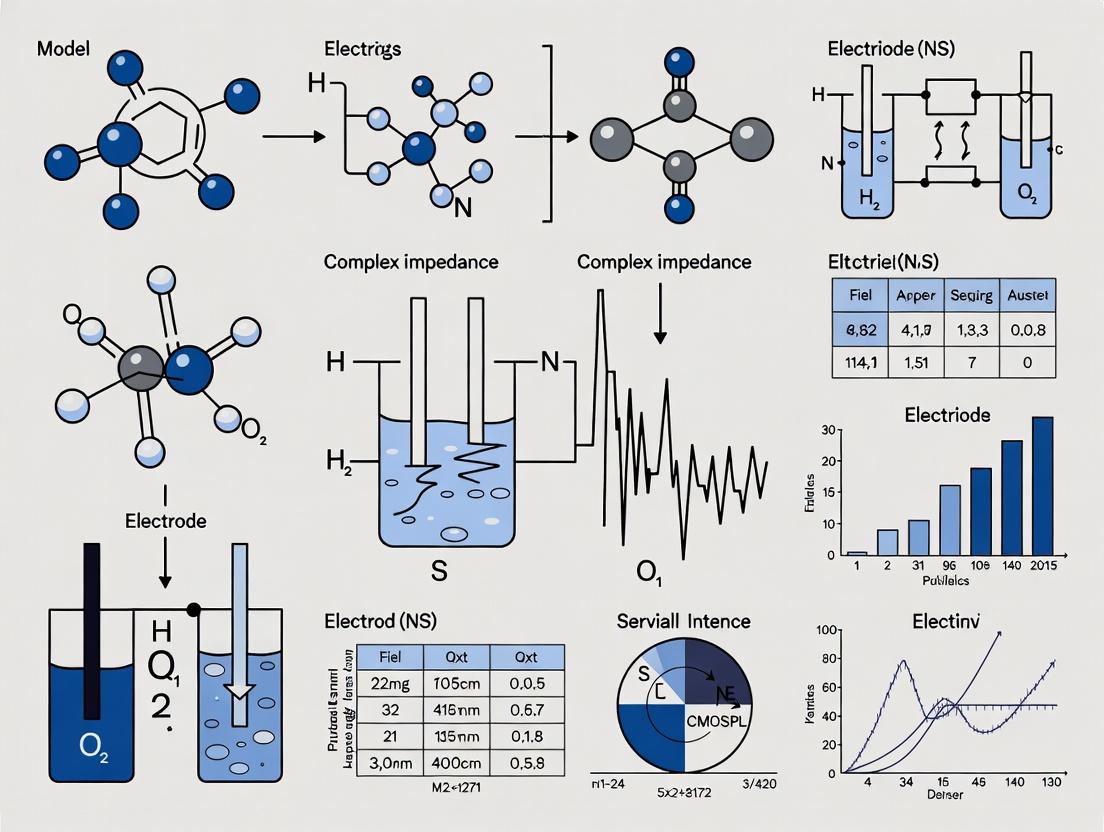
Within the framework of electrochemical impedance spectroscopy (EIS) data representation and complex impedance research, the concept of complex impedance serves as the fundamental mathematical and physical model for analyzing the frequency-dependent behavior of electrochemical systems. This guide provides an in-depth technical breakdown of complex impedance, its components, and its critical role in interpreting EIS data for applications in biosensing, material characterization, and drug development.
Core Definition and Mathematical Formalism
Complex impedance, denoted as Z(ω), is a frequency-domain representation of a system's opposition to alternating current (AC) flow. It extends simple resistance (R) to account for phase shifts between voltage and current caused by capacitive and inductive elements. The standard form is: Z(ω) = Z' + jZ" where:
- Z' is the real part, representing the resistive component.
- Z" is the imaginary part, representing the reactive component (capacitive or inductive).
- j is the imaginary unit (√-1).
- ω is the angular frequency (ω = 2πf).
This formulation allows impedance to be represented as a vector in the complex plane, characterized by its magnitude |Z| and phase angle φ, where Z' = |Z|cos(φ) and Z" = |Z|sin(φ).
Physical Interpretation of Components
The real and imaginary components correlate directly with physical processes within an electrochemical cell.
The Real Part (Z')
Z' represents energy dissipation. In an electrochemical system, this primarily corresponds to:
- Ohmic resistance of the electrolyte and electrodes.
- Charge transfer resistance (Rct) at the electrode-electrolyte interface, a critical parameter in kinetic studies.
- Dielectric losses within materials.
The Imaginary Part (Z")
Z" represents energy storage and release. Its sign indicates the dominant reactive element:
- Negative Z" (Capacitive Behavior): Arises from double-layer capacitance (Cdl), adsorption processes, or bulk material capacitance. The impedance of a pure capacitor is Z_C = 1/(jωC) = -j/(ωC).
- Positive Z" (Inductive Behavior): Less common in typical bio-electrochemistry, can arise from adsorbed intermediates, specific circuit configurations, or instrumentation artifacts.
Data Representation in EIS Research
EIS data is typically visualized in two primary plots, central to the thesis of data representation:
- Nyquist Plot: Plots -Z" vs. Z' across frequencies. Each point is a single frequency; the plot often reveals semicircles or features corresponding to different electrochemical processes.
- Bode Plot: Two separate plots of log |Z| vs. log(f) and φ vs. log(f), showing the frequency dependence of magnitude and phase.
These representations allow researchers to deconvolve overlapping processes based on their characteristic time constants (τ = RC).
Title: EIS Data Analysis Workflow for Parameter Extraction
Quantitative Data: Common Equivalent Circuit Elements
To translate complex impedance data into physical parameters, experimental data is fitted to an equivalent circuit model (ECM). The table below summarizes common circuit elements.
| Circuit Element | Symbol | Impedance Formula (Z) | Real Part (Z') | Imaginary Part (Z") | Physical Correlation |
|---|---|---|---|---|---|
| Resistor | R | R | R | 0 | Solution resistance (Rs), Charge transfer resistance (Rct) |
| Capacitor | C | 1/(jωC) | 0 | -1/(ωC) | Double-layer capacitance (Cdl), Coating capacitance |
| Constant Phase Element | Q | 1/(Y₀(jω)^α) | (cos(απ/2))/(Y₀ ω^α) | -(sin(απ/2))/(Y₀ ω^α) | Non-ideal capacitance, surface heterogeneity (0 ≤ α ≤ 1) |
| Warburg (Infinite Length) | W | (σ/√ω) * (1 - j) | σ/√ω | -σ/√ω | Semi-infinite linear diffusion (σ = Warburg coefficient) |
| Inductor | L | jωL | 0 | ωL | Parasitic inductance, adsorption process |
Experimental Protocol: Basic EIS Measurement for a Coated Electrode
This protocol outlines a standard EIS experiment to characterize a modified electrode surface, relevant to biosensor development.
1. Objective: To determine the charge transfer resistance (Rct) and double-layer capacitance (Cdl) of a bare and protein-coated gold electrode in a redox probe solution.
2. Materials & Equipment (Three-Electrode Setup):
- Potentiostat/Galvanostat with FRA capability.
- Working Electrode (WE): Gold disk electrode (e.g., 2 mm diameter).
- Counter Electrode (CE): Platinum wire.
- Reference Electrode (RE): Ag/AgCl (in saturated KCl).
- Electrolyte: 5 mM K₃[Fe(CN)₆]/K₄[Fe(CN)₆] (1:1) in 0.1 M PBS, pH 7.4.
- Coating Solution: Target protein or drug compound in PBS.
3. Procedure: 1. Electrode Preparation: Polish the WE with successive alumina slurries (1.0, 0.3, 0.05 µm). Rinse thoroughly with deionized water and ethanol. Sonicate for 5 minutes in water. 2. Electrochemical Cleaning: Place the electrodes in the redox electrolyte. Perform cyclic voltammetry (CV) from -0.2 V to +0.6 V vs. Ag/AgCl at 100 mV/s until stable peaks are obtained (~20 cycles). 3. Baseline EIS Measurement: Set the DC potential to the formal potential of the redox probe (~+0.22 V vs. Ag/AgCl). Apply a sinusoidal AC perturbation with amplitude of 10 mV rms. Measure impedance across a frequency range of 100 kHz to 0.1 Hz, collecting 10 points per decade. Record the Nyquist plot. 4. Surface Modification: Rinse the WE and dry under N₂. Incubate in the coating solution for 1 hour at room temperature. Rinse gently with PBS to remove unbound material. 5. Modified Electrode EIS Measurement: Return the coated WE to the same redox electrolyte. Repeat the EIS measurement under identical conditions (Step 3). 6. Data Fitting: Fit both Nyquist plots to the appropriate Randles equivalent circuit: [Rs(Cdl[RctW])] or a simplified [Rs(CdlRct)] if diffusion is negligible at high frequencies.
Title: Randles Circuit Mapping to Physical Electrode Interface
The Scientist's Toolkit: Key Research Reagent Solutions
| Item | Function in EIS Experiments |
|---|---|
| Redox Probe (e.g., [Fe(CN)₆]³⁻/⁴⁻) | Provides a reversible, well-characterized electron transfer reaction to probe interfacial changes (Rct). |
| Supporting Electrolyte (e.g., PBS, KCl) | Provides high ionic strength to minimize solution resistance (Rs) and ensure redox probe activity. |
| Blocking Agents (e.g., BSA, Casein) | Used to passivate non-specific binding sites on electrode surfaces, improving signal specificity in biosensing. |
| Self-Assembled Monolayer (SAM) Kits (e.g., alkanethiols) | Form well-defined, reproducible insulating or functional layers on gold electrodes for controlled interface studies. |
| Faradaic vs. Non-Faradaic Electrolytes | Choice between with-redox-probe (kinetics) and without-redox-probe (capacitive) measurements determines accessed information. |
| Electrode Polishing Kits (Alumina/Nanopaste) | Essential for reproducible, clean electrode surfaces prior to modification, ensuring consistent baseline impedance. |
| Equivalent Circuit Fitting Software (e.g., ZView, EC-Lab) | Enables quantitative modeling of complex impedance data to extract physical parameters via non-linear least squares fitting. |
Electrochemical Impedance Spectroscopy (EIS) is a cornerstone analytical technique for investigating the electrical properties of interfaces and materials. Within complex impedance research, particularly relevant to biosensing and drug development (e.g., characterizing cell-electrode interfaces or drug delivery systems), the choice of data representation is not merely aesthetic—it is analytical. The Nyquist and Bode plots are two fundamental, frequency-domain representations of the same complex impedance data (Z(ω) = Z' + jZ''), each offering unique insights into system dynamics. This whitepaper provides an in-depth comparison, rooted in the thesis that optimal interpretation of EIS data requires a deliberate, context-driven selection of visualization method.
Core Theoretical Foundation
Impedance is a complex, frequency-dependent quantity: Z(ω) = |Z|∠θ = Z' + jZ'', where Z' is the real part (resistance), Z'' is the imaginary part (reactance), |Z| is the magnitude, and θ is the phase shift. The two plots visualize this differently:
- Nyquist Plot: Plots -Z'' (imaginary) on the ordinate versus Z' (real) on the abscissa, with each point representing a specific frequency (ω). Frequency is an implicit parameter.
- Bode Plot: Uses two subplots: Magnitude (|Z| vs. frequency, log-log scale) and Phase (θ vs. frequency, semi-log scale), making frequency an explicit axis.
Comparative Analysis: Strengths, Weaknesses, and Applications
The following table summarizes the quantitative and qualitative differences that guide their use in complex impedance research.
Table 1: Comparative Analysis of Nyquist and Bode Plots for EIS Data Representation
| Feature | Nyquist Plot | Bode Plot | ||
|---|---|---|---|---|
| Axes & Variables | Real(Z) vs. -Imag(Z). Frequency is implicit. | Log | Z | vs. Log f; Phase θ vs. Log f. |
| Primary Strength | Intuitive visualization of time constants and relaxation processes. Ideal for identifying semicircular features corresponding to parallel RC circuits. | Clear display of frequency dependence across wide ranges. Direct readout of impedance magnitude and phase at any given frequency. | ||
| Primary Weakness | Frequency information is lost; can compress data, obscuring details at high and low extremes. | Less immediate for identifying the number of distinct relaxation processes in a system. | ||
| Optimal Use Case | Analyzing systems with well-separated time constants; model fitting to equivalent circuits. | Analyzing systems where magnitude response over frequency is critical (e.g., filter design, sensor operational range). | ||
| Typical EIS Application | Characterizing charge transfer resistance and double-layer capacitance in a standard Randles cell model. | Determining the frequency range for optimal sensitivity in a biosensor or the stability of a coating. |
Experimental Protocols for EIS Data Acquisition and Visualization
A standard protocol for generating comparable Nyquist and Bode plots from a biological sample (e.g., a monolayer of cultured cells on a working electrode) is detailed below.
Protocol 1: EIS Measurement of a Cell Monolayer for Impedance-Based Assays
Objective: To acquire complex impedance spectra of a biological barrier for subsequent visualization via Nyquist and Bode plots.
Materials: See "The Scientist's Toolkit" below. Procedure:
- Electrode Preparation & Cell Seeding: Sterilize gold or ITO interdigitated electrodes (IDEs). Coat with appropriate adhesion molecules (e.g., fibronectin, 5 µg/mL, 1 hr). Seed relevant cell type (e.g., Caco-2, MDCK) at confluence (e.g., 1 x 10^5 cells/cm²) in culture medium. Allow to form a tight monolayer (typically 5-7 days).
- Experimental Setup: Place the cell-coated IDE in a measurement chamber with culture medium (with/without treatment). Connect the IDE to a potentiostat capable of EIS. Use a standard three-electrode configuration if using a plate electrode, or two-electrode for most IDEs.
- EIS Parameter Configuration:
- DC Bias: 0 V (or open circuit potential).
- AC Amplitude: 10 mV (small signal to ensure linearity).
- Frequency Range: 0.1 Hz to 100,000 Hz.
- Data Points: 10 points per frequency decade (logarithmic spacing).
- Data Acquisition: Run the EIS sweep. Include a measurement of a bare electrode (no cells) and a blank (medium only) as controls.
- Data Processing: Export the data as a set of frequencies (f), real impedance (Z'), and imaginary impedance (Z'').
- Visualization:
- For Nyquist Plot: Plot -Z'' vs. Z'. Annotate key frequencies.
- For Bode Plot: Create two vertically stacked plots. Top: log10(|Z|) vs. log10(f). Bottom: Phase angle θ = arctan(Z''/Z') in degrees vs. log10(f).
Visualizing the Data Interpretation Workflow
The logical flow from raw EIS data to system insight through these plots is illustrated below.
Diagram 1: EIS Data Interpretation Workflow (91 chars)
The Scientist's Toolkit: Essential Reagents & Materials
Table 2: Key Research Reagent Solutions for Cell-Based EIS Experiments
| Item | Function in EIS Experiment |
|---|---|
| Interdigitated Electrodes (IDEs) | Gold or ITO microelectrodes that provide a large surface area for cell growth and sensitive impedance measurement. |
| Extracellular Matrix Proteins (e.g., Fibronectin, Collagen) | Coating applied to IDE surface to promote cell adhesion and formation of a confluent, polarized monolayer. |
| Cell Culture Medium (with serum/defined factors) | Provides nutrients for cell viability and maintenance of barrier function during measurement. |
| Phosphate Buffered Saline (PBS) | Used for rinsing electrodes and cells; provides a known, simple ionic background for control measurements. |
| Potentiostat/Galvanostat with FRA | The core instrument that applies a small AC potential and measures the resulting current to calculate impedance. |
| Paraformaldehyde (4%) | Used for fixing cell monolayers post-experiment for subsequent validation via microscopy. |
| Transepithelial Electrical Resistance (TEER) Meter | Complementary instrument to validate monolayer integrity independently of EIS measurements. |
Within the broader thesis on EIS (Electrochemical Impedance Spectroscopy) data representation and complex impedance research, equivalent circuit models serve as the critical bridge between raw spectral data and meaningful physicochemical interpretation. This guide details the evolution of these models from simple, discrete (lumped) elements to complex distributed representations, which are essential for accurate analysis in fields ranging from material science to biosensor development and drug discovery.
Foundational Models: Lumped Elements
The simplest equivalent circuits model electrochemical interfaces using discrete, ideal electrical components.
The Randles Circuit
The most fundamental model for a simple electrode-electrolyte interface.
- Components: Solution resistance (Rs) in series with a parallel combination of a double-layer capacitor (Cdl) and a charge-transfer resistor (Rct) often in series with a Warburg element (W) for diffusion.
- Application: Ideal, planar, homogeneous electrodes with a single, kinetically-controlled redox reaction.
Table 1: Common Lumped Equivalent Circuit Elements
| Circuit Element | Symbol | Impedance (Z) | Physical Meaning |
|---|---|---|---|
| Resistor | R | R | Solution resistance, charge transfer resistance. |
| Capacitor | C | 1/(jωC) | Double-layer capacitance, dielectric properties. |
| Constant Phase Element | Q | 1/(Y₀(jω)^n) | Imperfect capacitor (n=1→C, n=0.5→Warburg). |
| Warburg (Infinite) | W | σ/√ω * (1-j) | Semi-infinite linear diffusion. |
| Inductor | L | jωL | Adsorption processes, parasitic inductance. |
Evolution to Distributed Elements
Real-world systems (porous electrodes, rough surfaces, non-uniform coatings) exhibit frequency dispersion, where ideal elements fail. This necessitates distributed models.
The Role of the Constant Phase Element (CPE)
The CPE is a non-intuitive, distributed element with impedance ZCPE = 1/(Q(jω)n), where 0 ≤ n ≤ 1.
- n = 1: Ideal capacitor.
- n = 0.5: Warburg (diffusion) behavior.
- n ~ 0.8-0.9: Represents surface heterogeneity or roughness.
- Experimental Evidence: Studies on rough platinum and porous carbon electrodes show a depressed semicircle in the Nyquist plot, fittable only with a CPE replacing Cdl.
Transmission Line Models (TLM)
TLMs are the quintessential distributed circuit for porous electrodes, batteries, and coated metals, representing impedance distributed along a pore or channel.
Diagram 1: Transmission Line Model for a Porous Electrode
Experimental Protocol for TLM Validation (Porous Electrode):
- Electrode Fabrication: Create a well-defined porous electrode (e.g., by sintering metal particles, anodizing to create nanotubes, or using a commercial porous carbon).
- Cell Setup: Use a 3-electrode configuration with the porous electrode as working electrode, a large Pt mesh as counter electrode, and a stable reference electrode (e.g., Ag/AgCl).
- EIS Measurement: Immerse in a supporting electrolyte with a redox couple (e.g., 5 mM K3[Fe(CN)6]/K4[Fe(CN)6] in 1M KCl).
- Data Acquisition: Perform EIS from 100 kHz to 10 mHz with a 10 mV RMS perturbation at the open circuit potential.
- Model Fitting: Fit the acquired spectrum using software (e.g., ZView, EC-Lab) with a finite-length TLM circuit containing distributed Rion and distributed Zsurface (Rct in parallel with CPE).
- Validation: Compare fitted pore resistance/capacitance with values calculated from known pore geometry and electrolyte conductivity.
Hierarchical and Complex Biological Interfaces
Modeling biological systems (cell monolayers, tissue-engineered constructs) requires hierarchical circuits combining lumped and distributed concepts.
Diagram 2: Hierarchical Model for a Cell-Monolayer Biosensor
Table 2: Quantitative Parameters from Fitting Cell Monolayer EIS
| Parameter | Typical Range (MDCK or Caco-2 cells) | Represents | Method for Extraction |
|---|---|---|---|
| Rpara | 20 - 100 Ω·cm² | Tight junction integrity. | Fit low-frequency arc in Nyquist plot. |
| Cm | 1.0 - 2.0 μF/cm² | Apical/basolateral membrane capacitance. | Fit high-frequency semicircle. |
| Rm | 2,000 - 10,000 Ω·cm² | Ion channel activity in lipid bilayer. | Fit high-frequency semicircle. |
| α (CPE exponent) | 0.85 - 0.95 | Membrane roughness/heterogeneity. | Fit CPE replacing Cm. |
The Scientist's Toolkit: Research Reagent Solutions
Table 3: Essential Materials for EIS Model Validation Experiments
| Item | Function & Rationale | Example Product/ Specification |
|---|---|---|
| Potentiostat/Galvanostat with FRA | Applies potential/current perturbation and measures impedance response across frequency. Core instrument. | Biologic SP-300, Metrohm Autolab PGSTAT204 with FRA32M. |
| Faraday Cage | Shields electrochemical cell from external electromagnetic noise, critical for low-current/high-impedance measurements. | Custom-built or commercial shielded enclosure. |
| Low-Polarization Electrodes | Provide stable, non-polarizable interfaces for reference and counter electrodes. | Ag/AgCl (3M KCl) reference electrode; Platinum mesh counter electrode. |
| Standard Redox Couple | Well-defined, reversible kinetics for validating instrument and baseline circuit fitting. | 5 mM Potassium Ferri-/Ferrocyanide in 1M KCl. |
| CPE-Parameterized Software | Nonlinear least squares fitting software capable of handling distributed elements (CPE, TLM). | ZView (Scribner), EC-Lab (BioLogic), Equivalent Circuit (Boukamp). |
| Calibration Load Kit | Validates potentiostat impedance accuracy over full frequency range. | 100 Ω precision resistor, 1 μF low-loss capacitor. |
| Controlled-Geometry Cells | Enables modeling with known area and diffusion length (e.g., rotating disk electrode). | Glassy Carbon RDE (Pine Research), sealed measurement cell (e.g., from Tait Scientific). |
Physical Meaning of Real (Z') and Imaginary (Z") Components in Biological Systems
Within the broader thesis on Electrochemical Impedance Spectroscopy (EIS) data representation, this technical guide elucidates the biophysical interpretation of the real (Z', resistance) and imaginary (Z", reactance) components of complex impedance in biological systems. These components provide non-invasive, real-time insights into cellular and tissue-level phenomena, critical for biosensing, drug discovery, and pathophysiology research.
Core Biophysical Interpretation
In biological EIS, a small alternating current (AC) potential is applied across a cellular or tissue sample. The resulting current is measured, and the impedance (Z) is calculated as a complex number: Z(ω) = Z'(ω) + jZ"(ω), where ω is the angular frequency.
Z' (Real Component): Represents the resistive element. It quantifies energy dissipated as heat. In biological contexts, this primarily corresponds to the ionic conductivity of the extracellular fluid, cytoplasm, and nucleoplasm. Changes in Z' often reflect alterations in barrier integrity (e.g., tight junctions), cell membrane integrity, or overall ion concentration.
Z" (Imaginary Component): Represents the reactive/capacitive element. It quantifies energy stored and released per cycle. This is dominantly governed by the insulating lipid bilayer of cell membranes (acting as a dielectric) and the electrical double layers at electrode-electrolyte interfaces. Changes in Z" are sensitive to membrane morphology, cell-substrate adhesion, and intracellular organelle activity.
The frequency-dependent relationship between these components reveals system architecture.
Table 1: Interpretation of Z' and Z" Changes in Common Biological Events
| Biological Event / Condition | Low-Frequency Z' Trend | Low-Frequency Z" Trend | Primary Biophysical Cause |
|---|---|---|---|
| Cell Layer Formation | Increases | Increases (peak may shift) | Establishment of insulating barriers (tight junctions & cell membranes) restricting ion flow. |
| Membrane Permeabilization | Decreases | Decreases | Breakdown of capacitive lipid bilayer, allowing direct ionic current shunt. |
| Cell Detachment/Apoptosis | Decreases | Decreases | Loss of insulating cell body from electrode surface, reducing capacitive contribution. |
| Enhanced Cell Adhesion/Spreading | Minor Increase | Increases | Increased capacitive area as cell membrane closely apposes substrate. |
| Inflammation (Tissue Edema) | Decreases | Variable | Increased extracellular fluid volume and ion content lowering resistance. |
Table 2: Typical Impedance Magnitude Ranges for Common Experimental Models
| Experimental Model | Approx. | Z | Range (Ω) | Dominant Frequency Regime | Common Electrode Area |
|---|---|---|---|---|---|
| Confluent Epithelial Cell Layer (e.g., MDCK) | 1 kΩ - 10 kΩ | 100 Hz - 10 kHz | 0.1 - 0.3 cm² | ||
| Cardiomyocyte Monolayer (BEaC) | 500 Ω - 3 kΩ | 1 Hz - 1 kHz | 10⁻⁴ cm² (microelectrode) | ||
| 3D Tumor Spheroid | 100 Ω - 2 kΩ | 10 Hz - 100 kHz | Planar interdigitated electrodes | ||
| Skin Tissue (ex vivo) | 10 kΩ - 1 MΩ | 1 Hz - 1 MHz | 1 cm² |
Key Experimental Protocols
Protocol for Real-Time Cell Analysis (RTCA) using EIS
Objective: To monitor cell proliferation, adhesion, and barrier function in real time.
- Electrode Preparation: Use a commercial well plate (e.g., 16-well E-Plate) with integrated gold microelectrotes. Sterilize with UV light for 30 minutes.
- Baseline Measurement: Add 100 µL of culture medium (without cells) to each well. Perform a frequency sweep (e.g., 10 Hz to 100 kHz, 10 points per decade) at 37°C to establish a background impedance spectrum. This measures Z'₀ and Z"₀.
- Cell Seeding: Prepare a single-cell suspension. Add 100 µL of cell suspension to desired wells, creating a total volume of 200 µL. Gently agitate the plate to ensure even distribution.
- Impedance Monitoring: Place the plate in the incubator-integrated station. The system automatically performs a simplified sweep (often at a single, critical frequency) at set intervals (e.g., every 15 minutes).
- Data Normalization: The primary output is often the Cell Index (CI) = max (i=1,...,N) [Zᵢ(cellf) - Zᵢ(backgroundf)] / [Zᵢ(background_f)], where N is the number of frequency points used. This normalized parameter tracks changes dominated by Z' at the chosen frequency.
- Intervention: After establishing a confluent monolayer (CI plateau), introduce a drug compound or toxicant. Monitor changes in CI, which reflect alterations in Z' and Z" due to cytotoxicity, barrier disruption, or morphological change.
Protocol for Transepithelial/Transendothelial Electrical Resistance (TEER)
Objective: Specifically quantify the integrity of tight junction barriers.
- Setup: Grow cells on a permeable filter support (e.g., Transwell insert). Place insert into a matching well.
- Electrode Placement: Use a chopstick-style or integrated electrode pair. Position one electrode in the apical (top) compartment and the other in the basolateral (bottom) compartment.
- Measurement: Apply a small AC signal (typically a fixed frequency between 10-100 Hz). Measure the complex impedance.
- Calculation: The TEER is derived primarily from the Z' component at the measured frequency: TEER (Ω·cm²) = (Z'_sample - Z'_blank) × Effective Membrane Area. The blank is a cell-free insert. This value directly reflects the paracellular ionic resistance.
Visualizing EIS Data Interpretation in Biology
Title: EIS Data Interpretation Workflow
Title: Equivalent Circuit Model for a Cell Layer
The Scientist's Toolkit: Key Research Reagent Solutions
Table 3: Essential Materials for Biological EIS Experiments
| Item / Reagent | Function / Role in EIS Experiment |
|---|---|
| Microelectrode-Integrated Multiwell Plates (e.g., ACEA E-Plates, Applied BioPhysics ECIS Arrays) | Provide standardized, culture-compatible substrates with defined electrode geometries for reproducible impedance monitoring. |
| Transepithelial Permeable Supports (e.g., Corning Transwell, Millicell inserts) | Enable the growth of polarized cell layers and the specific measurement of transepithelial/transendothelial electrical resistance (TEER). |
| Low-Conductivity Culture Media (e.g., specialized EIS media, or PBS with low [Na+]) | Increase signal-to-noise ratio by reducing the background ionic current (lowering R_sol), making changes at the cell-electrode interface more pronounced. |
| Impedance-Tracing Cytotoxic Agents (e.g., Digitonin, Triton X-100) | Positive controls for cytotoxicity; rapidly permeabilize cell membranes, causing a characteristic steep drop in both Z' and Z". |
| Barrier-Enhancing Agents (e.g., Dexamethasone for endothelial cells) | Positive controls for barrier formation; increase tight junction complexity, leading to a sustained rise in Z' (TEER). |
| ECIS (Electric Cell-Substrate Impedance Sensing) Controller & Software | Dedicated hardware for applying AC signals and measuring precise impedance across a frequency spectrum, paired with software for data analysis and modeling. |
| Extracellular Matrix Coatings (e.g., Collagen I, Fibronectin, Matrigel) | Applied to electrode surfaces to promote specific cell adhesion and spreading, influencing the baseline capacitive (Z") signal. |
Electrochemical Impedance Spectroscopy (EIS) is a cornerstone analytical technique for probing interfacial processes in electrochemical systems. Within the broader thesis of representing complex impedance data, the accurate extraction and interpretation of three fundamental parameters—Solution Resistance (Rs), Charge Transfer Resistance (Rct), and Double Layer Capacitance (Cdl)—are critical. These parameters form the core of the ubiquitous Randles equivalent circuit model, enabling researchers to deconvolute contributions from bulk electrolyte, electrode kinetics, and the interfacial region. In fields like biosensing and drug development, monitoring changes in these parameters allows for the label-free detection of biomolecular interactions, such as antibody-antigen binding or cellular responses to pharmaceutical compounds.
Theoretical Foundation and Parameter Definitions
The Randles Circuit Model
The Randles circuit is the most common model for a simple electrode-electrolyte interface. It comprises:
- Rs (Solution Resistance): Represents the ohmic resistance of the ionic solution between the working and reference electrodes. It is frequency-independent and appears as the high-frequency intercept on the real Z' axis in a Nyquist plot.
- Cdl (Double Layer Capacitance): Models the dielectric behavior of the charged interface between the electrode and the electrolyte, where ions and solvent molecules orient in response to the electrode potential. It is non-ideal, often represented as a Constant Phase Element (CPE).
- Rct (Charge Transfer Resistance): Represents the kinetic barrier to faradaic charge transfer across the electrode interface. It is inversely proportional to the rate of the electrochemical reaction (k). Changes in Rct are directly correlated with surface-modifying events in biosensing.
Relationship to Complex Impedance
In the Nyquist representation, a simple Randles circuit produces a semicircle. Rs is the left intercept, the diameter of the semicircle is Rct, and the characteristic frequency at the top of the semicircle (ωmax = 1/(RctCdl)) defines the time constant of the interface. Accurate fitting of the EIS spectrum to this model is essential for reliable parameter extraction.
Quantitative Parameter Ranges in Research Contexts
The following table summarizes typical values for these key parameters across common experimental systems in biomedical research.
Table 1: Typical Ranges for EIS Parameters in Bioelectrochemical Systems
| System / Condition | Rs (Ω) | Rct (kΩ) | Cdl (nF) | Notes |
|---|---|---|---|---|
| Baseline (Clean Gold Electrode in PBS) | 50 - 200 | 1 - 10 | 10 - 100 | Depends on electrode area (A), [PBS] |
| After SAM Formation (e.g., MCH on Au) | 50 - 200 | 50 - 500 | 5 - 20 | Rct increases, Cdl decreases due to insulating layer |
| After Protein Capture (e.g., Antibody) | 50 - 200 | 100 - 1000 | 1 - 10 | Further increase in Rct due to biomolecular insulation |
| Cell Layer on ECIS Electrode | 100 - 500 | 2 - 20 | 10 - 50 | Rct correlates with barrier integrity; Cdl with cell coverage |
| Low Ionic Strength Buffer (e.g., 1 mM PBS) | 1000 - 5000 | Higher baseline | Lower baseline | Higher Rs impacts signal-to-noise and potential control |
Experimental Protocols for Parameter Measurement
Standard EIS Protocol for Faradaic Biosensing
This protocol outlines the steps for measuring Rct changes upon biomolecular binding using a redox probe like [Fe(CN)6]3−/4−.
Materials: Potentiostat/Galvanostat with EIS module, 3-electrode cell (Working: functionalized gold; Counter: Pt wire; Reference: Ag/AgCl), Phosphate Buffered Saline (PBS, 10 mM, pH 7.4), potassium ferri/ferrocyanide (5 mM each).
Procedure:
- Electrode Pretreatment: Clean the gold working electrode via mechanical polishing (0.05 µm alumina) or electrochemical cycling in 0.5 M H2SO4.
- Baseline EIS Measurement:
- Immerse the electrode cell in a solution of 5 mM [Fe(CN)6]3−/4− in 10 mM PBS.
- Apply the open circuit potential (OCP) or a DC potential near the probe's formal potential (typically ~0.22 V vs. Ag/AgCl).
- Superimpose an AC sinusoidal perturbation with an amplitude of 5-10 mV rms.
- Record impedance spectra over a frequency range of 100 kHz to 0.1 Hz.
- Surface Functionalization: Immerse the electrode in a thiolated probe molecule (e.g., aptamer or capture antibody) solution (1-10 µM in PBS) for 1-2 hours. Rinse thoroughly with PBS.
- Backfilling: Treat with a backfiller (e.g., 1 mM 6-mercapto-1-hexanol) for 30 minutes to passivate unbound gold sites. Rinse.
- Post-Functionalization EIS: Repeat step 2 in the same redox probe solution.
- Target Incubation: Incubate the functionalized electrode with the target analyte (e.g., protein, drug candidate) for a specified time (30-60 min). Rinse gently.
- Post-Binding EIS: Repeat step 2 again.
- Data Analysis: Fit all Nyquist plots to the Randles equivalent circuit using the potentiostat's software. Monitor the increase in Rct and decrease in Cdl as the insulating layer forms.
Non-Faradaic EIS for Cell Monitoring (ECIS)
This protocol measures changes in Rs and Cdl (often at a fixed high frequency) to monitor cell behavior without a redox probe.
Materials: ECIS instrument array, 8-well culture slide with integrated gold film electrodes, cell culture medium, trypsin-EDTA.
Procedure:
- Electrode Conditioning: Add 400 µL of cell culture medium to each well. Perform a background scan (e.g., 10 kHz to 1 Hz) to establish baseline Rs and Cdl.
- Cell Seeding: Trypsinize and count cells. Seed at a desired density (e.g., 50,000 cells/well) onto the electrode array.
- Continuous Monitoring: Place the array in the ECIS station (37°C, 5% CO2). The instrument automatically applies a small AC signal (often 1 V, 1-64 kHz) at regular intervals (e.g., every minute).
- Data Interpretation: As cells attach and spread, they act as insulating particles, increasing the measured impedance at high frequencies (related to Rs and constricted current flow). Morphological changes and barrier function are tracked via modeled Rct (barrier resistance) and Cdl (membrane capacitance).
Visualization of Core Concepts
Title: EIS Data Analysis Pathway from Measurement to Insight
Title: Standard EIS Biosensing Experimental Workflow
The Scientist's Toolkit: Essential Research Reagents & Materials
Table 2: Key Reagent Solutions for EIS-based Bioassays
| Item | Function in Experiment | Typical Specification / Notes |
|---|---|---|
| Redox Probe | Provides a faradaic current for sensitive Rct measurement. | Potassium ferri/ferrocyanide, 5 mM equimolar in PBS. Must be prepared fresh to avoid photodecomposition. |
| Self-Assembled Monolayer (SAM) Thiols | Forms an ordered, insulating layer on gold electrodes for probe immobilization. | Thiolated DNA/RNA aptamers (1-10 µM) or capture antibodies (via thiol-PEG linkers). |
| Backfiller | Passivates exposed gold to reduce non-specific binding and orient probe molecules. | 6-Mercapto-1-hexanol (MCH), 1 mM in PBS or ethanol. |
| Blocking Agent | Further minimizes non-specific adsorption of proteins or cells. | Bovine Serum Albumin (BSA, 1% w/v) or casein in PBS. |
| High-Purity Buffer | Maintains pH and ionic strength; defines Rs. | Phosphate Buffered Saline (PBS, 10 mM, pH 7.4), 0.22 µm filtered. |
| Electrode Cleaning Solution | Removes organic contaminants and oxide layers to ensure reproducible surfaces. | Piranha solution (H2SO4:H2O2 3:1) OR 0.5 M H2SO4 for electrochemical cycling. (CAUTION: Piranha is extremely hazardous). |
| Cell Culture Medium | Environment for non-faradaic cell monitoring assays (ECIS). | Serum-containing medium (e.g., DMEM + 10% FBS) appropriate for the cell line. |
In electrochemical impedance spectroscopy (EIS) research for biomedical systems, the core assumption of ideal frequency-independent behavior is routinely violated. This phenomenon, termed frequency dispersion, manifests as a deviation from the ideal semicircle in Nyquist plots and a broadening of the phase angle peak in Bode plots. Within the broader thesis on EIS data representation, this whitepaper examines the physical origins, quantification methods, and critical implications of frequency dispersion for interpreting complex impedance in real biological interfaces, such as electrode-tissue interfaces, cell monolayers, and biosensor surfaces.
Physical Origins of Dispersion in Biomedical Interfaces
Frequency dispersion arises from intrinsic heterogeneity and non-ideal capacitive behavior at biomedical electrodes.
Key Mechanisms:
- Geometric/Crystallographic Heterogeneity: Non-uniform current distribution due to rough or fractal electrode surfaces.
- Time-Constant Distributions: The presence of multiple, similar electrochemical processes with distributed relaxation times, common in porous tissue or cell scaffolds.
- Dynamic System Changes: Time-varying interfacial properties due to adsorption, biofilm formation, or cellular activity during measurement.
- Non-Ideal Capacitance (Constant Phase Element - CPE): The capacitance of biological double layers often behaves as a CPE with impedance ( Z_{CPE} = 1/[Q(j\omega)^n] ), where ( n ) is the dispersion index (( 0 \leq n \leq 1 )).
Quantifying Dispersion: Models and Data Representation
Equivalent Circuit Models Incorporating Dispersion
Ideal Randles circuits are modified to replace ideal capacitors with Constant Phase Elements (CPEs).
Table 1: Comparison of Ideal vs. Dispersion-Inclusive Equivalent Circuit Models
| Circuit Model | Ideal Element (Nyquist Plot) | Dispersion Element | Impedance Formula | Typical n Value in Bio-Systems |
|---|---|---|---|---|
| Pure Capacitance | Perfect vertical line | Constant Phase Element (CPE) | ( Z = 1/(Q(j\omega)^n) ) | 0.9 - 1.0 (near ideal) |
| Single Time Constant | Perfect semicircle | CPE in parallel with R | ( Z = R/(1+ RQ(j\omega)^n) ) | 0.7 - 0.9 (moderate dispersion) |
| Porous/ Rough Electrode | Ambiguous | Dual CPE with distributed elements | Multiple formulations | 0.5 - 0.8 (strong dispersion) |
Quantitative Metrics for Dispersion
- CPE Exponent n: Primary indicator. n=1 indicates ideal capacitor; lower values indicate increasing dispersion.
- Depressed Semicircle Center: The degree of depression below the real axis in a Nyquist plot, related to n.
- Full Width at Half Maximum (FWHM) of phase angle peak in Bode plots.
Table 2: Measured Dispersion Parameters in Representative Biomedical Studies
| Biological System | Electrode Type | Reported CPE n value | FWHM of Phase Peak (decades) | Probable Dispersion Source |
|---|---|---|---|---|
| Cardiac Tissue | Pt-Ir Pacemaker | 0.76 ± 0.04 | ~1.8 | Fibrous tissue heterogeneity |
| Neuronal Culture | Microelectrode Array (MEA) | 0.82 ± 0.07 | ~1.5 | Distributed cell-electrode gaps |
| Blood Coagulation | Interdigitated Au Sensor | 0.65 ± 0.10 | >2.0 | Dynamic protein adsorption |
| Skin Impedance | Ag/AgCl ECG electrode | 0.71 ± 0.05 | ~1.9 | Stratified corneal layers |
Experimental Protocols for Characterizing Dispersion
Protocol: EIS Measurement for Dispersion Analysis on Cell Monolayers
Objective: To quantify frequency dispersion at the electrode-cell monolayer interface. Materials: (See Scientist's Toolkit). Procedure:
- Baseline Measurement: In cell culture medium, acquire EIS spectrum (e.g., 100 kHz to 0.1 Hz, 10 mV RMS) at open circuit potential.
- Cell Seeding: Seed electrode area with adherent cells (e.g., HEK293, endothelial cells) at standard density.
- Monitoring: Perform daily EIS measurements at consistent temperature and CO₂ levels.
- Model Fitting: Fit spectra to a modified Randles circuit with a CPE replacing the double-layer capacitor. Use complex nonlinear least squares (CNLS) algorithm.
- Data Analysis: Track temporal evolution of CPE parameters Q and n, and charge transfer resistance (R_ct).
Protocol: Assessing the Impact of Surface Roughness on Dispersion
Objective: To correlate controlled electrode topography with CPE exponent n. Procedure:
- Surface Fabrication: Create a series of gold electrodes with defined roughness (via electrochemical deposition or plasma etching). Quantify roughness via AFM (Ra, Rq).
- Electrochemical Characterization: In a simple redox couple (e.g., [Fe(CN)₆]³⁻/⁴⁻), perform EIS at formal potential.
- Modeling: Fit data to a circuit with a CPE. Use Brug's formula: ( C{dl} = (Q * R{ct}^{1-n})^{1/n} ) to estimate true capacitance.
- Correlation: Plot CPE exponent n versus surface roughness metrics (Ra) and fractal dimension.
The Scientist's Toolkit: Research Reagent Solutions
Table 3: Essential Materials for EIS Dispersion Research in Biomedicine
| Item | Function & Relevance to Dispersion Studies |
|---|---|
| CPE-based Equivalent Circuit Software | (e.g., ZView, EC-Lab, MEISP). Essential for accurate fitting of non-ideal, dispersive data. |
| Low-High Impedance Potentiostat | Required for the broad frequency range needed to characterize distributed processes. |
| Microelectrode Arrays (MEAs) | Planar, multi-electrode setups for spatial mapping of dispersion across biological samples. |
| Defined Redox Probes | (e.g., Potassium Ferri/Ferrocyanide). Used to deconvolute dispersion sources (surface vs. biological). |
| ECM-Coated Electrodes | (e.g., Fibronectin, Matrigel). Standardizes cell adhesion, reducing dispersion from variable attachment. |
| Reference Electrodes with Stable Junction | (e.g., Ag/AgCl with ceramic frit). Minimizes instrumental dispersion from unstable reference potential. |
Implications for Drug Development and Biosensing
Misinterpreting dispersion as a Faradaic process can lead to false conclusions. A decreasing CPE n value over time may indicate progressive cell coverage or biofilm formation, not necessarily an increase in charge transfer resistance. Accurate CPE analysis is critical for label-free cell-based assays and corrosion stability of implantable biosensors.
Frequency dispersion is not an artifact but a rich source of information about the structural and dynamic complexity of biomedical interfaces. Moving beyond ideal models to embrace and quantify dispersion is essential for advancing the accuracy of EIS data representation in complex impedance research, leading to more reliable diagnostic biosensors and a deeper understanding of electrode-tissue integration.
Visualizations
Diagram Title: Origins and Modeling of Frequency Dispersion
Diagram Title: Workflow for Measuring Dispersion in Bio-EIS
Practical EIS Protocols: From Electrode Design to Data Acquisition in Biomedical Research
This guide provides a technical framework for selecting and implementing two-electrode (2E) and three-electrode (3E) configurations in electrochemical impedance spectroscopy (EIS) studies of biological samples. The context is a broader thesis on representing complex impedance data, focusing on extracting meaningful electrical models of biological interfaces, such as cells, tissues, and biofilms, for applications in biosensing and drug development.
Core Principles & Comparative Analysis
The fundamental difference lies in the control and measurement of potential. A 3E system (Working, Counter, Reference) independently controls the working electrode potential versus a stable reference, accurately measuring the impedance of the working electrode interface alone. A 2E system combines the counter and reference functions, measuring the total impedance of the entire cell, including the counter electrode interface.
Table 1: Comparative Analysis of 2-Electrode vs. 3-Electrode Configurations
| Parameter | 2-Electrode Configuration | 3-Electrode Configuration |
|---|---|---|
| Primary Use Case | High-impedance samples (e.g., tissues, skin, low-conductivity media), in vivo measurements, sealed systems. | Low-impedance samples (e.g., cell monolayers, electrolytes), precise interface studies, corrosion, battery research. |
| Measured Impedance | Total impedance between WE and CE/RE: Ztotal = ZWE + ZCE + Zsolution. | Impedance primarily of the Working Electrode (WE) interface alone (ZWE), assuming ideal reference. |
| Potential Control | Poor. Applied potential is split between WE and CE interfaces. Potential at WE is undefined vs. a true reference. | Excellent. Potentiostat maintains precise potential at WE versus the stable Reference Electrode (RE). |
| Current Path | Same path for current supply (CE) and potential measurement. | Current supplied via CE; potential sensed by RE (high-impedance) without current flow. |
| Key Advantage | Simpler setup, minimal sample volume, suitable for very high impedance where CE impedance is negligible. | Accurate, site-specific characterization of the WE interface, eliminates CE contribution. |
| Key Limitation | Counter electrode impedance contributes to measurement, making data ambiguous for low-impedance samples. | Requires stable RE placement and higher sample conductivity. More complex setup. |
| Optimal Sample Impedance | > 1 MΩ (where ZCE << Zsample). | < 100 kΩ (where solution resistance is low for stable RE potential). |
| Typical EIS Frequency Range | Often limited at high frequency by cable/interface capacitance. | Broader, more reliable frequency range. |
Experimental Protocols for Biological EIS
Protocol 3.1: Standardized 3-Electrode Setup for Adherent Cell Monolayer Assessment
Objective: To obtain precise, reproducible impedance data from a cultured endothelial or epithelial cell monolayer (e.g., for barrier function or drug response studies).
Electrode Preparation:
- Working Electrode (WE): Use a commercially available cell culture-insert system with integrated gold, platinum, or indium tin oxide (ITO) electrodes. Clean via UV-ozone treatment for 15 minutes.
- Counter Electrode (CE): A large platinum wire or mesh, placed symmetrically in the bulk electrolyte.
- Reference Electrode (RE): Ag/AgCl (in 3M KCl) with a porous frit, placed close to the WE (∼2 mm) using a Luggin capillary arrangement to minimize solution resistance (Rs).
Cell Seeding & Culture: Seed cells at confluent density directly onto the WE. Culture until a stable, tight monolayer forms (typically 3-7 days, monitoring with transepithelial electrical resistance, TEER).
Electrolyte & Setup: Use standard cell culture medium (e.g., PBS with Ca2+/Mg2+) or a low-redox HEPES-buffered saline. Ensure no air bubbles are trapped. Place the RE in a fixed position.
EIS Measurement Parameters:
- Instrument: Potentiostat with FRA module.
- DC Potential: Open circuit potential (OCP) of the cell-covered WE vs. RE. Monitor for stability (±2 mV over 60s).
- AC Amplitude: 10 mV RMS (to avoid perturbing cells).
- Frequency Range: 100 kHz to 0.1 Hz.
- Points/Decade: 10.
- Equilibration: 300 seconds at OCP before scan.
Data Validation: Record impedance of a bare electrode (no cells) and a fully confluent monolayer. Fit to a validated equivalent circuit (e.g., modified Randles circuit with constant phase element, CPE).
Protocol 3.2: Optimized 2-Electrode Setup for Ex Vivo Tissue Impedance
Objective: To measure the bulk impedance of a high-resistance tissue sample, such as skin or cartilage.
- Electrode Configuration: Use two identical, non-polarizable electrodes (e.g., Ag/AgCl pellet electrodes or large surface area platinum black) to minimize electrode polarization impedance.
- Sample Preparation: Mount tissue in a sealed measuring chamber (e.g., Ussing chamber) to prevent dehydration. Ensure full contact between tissue surface and electrodes, using conductive gel if appropriate.
- System Calibration: Measure the impedance of the chamber filled only with conductive saline (e.g., PBS) to characterize the baseline system impedance (Zsystem).
- EIS Measurement Parameters:
- Instrument: Impedance Analyzer or Potentiostat in 2E mode.
- DC Bias: 0 V.
- AC Amplitude: 50 mV RMS (may be increased for very high-impedance tissues).
- Frequency Range: 1 MHz to 1 Hz.
- Averaging: 3 measurements per frequency to improve SNR.
- Data Processing: The tissue impedance (Ztissue) is derived from the measured total impedance (Ztotal) by subtracting the pre-characterized Zsystem (primarily from wiring and electrolyte), though this is an approximation: Ztissue ≈ Ztotal - Zsystem.
Data Interpretation & Equivalent Circuit Modeling
For biological samples, data is fitted to physicochemical models using equivalent electrical circuits.
Table 2: Common Equivalent Circuit Models for Biological Samples
| Sample Type | Recommended Circuit Model | Circuit Elements & Biological Correlate |
|---|---|---|
| Cell Monolayer (3E) | R_s([R_par CPE_par](CPE_dl)) |
Rs: Solution resistance. Rpar: Paracellular resistance (barrier integrity). CPEpar: Capacitance of cell monolayer. CPEdl: Double-layer capacitance at electrode. |
| Bulk Tissue (2E) | R_s([R1 CPE1]([R2 CPE2])) |
Rs: Electrode/contact resistance. R1//CPE1: Resistance and constant phase element of extracellular matrix. R2//CPE2: Resistance and CPE of cell membranes/intracellular space. |
| Biofilm (3E/2E) | R_s([R_bf CPE_bf](CPE_dl)) |
Rs: Solution resistance. Rbf: Resistance to ion flow through biofilm matrix. CPEbf: Biofilm capacitance. |
The Scientist's Toolkit: Essential Research Reagents & Materials
Table 3: Key Reagent Solutions and Materials for Biological EIS
| Item | Function & Rationale |
|---|---|
| Ibidi Cell Culture-Inserts with Electrodes | Provides ready-to-use, biocompatible gold film working electrodes in standard well-plate format for barrier function assays. |
| Ag/AgCl Reference Electrode (3M KCl) | Provides a stable, non-polarizable potential reference in 3E setups. The porous frit minimizes junction potential shifts. |
| Platinum Black Electrodes | Used as counter or 2E electrodes; high surface area minimizes polarization impedance, critical for 2E measurements. |
| ECIS (Electric Cell-Substrate Impedance Sensing) Arrays | Specialized 8-well slides with microfabricated gold electrodes for real-time, label-free cell monitoring. |
| BioLogic Science Instruments Potentiostat/FRA | High-performance instrument with low-current capabilities and optimized software for modeling biological impedance. |
| Electrolyte: HEPES-Buffered Saline (Low Redox) | Provides stable pH and ionic strength without the redox activity of cell culture media (e.g., phenol red, amino acids). |
| Conductive Gel (e.g., SignaGel) | Used for 2E measurements on tissue; ensures stable, low-resistance contact between solid electrode and irregular tissue surface. |
| Equivalent Circuit Fitting Software (e.g., ZView, EC-Lab) | Used to fit complex EIS data to physioelectrical models, extracting quantitative parameters (R, C, CPE). |
Decision Framework & Visual Guides
Title: Decision Flow for Electrode Configuration Selection
Title: 3-Electrode Setup Workflow for Cell Monolayers
Title: Experimental Setup Role in EIS Data Representation Thesis
Within the broader thesis on Electrochemical Impedance Spectroscopy (EIS) data representation for complex impedance research, the selection of an appropriate frequency range stands as a critical, non-trivial experimental design parameter. This choice directly dictates the temporal and spatial resolution of the measured electrochemical system, impacting the ability to resolve time constants and differentiate interfacial processes. For researchers in drug development, particularly in biosensor characterization, membrane transport studies, and nanoparticle-biomolecule interaction analysis, optimizing this balance is essential for generating reliable, interpretable data within practical experimental timelines.
Theoretical Foundations: The Frequency-Information Relationship
The impedance of an electrochemical system is a function of frequency (f). Different physical and chemical processes dominate within specific frequency bands:
- High Frequencies (>10 kHz): Probe solution resistance, lead inductance, and double-layer charging. Fast processes.
- Mid Frequencies (1 Hz - 10 kHz): Often reveal charge transfer kinetics at electrode interfaces and bulk dielectric properties.
- Low Frequencies (<1 Hz): Characterize slow diffusion-limited processes, adsorption, and corrosion.
The total measurement time (Tmeas) for a logarithmically spaced frequency sweep with N points is approximately Tmeas ≈ N * (5/fmin), where fmin is the lowest frequency, and 5 cycles is a typical averaging constant. This creates the fundamental trade-off: extending to lower frequencies increases the window of observable time constants but exponentially increases measurement duration.
Quantitative Framework for Range Selection
Table 1: Typical Frequency Ranges and Resolved Processes in Bio-Electrochemistry
| Frequency Range | Approx. Measurement Time (N=10 pts/dec) | Dominant Processes Resolved | Typical Application in Drug Development |
|---|---|---|---|
| 1 MHz - 1 Hz | ~5.5 hours | Solution resistance, double-layer capacitance, charge-transfer kinetics, Warburg diffusion. | Fundamental characterization of novel electrode materials for biosensing. |
| 100 kHz - 100 mHz | ~55 hours | Above, plus very slow diffusion, adsorption/desorption, coating degradation. | Long-term stability studies of drug-release polymer coatings. |
| 10 kHz - 1 Hz | ~5.5 minutes | Double-layer charging and fast charge-transfer. | Real-time monitoring of rapid protein binding on functionalized surfaces. |
| 1 kHz - 100 mHz | ~8.3 hours | Charge-transfer, finite diffusion. | Studying transporter kinetics in vesicle or cell layer models. |
Table 2: Impact of Frequency Points per Decade on Data Quality & Time
| Points per Decade (Log Spacing) | Total Points (1 mHz - 100 kHz) | Est. Measurement Time (5 cyc avg.) | Data Resolution | Risk of Artifacts |
|---|---|---|---|---|
| 5 | 30 | ~27.8 hours | Low. May miss subtle features. | High. Under-sampling can distort peaks. |
| 10 | 60 | ~55.6 hours | Medium. Standard for many systems. | Medium. |
| 15 | 90 | ~83.3 hours | High. Good for complex equivalent circuits. | Low. Provides clear shape. |
Experimental Protocol for Optimal Range Determination
Protocol: Iterative Frequency Window Optimization for a Novel Biosensor
Objective: Determine the minimum frequency range required to accurately model the charge-transfer resistance (R_ct) and diffusion impedance of a label-free immunosensor.
Materials: See "The Scientist's Toolkit" below.
Method:
- Initial Broad Scan: Perform a full-range scan from 100 kHz to 10 mHz (10 pts/decade) on the stabilized sensor in buffer alone. Record total time.
- Identify Time Constants: Fit the data to a preliminary model (e.g., Randles cell with CPE). Use a Bode plot to identify frequency regions where phase angle peaks occur (τ = 1/(2πf_peak)).
- Define High-Frequency Cutoff: Set f_max to one decade above the highest frequency feature of interest (e.g., the double-layer time constant). This removes unnecessary high-speed data points.
- Define Low-Frequency Limit via Pilot Experiment: Conduct a short kinetic experiment by introducing the target analyte. Acquire sequential EIS measurements at a single, low frequency (e.g., 1 Hz) known to be sensitive to Rct. Observe the time for the signal to stabilize post-addition (tstable).
- Set fmin: Set the lowest frequency such that its characteristic measurement time (5/fmin) is significantly less than tstable. For example, if tstable = 300s, then f_min should be >> 5/300 ≈ 0.017 Hz. A choice of 0.1 Hz is appropriate.
- Validation: Perform a final EIS measurement using the optimized range (e.g., 10 kHz to 0.1 Hz). Compare the extracted R_ct and CPE values to those from the full-range scan. Agreement within 5% validates the shortened range.
Visualizing the Decision Workflow
Decision Workflow for Frequency Range Selection
The Scientist's Toolkit: Key Reagent Solutions
Table 3: Essential Materials for Bio-Electrochemical EIS Studies
| Item | Function & Relevance to Frequency Selection |
|---|---|
| Potentiostat/Galvanostat with FRA | Core instrument. Must have low-current noise and ability to apply frequencies from mHz to MHz. Stability at low f is critical. |
| Faraday Cage | Minimizes 50/60 Hz mains interference and environmental noise, essential for reliable low-frequency measurements. |
| Redox Probe (e.g., [Fe(CN)₆]³⁻/⁴⁻) | A well-characterized, reversible redox couple used to benchmark electrode kinetics and validate frequency range. |
| Phosphate Buffered Saline (PBS) with KCl | Standard electrolyte. Consistent ionic strength ensures stable double-layer capacitance (C_dl) across scans. |
| Electrochemical Cell (3-electrode) | Includes working, counter, and stable reference electrode (e.g., Ag/AgCl). Small cell volume reduces solution resistance. |
| Data Fitting Software (with CPE) | For modeling complex impedance and quantifying error. Use of Constant Phase Element (CPE) is often more accurate than ideal capacitor. |
| Temperature Controller | Temperature fluctuations cause drift, devastating for long, low-frequency measurements. |
Advanced Considerations: The Kramers-Kronig Relations
A final check for data validity within the chosen range is compliance with the Kramers-Kronig (K-K) relations. These transformations demand that the data are causal, linear, and stable. Modern potentiostat software often includes K-K validation tools. A failed K-K test indicates the chosen range may include frequencies where the system was non-stationary (e.g., due to corrosion or adsorption), necessitating range adjustment or experimental redesign.
This whitepaper, situated within a broader thesis on Electrochemical Impedance Spectroscopy (EIS) data representation and complex impedance research, addresses the critical challenge of signal amplitude optimization. When applying electrical stimuli to sensitive biological interfaces—such as cultured neurons, epithelial cell layers, or in vivo neural implants—excessive amplitude induces nonlinear electrochemical phenomena that corrupt impedance data and risk biological damage. Optimizing amplitude is therefore paramount for accurate, repeatable, and safe bio-impedance measurements.
Core Nonlinear Effects & Amplitude Thresholds
Nonlinear electrochemical responses arise when the stimulus amplitude exceeds system limits. Key effects and their approximate onset thresholds for typical physiological saline (0.1-0.15 M PBS) at room temperature are summarized below.
Table 1: Nonlinear Effects and Their Amplitude Onset Thresholds
| Nonlinear Effect | Description | Primary Consequence for EIS | Typical Onset (vs. Ag/AgCl Ref.) | Key Determinants |
|---|---|---|---|---|
| Faradaic Charge Transfer | Electron exchange at electrode; redox reactions. | Introduces Warburg impedance; distorts low-freq phase. | > ±200-400 mV | Electrode material, surface area, solution redox species. |
| Electrode Double-Layer Saturation | Exceeding charge capacity of Helmholtz double layer. | Capacitance depression; highly non-linear current response. | > ±500-800 mV | Electrode capacitance, electrolyte concentration. |
| Gassing (Hydrolysis) | Water electrolysis producing H₂ and O₂ bubbles. | Unstable impedance; mechanical disruption of interface. | > ±1.2 V (thermodynamic) | Overpotential, electrode catalysis, pulse duration. |
| Bio-Interface Disruption | Membrane electroporation; protein denaturation. | Irreversible impedance changes; cell death. | Cell-dependent: ~ ±100-300 mV across membrane. | Cell type, frequency (lower freq = higher risk). |
Experimental Protocol for Determining Optimal Amplitude
The following stepwise protocol is recommended for empirically determining the maximum linear amplitude (MLA) for any novel biological interface.
Protocol: Maximum Linear Amplitude (MLA) Determination via Harmonic Analysis
- Setup: Place the biological sample (e.g., cell monolayer on electrode, tissue slice) in standard perfusion chamber. Configure potentiostat/EIS analyzer with appropriate 2-, 3-, or 4-electrode setup.
- Baseline Measurement: Acquire a reference EIS spectrum (e.g., 100 kHz to 1 Hz) at a very low, known-safe amplitude (e.g., 10 mV RMS). Record the magnitude and phase.
- Amplitude Sweep: At a single, representative frequency (e.g., 1 kHz), perform a series of EIS measurements while monotonically increasing the applied sinusoidal amplitude (e.g., from 10 mV to 500 mV RMS). Maintain all other parameters constant.
- Harmonic Detection: For each amplitude step, use the analyzer's Fast Fourier Transform (FFT) capability to measure the magnitude of the 3rd harmonic (3f) component in the current response. A purely linear system produces no harmonics.
- MLA Identification: Plot the 3rd harmonic magnitude (or total harmonic distortion, THD) against applied amplitude. The MLA is defined as the amplitude at which the harmonic distortion exceeds a predefined noise floor threshold (typically 1% THD or when harmonic exceeds 3 standard deviations of baseline noise).
- Validation: Perform a final full-spectrum EIS measurement at the determined MLA. Compare to the low-amplitude baseline. The spectra (particularly phase) should be superimposable, confirming operation within the linear regime.
Diagram Title: MLA Determination via Harmonic Analysis Workflow
Key Signaling Pathways Impacted by Nonlinear Stimulation
Excessive electrical amplitude can activate unintended biological pathways, confounding research where EIS is used to monitor specific cellular responses.
Diagram Title: Unintended Pathways from Excessive Stimulation Amplitude
The Scientist's Toolkit: Essential Research Reagent Solutions
Table 2: Key Reagents & Materials for Reliable Bio-EIS
| Item | Function/Benefit | Example/Criteria |
|---|---|---|
| Low-Polarization Electrodes | Minimizes overpotential, raises threshold for Faradaic reactions. | Platinized platinum or PEDOT:PSS-coated electrodes. |
| Stable Reference Electrode | Provides stable potential point for amplitude control. | Ag/AgCl (with KCl gel bridge) for in-vitro; leakless miniaturized for in-vivo. |
| Redox-Inert Electrolyte | Eliminates Faradaic currents from solution species. | Phosphate Buffered Saline (PBS) without ascorbate/cysteine. |
| Electrode Coating (Blocking) | Forms capacitive, ion-permeable barrier to block DC. | Nafion or poly-L-lysine-polyethylene glycol coatings. |
| Validated Cell Culture Insert | Reproducible, electrode-integrated biological barrier. | CellQART or Applied BioPhysics ECIS electrode arrays. |
| Nonlinear Validation Software | Harmonic analysis and THD calculation. | Modulab XM EIS or custom LabVIEW/Python scripts with FFT. |
Advanced Protocol: Multi-Frequency Amplitude Test (MFAT)
For a comprehensive map of linearity across the frequency spectrum, the MFAT protocol is employed.
Protocol: Multi-Frequency Amplitude Test (MFAT)
- Select a discrete set of frequencies spanning the range of interest (e.g., 1 MHz, 10 kHz, 100 Hz, 10 Hz, 1 Hz).
- At each frequency (f₀), apply a multi-sine stimulus containing f₀ and several odd harmonics (e.g., 3f₀, 5f₀) at low, matched amplitude.
- Record the current response and perform FFT.
- Calculate the Linearity Ratio (LR) = (Magnitude at f₀) / (Sum of Magnitudes at Harmonics).
- Repeat steps 2-4 while increasing the fundamental amplitude (f₀).
- The amplitude at which LR drops below a critical value (e.g., 100) indicates the onset of nonlinearity for that specific frequency. Lower frequencies typically have a lower MLA.
Diagram Title: Multi-Frequency Amplitude Test Protocol Flow
Table 3: Example MFAT Results for a Model Epithelial Layer
| Frequency (Hz) | MLA (mV RMS) | Primary Nonlinearity Source (at MLA exceedance) |
|---|---|---|
| 1,000,000 | 450 | Instrumentation amplifier saturation. |
| 10,000 | 250 | Double-layer distortion. |
| 100 | 80 | Onset of ion channel gating / membrane polarization. |
| 1 | 25 | Significant Faradaic contribution and cell membrane stress. |
Optimizing signal amplitude is a foundational step in acquiring valid, high-fidelity EIS data from sensitive biological interfaces. By integrating harmonic distortion analysis and multi-frequency testing into standard protocols, researchers can rigorously define the linear operating regime. This practice prevents the confounding effects of nonlinear electrochemical artifacts and unintended biological activation, thereby ensuring that observed impedance changes accurately reflect the underlying biological phenomena under study—a critical consideration for all complex impedance research within drug development and biosensing.
Step-by-Step Protocol for EIS Measurement of Cell Monolayers and Tissue Constructs
Within the broader context of research on EIS data representation and complex impedance analysis, this guide provides a standardized, detailed protocol for the application of Electrochemical Impedance Spectroscopy (EIS) to in vitro biological systems. This technique is pivotal for non-invasively monitoring barrier integrity, cellular adhesion, and differentiation in real-time, providing quantitative data that complements traditional endpoint assays.
Core Principles and Data Representation
The impedance of a cellular monolayer or tissue construct is typically modeled using an equivalent electrical circuit. The most common model for a barrier-forming cell layer (e.g., endothelial or epithelial) on a permeable support is the "Resistance-Capacitance (RC)" or "Randles-like" circuit. The measured impedance (Z) is a complex number: Z = Z' + jZ'', where Z' is the real part (resistance), Z'' is the negative imaginary part (capacitive reactance), and j is the imaginary unit. Data is often visualized on a Nyquist plot (-Z'' vs. Z') or a Bode plot (|Z| and Phase vs. Frequency).
Table 1: Common Equivalent Circuit Models for Biological Systems
| Biological System | Recommended Equivalent Circuit | Key Circuit Elements & Biological Correlates |
|---|---|---|
| Confluent Cell Monolayer | (Rs(Cdl(RctW))) | Rs: Solution resistance; Cdl: Non-ideal membrane capacitance (CPE); Rct: Transcellular/paracellular resistance; W: Warburg diffusion element. |
| 3D Tissue Construct | (Rs(Cg(Rg(Cc(Rc))))) | Rs: Solution resistance; Cg/Rg: Gel/ECM matrix capacitance & resistance; Cc/Rc: Cell layer capacitance & resistance. |
| Cell Suspension / Attachment | (Rs(CdlRct)) | Rct correlates with degree of cell attachment and spreading on electrode surface. |
Detailed Experimental Protocol
Phase 1: Pre-Measurement Setup and Calibration
Step 1: Electrode System Selection and Preparation
- Electrodes: Use commercially available cell culture-insert integrated electrode arrays (e.g., 8W10E+ from Applied Biophysics or similar). For custom setups, use evaporated gold or platinum film electrodes on culture substrates.
- Sterilization: Soak electrodes in 70% ethanol for 20 minutes. Rinse thoroughly with sterile phosphate-buffered saline (PBS) or culture medium. For non-autoclavable systems, UV sterilize for 30 minutes in a biosafety cabinet.
- Coating (if required): Coat electrodes with appropriate extracellular matrix (e.g., 5-20 µg/mL collagen I, fibronectin) for 1-2 hours at 37°C. Aspirate excess and air dry briefly before cell seeding.
Step 2: Cell Seeding and Culture on Electrodes
- Seed cells at a defined, optimized density to achieve rapid, uniform confluence (e.g., 50,000 - 100,000 cells/cm² for many epithelial lines).
- Allow cells to adhere for 24-48 hours, then replace medium to remove non-adherent cells.
- Culture until a stable transepithelial/transendothelial electrical resistance (TEER) is reached, typically 3-7 days, with regular medium changes.
Step 3: Instrumentation and Baseline Setup
- Connect the electrode array to the impedance analyzer (e.g., from Gamry, Metrohm, or BioLogic) within the culture hood using sterile cables.
- Fill the apical and basolateral compartments with pre-warmed, fresh culture medium (typically 400 µL apical, 800 µL basolateral for standard inserts).
- Equilibrate the system for 15-30 minutes in the incubator (37°C, 5% CO₂) with the lid closed to allow temperature and pH stabilization.
Phase 2: EIS Measurement Execution
Step 4: Parameter Configuration
- Frequency Range: A broad spectrum from 1 Hz to 100 kHz or 1 MHz is standard. The low frequency (<100 Hz) informs on resistive barriers, while high frequency (>10 kHz) informs on capacitive and solution properties.
- AC Stimulus Amplitude: Set to a non-perturbative level, typically 10-25 mV (rms). Always verify amplitude does not induce a nonlinear response by checking for consistency at half/double amplitude.
- DC Bias: Typically 0 V vs. open circuit potential. For some redox systems, a specific bias may be needed.
- Data Points per Decade: Acquire at least 8-10 points per frequency decade for sufficient resolution.
- Integration Time / Averaging: Set based on signal-to-noise requirements; 3-5 cycles per frequency with 3 measurement replicates is typical.
Step 5: Data Acquisition
- Initiate measurement from the software interface. The system automatically sweeps through the defined frequency range.
- After the sweep, immediately inspect the raw Nyquist and Bode plots for obvious artifacts (e.g., open circuits, significant noise).
- For time-course studies, program the instrument to take measurements at defined intervals (e.g., every 15-60 minutes).
Phase 3: Post-Measurement and Data Analysis
Step 6: Data Validation and Pre-processing
- Validation: Check that the high-frequency intercept on the Nyquist plot (solution resistance, Rs) is stable and reasonable for your medium (~20-50 Ω·cm for typical saline-based media).
- Normalization: Normalize impedance magnitude (|Z|) or resistance (R) values to the effective surface area of the electrode (Ω·cm²).
- Correction: Subtract the impedance spectrum of a cell-free, coated electrode (blank control) if necessary, particularly for thin, low-resistance monolayers.
Step 7: Equivalent Circuit Modeling
- Import the validated data (Z', -Z'') into fitting software (e.g., Gamry Echem Analyst, ZView, or BioLogic EC-Lab).
- Select an appropriate initial equivalent circuit model (see Table 1).
- Assign initial parameter estimates (e.g., Rs from high-frequency intercept, Rct from low-frequency intercept).
- Perform a complex non-linear least squares (CNLS) fit. Evaluate goodness-of-fit using chi-squared (χ²) values and visual alignment of the fitted curve with data points.
- Iterate with different circuit models if the fit is poor. Use the simplest model that adequately describes the data.
Step 8: Data Representation and Interpretation
- Generate final plots: Include Nyquist, Bode magnitude, and Bode phase plots.
- Report key fitted parameters (Rs, CPE magnitude (Y0) and exponent (n), Rct) with their confidence intervals.
- Correlate changes in Rct (often reported as TEER) with biological events (e.g., barrier disruption, toxic insult, differentiation).
The Scientist's Toolkit
Table 2: Essential Research Reagent Solutions & Materials
| Item | Function / Purpose | Typical Example / Specification |
|---|---|---|
| Impedance Analyzer | Applies AC potential and measures current response to calculate complex impedance. | Gamry Interface 1010E, Metrohm Autolab PGSTAT204, BioLogic SP-300. Must have µHz to MHz range. |
| Cell Culture Electrode Array | Provides sterile, biocompatible electrodes integrated into a cell culture platform. | Applied Biophysics 8W10E+ (8 wells, 10 electrodes/well). Ibidi µ-Slide for cell migration. |
| Electrode Coating Reagent | Promotes cell adhesion, spreading, and differentiation on the electrode surface. | Collagen Type I (rat tail), Fibronectin (human), Matrigel (for 3D cultures). |
| Cell Culture Medium | Maintains cell viability and phenotype during extended EIS measurements. | Phenol-red free medium (to avoid optical interference if combined with imaging), with defined serum or growth factors. |
| Positive/Negative Control Agents | Validate assay sensitivity and cellular response. | Barrier Disruptor: Histamine (10-100 µM), EGTA (2-5 mM). Barrier Enhancer: Dexamethasone (1 µM). |
| Data Fitting Software | Performs CNLS fitting of impedance data to equivalent circuit models. | ZView (Scribner Associates), EC-Lab (BioLogic), Equivalent Circuit (B. A. Boukamp). |
| Reference Electrode (for 3-electrode setups) | Provides a stable, known potential reference in the electrochemical cell. | Ag/AgCl (in 3 M KCl) miniature electrode, sterilizable. |
Visualizing the Workflow and Data
Title: Three-Phase EIS Experimental Workflow for Cell Layers
Title: From Impedance Data to Biological Parameters
This in-depth technical guide examines the application of Electrical Impedance Spectroscopy (EIS) within a broader thesis on complex impedance data representation for monitoring in vitro cellular barrier integrity in response to pharmaceutical compounds. The non-invasive, real-time nature of EIS provides a quantitative biophysical readout of barrier function, making it a critical tool in preclinical toxicology and drug development workflows.
The integrity of cellular barriers—such as the gastrointestinal epithelium, the blood-brain barrier, and vascular endothelium—is paramount for homeostasis and a key consideration in drug safety. EIS measures the complex impedance (Z = Z' + jZ'') across a cell monolayer cultured on a permeable support fitted with integrated electrodes. Changes in impedance, particularly at low frequencies, correlate directly with paracellular ionic flux, which is governed by tight junction integrity. This study frames EIS data not as a singular parameter (e.g., transepithelial electrical resistance, TEER) but as a rich, frequency-dependent complex dataset whose representation and analysis can reveal nuanced, drug-induced physiological changes.
Core Experimental Protocol
Cell Culture and Barrier Formation
- Cell Line: Human colonic epithelial cells (e.g., Caco-2) are a standard model.
- Culture Surface: Cells are seeded onto a transwell insert with a porous (0.4 µm) membrane and integrated gold film electrodes (apical and basolateral).
- Medium: High-glucose DMEM supplemented with 10% FBS, 1% Non-Essential Amino Acids, and 1% penicillin-streptomycin.
- Protocol: Cells are seeded at a density of 1.0 x 10^5 cells/cm². The culture medium is replaced every 48 hours. The monolayer is cultured for 18-21 days to ensure full differentiation and mature tight junction formation. Impedance is monitored periodically to confirm stable, high barrier resistance (>1000 Ω·cm²) prior to experimentation.
Impedance Measurement Protocol
- Instrumentation: An impedance analyzer or dedicated cell monitoring system (e.g., ECIS, xCELLigence RTCA) is used.
- Measurement Settings: A small AC excitation signal (typically 10-20 mV) is applied across a frequency spectrum (e.g., 10 Hz to 100 kHz). A reference frequency (often 4000 Hz) is used for continuous real-time monitoring. Measurements are taken at regular intervals (e.g., every 5-60 minutes).
- Baseline Phase: Impedance is monitored for 1-2 hours pre-treatment to establish a stable baseline.
- Treatment Phase: Test compounds (drug candidates, toxins, positive controls) are applied to the apical, basolateral, or both compartments. Vehicle controls are run in parallel.
- Data Acquisition: Complex impedance (Z', Z'') is recorded for the full spectrum at specified time points and at the reference frequency continuously.
Data Analysis Workflow
- Normalization: Impedance values at time t are normalized to the average baseline value pre-treatment.
- Model Fitting: Data is fitted to an equivalent electrical circuit model (e.g., the "Cellensor" model: a paracellular resistor (Rb) in parallel with the transcellular capacitor (Cm) and series electrode resistance (Re)).
- Parameter Extraction: Key parameters like Rb (barrier resistance) and α (a constant related to Cm) are extracted.
- Multivariate Representation: Data is represented in Nyquist (Z'' vs. Z') and Bode (|Z| and Phase vs. Frequency) plots for comprehensive analysis.
Diagram Title: EIS Barrier Integrity Assay Workflow
Key Signaling Pathways Affecting Barrier Integrity
Drug-induced barrier disruption or enhancement often occurs via modulation of specific intracellular signaling cascades, which alter tight junction protein expression, localization, and phosphorylation.
Diagram Title: Signaling Pathways Linking Drugs to Tight Junctions
Quantitative Data Representation
Table 1: EIS-Derived Parameters for Common Barrier-Affecting Compounds
| Compound (Example) | Mechanism of Action | Typical Concentration | Effect on Rb (Normalized) | Key Frequency Shift in Nyquist Plot | Time to Onset (mins) |
|---|---|---|---|---|---|
| Histamine | Agonist of H1 receptor, ↑ intracellular Ca2+, activates MLCK | 100 µM | Decrease to ~0.2-0.4 | Low-frequency arc contraction | 5-15 |
| DMSO (Vehicle Control) | Solvent, mild membrane fluidizer | 0.1-1% | Minimal change (~0.9-1.1) | Negligible | - |
| Cytochalasin D | Actin polymerization inhibitor | 1-5 µM | Decrease to ~0.1-0.3 | Significant contraction across all frequencies | 30-60 |
| Dexamethasone | Glucocorticoid, induces tight junction protein synthesis | 100 nM | Increase to ~1.3-1.6 | Expansion of low-frequency arc | 360+ (hours) |
| TNF-α | Pro-inflammatory cytokine, activates NF-κB & MLCK | 10-100 ng/mL | Decrease to ~0.3-0.6 | Contraction, increased dispersion | 60-180 |
Table 2: Equivalent Circuit Model Parameter Changes
| Condition | Paracellular Resistance (Rb) | Transcellular Capacitance (α) | Electrode Resistance (Re) | Interpretation |
|---|---|---|---|---|
| Healthy Barrier | High (>1000 Ω·cm²) | Low (~1 µF/cm²) | Constant | Intact tight junctions. |
| Tight Junction Disruption | Sharply Decreases | May increase slightly | Constant | Increased paracellular leak. |
| Cell Detachment/Death | Decreases | Dramatically Decreases | May change | Loss of insulating cell body. |
| Barrier Enhancement | Increases | Stable or slight decrease | Constant | Improved junctional sealing. |
The Scientist's Toolkit: Key Research Reagent Solutions
| Item | Function in EIS Barrier Assays |
|---|---|
| Impedance Analyzer & Cell Culture Stations | Provides hardware for applying AC signals and measuring complex impedance across cell-covered electrodes in a controlled incubator environment. |
| Electrode-Integrated Cell Culture Inserts | Polycarbonate or PET membranes with microfabricated gold electrodes. The foundational substrate for growing barriers and taking measurements. |
| Polarized Epithelial Cell Lines (Caco-2, MDCK, hCMEC/D3) | Standardized in vitro models of intestinal, renal, and blood-brain barriers, respectively, capable of forming high-resistance monolayers. |
| Paracellular Flux Tracers (FITC-Dextran, Lucifer Yellow) | Fluorescent molecules used in endpoint assays to validate and correlate impedance changes with permeability coefficients. |
| Positive Control Compounds (Histamine, EGTA, Cytochalasin D) | Pharmacological agents with known rapid barrier-disrupting mechanisms, used for assay validation and system calibration. |
| Tight Junction Protein Antibodies (Anti-ZO-1, Occludin, Claudin) | For immunofluorescence staining, providing spatial and morphological correlation to impedance data post-experiment. |
| Equivalent Circuit Modeling Software | Enables the deconvolution of complex impedance spectra into biologically relevant electrical parameters (e.g., Rb, Cm). |
This guide details the application of Faradaic Electrochemical Impedance Spectroscopy (EIS) for label-free biosensing, framed within the broader thesis of EIS Data Representation Complex Impedance Research. The core challenge in this field is the accurate deconvolution of the heterogeneous charge transfer resistance (Rct), the critical analytical signal, from the total measured impedance spectrum, which is convoluted with contributions from double-layer capacitance (Cdl), solution resistance (Rs), and mass transport (Warburg element, Zw). Representing and interpreting the complex impedance (Z(ω) = Z' + jZ'') is fundamental to transforming raw data into quantitative biomarker information.
Fundamental Principles & Sensing Mechanism
In Faradaic EIS biosensing, a redox probe (e.g., [Fe(CN)6]3−/4−) in solution undergoes electron transfer at a functionalized electrode surface. Biomarker binding (e.g., antigen-antibody interaction) modulates the interfacial electron transfer kinetics, primarily increasing Rct. This change is monitored in a label-free manner. The equivalent circuit model, typically a modified Randles circuit, is fitted to the impedance data to extract Rct with high specificity.
Experimental Protocol: Standard Sandwich Assay for Protein Detection
Objective: Quantify a target protein biomarker (e.g., PSA, IL-6) in buffer or diluted serum.
Materials & Electrode Preparation
- Working Electrode: Gold disk electrode (2 mm diameter).
- Redox Probe: 5 mM Potassium ferri/ferrocyanide ([Fe(CN)6]3−/4−, 1:1) in 1X PBS (pH 7.4).
- Capture Layer: Thiolated antibody (e.g., anti-PSA) solution (50 µg/mL in PBS).
- Blocking Agent: 1 mM 6-mercapto-1-hexanol (MCH) in PBS.
- Target Analyte: Serial dilutions of the recombinant protein in assay buffer.
- Detection Layer: Biotinylated detection antibody (50 µg/mL) followed by Streptavidin-conjugated Alkaline Phosphatase (SA-ALP, 1 µg/mL). Note: For purely label-free Faradaic EIS, this step is omitted. Its inclusion here enables amplified detection via ALP-generated precipitates for ultra-low LOD.
Step-by-Step Procedure
- Electrode Cleaning: Polish Au electrode with 0.3 µm and 0.05 µm alumina slurry. Sonicate in ethanol and water. Electrochemically clean in 0.5 M H2SO4 via cyclic voltammetry.
- Baseline EIS: Record impedance spectrum in redox probe solution (Frequency: 0.1 Hz to 100 kHz, Amplitude: 10 mV). Fit data to Randles circuit to obtain initial Rct(0).
- Capture Probe Immobilization: Incubate electrode in thiolated antibody solution for 1 hour at 25°C. Rinse with PBS.
- Surface Blocking: Incubate in MCH solution for 30 minutes to passivate unbound Au sites.
- Post-Functionalization EIS: Record impedance spectrum again (Rct(1)).
- Target Incubation: Incubate electrode in sample/target solution for 40 minutes. Rinse.
- Faradaic EIS Measurement: Record impedance spectrum in redox probe (Rct(2)).
- Amplification (Optional): Incubate with biotinylated detection Ab (30 min), then SA-ALP (20 min). Add enzyme substrate (e.g., AAP/BCIP) to form insoluble precipitate on electrode for 5 min.
- Final Amplified EIS Measurement: Record final impedance spectrum (Rct(3)).
The Scientist's Toolkit: Key Research Reagent Solutions
| Item | Function in Faradaic EIS Biosensing | Example/Notes |
|---|---|---|
| Gold Electrode | Provides a stable, clean, and easily functionalizable (via Au-S chemistry) sensing surface. | Polycrystalline Au disk, 1-3 mm diameter. Requires meticulous cleaning. |
| Redox Probe | Provides the Faradaic current. Its electron transfer rate is modulated by surface binding events. | [Fe(CN)6]3−/4− is standard. Ru(NH3)63+/2+ is used for charged monolayer studies. |
| Thiolated Capture Probe | Forms a self-assembled monolayer (SAM) on Au, presenting biorecognition elements. | Thiolated antibody, aptamer, or peptide. Concentration & time control monolayer density. |
| Alkanethiol Backfiller | (e.g., 6-Mercapto-1-hexanol, MCH). Passivates bare gold sites, displaces non-specifically adsorbed probes, and orientates capture probes. | Critical for reproducibility. Reduces non-specific binding and electrical leakage. |
| Target Analyte | The biomarker of interest that binds to the capture layer, causing the detectable signal change. | Proteins, nucleic acids, small molecules. Must be in a compatible buffer (low salt can distort EIS). |
| Detection Ab & Enzyme | For signal-amplified formats. Enzymatic precipitation locally insulates the electrode, dramatically increasing Rct. | Biotinylated Ab + Streptavidin-Alkaline Phosphatase (SA-ALP) is a common cascade. |
| Equivalent Circuit Fitting Software | Deconvolutes the complex impedance spectrum to extract the quantitative parameter Rct. | ZView, EC-Lab, or custom scripts (e.g., Python's SciPy). Accurate fitting is essential. |
Data Representation & Analysis
The core of the thesis context lies in the representation and interpretation of complex impedance data. The Nyquist plot (‑Z'' vs. Z') is most informative for Faradaic systems, typically showing a semicircle (kinetic control) at high frequency and a linear region (diffusion control) at low frequency. The diameter of the semicircle corresponds to Rct. The table below summarizes typical quantitative data extracted from a fitted Randles circuit during a simulated PSA detection assay.
Table 1: Simulated EIS Data for PSA Detection Using Faradaic EIS (Fitted Randles Circuit Parameters)
| Assay Step | Target [PSA] (pg/mL) | Rs (Ω) | Rct (kΩ) | Cdl (nF) | Zw (kΩ·s−½) | Notes |
|---|---|---|---|---|---|---|
| Baseline (Clean Au) | N/A | 110 | 1.2 ± 0.1 | 45.2 | 0.5 | Low Rct indicates fast electron transfer. |
| After Ab/MCH SAM | N/A | 112 | 8.5 ± 0.3 | 12.8 | 3.8 | Rct increases due to insulating monolayer. |
| Label-Free Detection | 10 | 115 | 10.1 ± 0.4 | 12.5 | 3.9 | Small ΔRct (~1.6 kΩ). |
| Label-Free Detection | 1000 | 113 | 15.7 ± 0.6 | 12.1 | 4.0 | Clear concentration-dependent increase. |
| After Amplified Detection | 10 | 118 | 45.3 ± 1.8 | 10.5 | 4.5 | Enzymatic precipitation causes large ΔRct, enhancing sensitivity. |
Advanced Considerations & Pathway Integration
For cellular or pathway biomarkers, Faradaic EIS can monitor secretion or cell surface expression in real-time. The signaling pathway leading to biomarker release integrates logically with the EIS measurement workflow.
Solving Common EIS Challenges: Noise Reduction, Fitting Errors, and Data Integrity
Electrochemical Impedance Spectroscopy (EIS) is a powerful, non-invasive analytical technique extensively used in biological research, from monitoring cell behavior in real-time to developing sensitive biosensors for drug discovery. Within the broader thesis on EIS data representation and complex impedance research, accurate data is paramount. Noise contamination corrupts the impedance spectrum, leading to misinterpretation of equivalent circuit models and flawed conclusions regarding cellular or molecular processes. This guide details the identification and systematic mitigation of prevalent noise sources in biological EIS measurements.
Noise in biological EIS can be categorized by its origin. The table below summarizes common sources and their characteristic signatures in Nyquist and Bode plots.
Table 1: Common Noise Sources in Biological EIS and Their Signatures
| Noise Category | Specific Source | Typical Frequency Manifestation | Impact on Data | Visual Signature in Nyquist Plot |
|---|---|---|---|---|
| Instrumental | Potentiostat Current/Voltage Noise | Broadband | Reduced accuracy at low & high frequencies | Scatter across all points, enlarged semicircle. |
| Instrumental | Stray Capacitance (Cabling) | High Frequency (>10 kHz) | Depression of semicircle, phase angle errors | Leftward skew of high-frequency intercept. |
| Electrode | Unstable Reference Electrode Potential | Low Frequency (<1 Hz) | Drift and large scatter in low-frequency data | Extreme scatter in tail of semicircle. |
| Electrode | Non-Stationary Electrode Interface | All Frequencies, time-dependent | Hysteresis, non-reproducible measurements | Successive scans do not overlap. |
| Experimental Setup | Uncontrolled Temperature Fluctuations | Low Frequency (<0.1 Hz) | Drift in impedance magnitude | Vertical or horizontal drift in low-freq tail. |
| Experimental Setup | Mechanical Vibration / Solution Flow | Mid-Frequency (1 Hz - 1 kHz) | Increased stochastic scatter | Random scatter across mid-frequency region. |
| Biological Sample | Cell Proliferation/Migration (in vitro) | Very Low Frequency (mHz) | Time-dependent baseline drift | Evolving shape over time, not stable. |
| Biological Sample | Evaporation/Osmolarity Change | DC & Very Low Frequency | Systematic increase in solution resistance | Rightward shift of high-frequency intercept. |
Experimental Protocols for Noise Identification and Mitigation
Protocol 2.1: Baseline System Characterization
Objective: To isolate and quantify intrinsic instrumental and setup noise before introducing the biological sample.
- Setup: Fill measurement chamber (e.g., well of a cell culture plate) with standard phosphate-buffered saline (PBS) or relevant culture media without cells.
- Electrode Conditioning: Soak and electrochemically clean electrodes (e.g., gold, platinum) per manufacturer protocol. For Ag/AgCl reference electrodes, confirm KCl filling level.
- Shielding & Grounding: Enclose the entire setup in a Faraday cage. Ensure a single-point ground connecting the potentiostat, microscope (if used), and cage.
- Control Measurement: Perform a full EIS sweep (e.g., 100 kHz to 0.1 Hz, 10 points per decade, 10 mV RMS AC amplitude) over 3-5 consecutive runs.
- Analysis: Overlay Nyquist plots. A perfect system shows overlapping lines with minimal scatter. Scatter indicates instrumental noise; drift indicates thermal or reference electrode instability.
Protocol 2.2: Stability Test for Long-Term Cell Monitoring
Objective: To assess low-frequency noise contributions from a living biological system.
- Cell Preparation: Seed electrodes (e.g., on a commercially available cell chip) with the relevant cell line at confluence.
- Environmental Control: Place the system in a temperature-controlled, humidified incubator with 5% CO₂. Use long, shielded cables to allow the potentiostat to remain outside.
- Time-Lapse EIS: Set the instrument to repeatedly measure at a single, biologically relevant low frequency (e.g., 100 Hz for cell layer integrity) and at the solution resistance frequency (e.g., 50 kHz) every 5 minutes for 24-72 hours.
- Data Processing: Plot impedance magnitude at both frequencies versus time. The high-frequency signal should be stable. Sharp, transient changes in low-frequency signal may indicate environmental noise (vibration), while gradual trends indicate biological activity or evaporation.
Protocol 2.3: Diagnostic Test for Stray Capacitance
Objective: To identify and minimize parasitic capacitance from cables and connectors.
- Setup: Configure system with the shortest possible cables.
- Measurement 1: Perform EIS on a dummy cell (known precision resistor, e.g., 1 kΩ) or a simple PBS solution.
- Measurement 2: Add a 1-meter extension to the working electrode cable and repeat.
- Analysis: Compare the high-frequency (>10 kHz) phase angle. A significant deviation towards more negative phase angles (increased capacitive behavior) in Measurement 2 confirms cable capacitance. Mitigate by using shielded, low-capacitance cables and keeping them rigidly fixed.
The Scientist's Toolkit: Key Research Reagent Solutions
Table 2: Essential Materials for Low-Noise Biological EIS
| Item | Function & Rationale |
|---|---|
| Faraday Cage | Metallic enclosure that blocks external electromagnetic interference (EMI) from lights, computers, and motors. |
| Vibration Isolation Table | Pneumatic or passive isolator that decouples the experimental setup from building vibrations. |
| Temperature-Controlled Chamber | Maintains sample at constant temperature (±0.2°C) to prevent thermally induced drift in kinetics and solution resistance. |
| Low-Capacitance, Shielded Cables | Minimize parasitic capacitance and act as a coaxial shield to prevent EMI pickup. |
| Electrochemical Impedance Analyzer | A potentiostat with FRA (Frequency Response Analyzer) capable of applying low-amplitude (1-10 mV) AC signals. |
| Ag/AgCl Reference Electrode with Vycor Frit | Provides a stable, non-polarizable reference potential. The Vycor frit minimizes chloride leakage into biological samples. |
| Platinum or Gold Microelectrodes | Inert, reproducible working and counter electrodes. Micro-scale designs reduce solution resistance and current. |
| Commercially Available Cell-Culture EIS Plates | (e.g., 8-well plates with integrated electrodes). Ensure electrode surface treatment (e.g., fibronectin coating) for consistent cell adhesion. |
| Redox Probe (e.g., [Fe(CN)₆]³⁻/⁴⁻) | Used in system validation. A reversible probe gives a known, predictable Nyquist semicircle to test data quality. |
| Electrochemical Noise Filter (Software/Hardware) | Digital filters (e.g., moving average, Kalman) applied post-measurement to smooth stochastic noise without altering underlying signal. |
Visualizing the Noise Identification Workflow
Data Representation and Impact on Complex Impedance Research
The integrity of complex impedance research hinges on noise-free data. For instance, analyzing the distribution of relaxation times (DRT) or constructing accurate constant phase element (CPE) parameters for modeling cell membranes is highly sensitive to spectral artifacts. The table below quantifies how noise levels impact derived parameters in a simple R(CPE) circuit, a common model for a cell-covered electrode.
Table 3: Impact of Added Noise on Derived Circuit Parameters (Simulated Data for R(CPE) Model)
| Added Gaussian Noise (% of | Z | ) | Error in R_sol (Ω) | Error in CPE-T (µF·s^(α-1)) | Error in CPE-α (Unitless) | Quality of DRT Peak |
|---|---|---|---|---|---|---|
| 0.1% (Baseline) | ± 0.5 | ± 0.03 | ± 0.002 | Sharp, well-resolved. | ||
| 1.0% (Moderate) | ± 4.2 | ± 0.31 | ± 0.018 | Broadened, slight shift. | ||
| 5.0% (High) | ± 21.5 | ± 1.85 | ± 0.091 | Poorly resolved, multi-peaked artifacts. |
Assumptions: Simulation based on typical cell-electrode parameters: R_sol = 100 Ω, CPE-T = 1.5 µF·s^(α-1), CPE-α = 0.85, R_ct = 1000 Ω.
Implementing the noise elimination strategies outlined herein—rigorous system validation, environmental control, and intelligent experimental design—is not merely a preparatory step but a foundational component of robust EIS data representation. It ensures that the complex impedance data reflects true biological phenomena, enabling reliable insights in biosensing, toxicology, and real-time cell analysis for drug development.
In the broader thesis of Electrochemical Impedance Spectroscopy (EIS) data representation, the choice and fitting of an equivalent circuit model (ECM) is a critical act of translation. It transforms complex physico-chemical phenomena into a quantitative, interpretable language of resistors, capacitors, and distributed elements. A non-converging fit represents a fundamental breakdown in this translation, indicating a mismatch between the proposed model narrative and the experimental data's reality. For researchers in biosensing and drug development, where EIS monitors cell behavior or target binding, a robust fitting process is paramount for deriving accurate, reproducible parameters like charge transfer resistance (Rct) or membrane capacitance.
Core Reasons for Non-Convergence and Diagnostic Data
Poor fitting typically stems from a few key issues. The following table summarizes primary causes, their signatures in the data, and quantitative diagnostic checks.
Table 1: Root Causes and Diagnostics for Poor ECM Convergence
| Root Cause | Manifestation in EIS Data (Nyquist/Bode) | Key Diagnostic Checks & Quantitative Thresholds | ||
|---|---|---|---|---|
| Incorrect Model Selection | Systematic residuals, poor fit across entire frequency range. Physical plausibility errors (e.g., negative R/C). | Use model selection criteria: Compare corrected Akaike Information Criterion (AICc) values. A ΔAICc > 10 suggests the model with lower score is strongly preferred. | ||
| Poor Initial Parameter Estimates | Fit fails to start or converges to a local, unrealistic minimum. | Parameter sanity check: Initial R estimates should be within order of magnitude of data intercepts (e.g., Rs ~ high-frequency Z'). C estimates for bio-interfaces typically 10-6 to 10-9 F. | ||
| Noisy or Low-Quality Data | Excessive scatter, particularly at low frequencies where signal-to-noise ratio drops. Violation of Kramers-Kronig relations. | Kramers-Kronig (KK) test: A root-mean-square (RMS) percent error between measured and KK-transformed data > 5-10% indicates invalid, non-stationary data. | ||
| Over-parameterization | Unrealistically small confidence intervals for parameters, high parameter correlation (> 0.95). | Parameter correlation matrix: Values > | 0.95 | indicate redundancy. Use F-test for nested models: p-value > 0.05 suggests added element is not statistically justified. |
| Under-parameterization | Large, non-random residuals, especially in specific frequency regions (e.g., mid-frequency semicircle distortion). | Residual analysis: Normalized residuals should be randomly distributed around zero with magnitude < 3%. |
Detailed Experimental Protocol for Systematic Troubleshooting
Adherence to a rigorous pre-fitting experimental and analytical protocol is essential.
Protocol 1: Pre-Fitting Data Validation and KK Analysis
- Experiment: Perform triplicate EIS measurements on the system of interest (e.g., electrode in buffer, cell monolayer on electrode).
- Data Acquisition: Acquire data across a biologically relevant frequency range (e.g., 100 kHz to 0.1 Hz) with 10 points per decade, using a perturbation voltage of 10 mV (typical for linearity).
- Stability Test: Record impedance at a single mid-range frequency (e.g., 1 kHz) for 60 seconds prior to full scan. Accept standard deviation < 2%.
- KK Transformation: Process raw data using KK validation software (e.g., integrated in EC-Lab, ZView, or custom Python/R scripts).
- Criteria: Reject datasets where RMS error between measured and KK-calculated imaginary impedance exceeds 5%.
Protocol 2: Iterative Model Identification and Reduction
- Start Simple: Begin fitting with a simple model (e.g., Rs + [Rct//Cdl]).
- Visualize Residuals: Plot complex residuals (real vs. imaginary) versus frequency. Non-random patterns indicate model deficiency.
- Add Elements Judiciously: To address a low-frequency diffusion tail, add a Warburg element (W). To account for surface heterogeneity, replace Cdl with a Constant Phase Element (CPE).
- Statistical Justification: After each addition, perform an F-test comparing the weighted sum of squares of the errors. Adopt the more complex model only if p < 0.05.
- Constraint Application: Apply physically meaningful constraints (e.g., CPE exponent n between 0.7 and 1.0 for a rough capacitor).
Visualization of the Troubleshooting Workflow
Troubleshooting Poor Fit Decision Tree
The Scientist's Toolkit: Key Research Reagent & Solutions
Table 2: Essential Materials for Robust EIS Bio-experimentation
| Item | Function & Rationale |
|---|---|
| Redox Probe (e.g., [Fe(CN)₆]³⁻/⁴⁻) | A reversible, well-characterized redox couple used to probe charge transfer kinetics. Changes in Rct upon bio-functionalization/binding are a primary metric. |
| High-Purity Buffer (e.g., PBS, HEPES) | Provides stable ionic strength and pH. Impurities can adsorb to the electrode, causing drift and unstable baselines. |
| Blocking Agents (e.g., BSA, Casein) | Used to passivate unmodified electrode surfaces to minimize non-specific binding, which can introduce spurious capacitive elements. |
| Potentiostat/Galvanostat with FRA | The core instrument. Must have a Frequency Response Analyzer (FRA) module capable of low-current, low-frequency measurement with minimal phase error. |
| Faradaic Cage | A grounded metal enclosure to shield the electrochemical cell from ambient electromagnetic noise, crucial for reliable low-frequency data. |
| Validated Equivalent Circuit Fitting Software (e.g., ZView, EC-Lab, PyEIS) | Software that implements robust fitting algorithms (e.g., Levenberg-Marquardt) and allows for weighting, constraints, and residual analysis. |
Advanced Considerations: The Constant Phase Element (CPE) Dilemma
In biological EIS, the ideal capacitor (C) is often replaced by a CPE, defined as ZCPE = 1/[Q(jω)n], where Q is a pseudo-capacitance and n (0 < n ≤ 1) quantifies dispersion. While the CPE improves fit numerically, its physical interpretation within the thesis of representation is critical.
Protocol 3: Converting CPE to Effective Capacitance for Biological Reporting For a circuit element Rct in parallel with a CPE, the effective capacitance can be calculated using the Brug formula: Ceff = (Q * Rs(1-n) * Rct(n-1))1/n This calculated Ceff provides a more comparable metric for reporting changes in membrane properties or dielectric layer formation in drug binding studies.
CPE Physical Interpretation Pathway
Troubleshooting a non-converging ECM is not merely a technical exercise but a core part of the EIS data representation thesis. It demands a rigorous, iterative dialogue between hypothesis (the model), experimental quality (the data), and statistical validation. By employing systematic protocols, diagnostic checks, and clear reporting standards—particularly for distributed elements like the CPE—researchers in drug development can ensure their impedance-derived conclusions about cellular response or binding kinetics are both mathematically sound and physiologically relevant.
Dealing with Drift and Non-Stationarity in Living Biological Systems
Abstract This technical guide addresses the critical challenge of signal drift and non-stationarity in dynamic biological systems, with a specific focus on impedance-based assays, particularly Electrical Impedance Spectroscopy (EIS). Within the broader thesis of complex impedance research for data representation, we detail methodologies to detect, model, and compensate for these confounding factors to ensure robust, reproducible data in long-term live-cell monitoring for drug discovery and basic research.
1. Introduction: The Core Challenge Living biological systems are inherently non-stationary. Processes such as cell proliferation, differentiation, death, metabolic cycling, and adaptation to stimuli cause the system's statistical properties to change over time. In EIS-based monitoring—where impedance (Z) is measured across a spectrum of frequencies to infer cell status—this manifests as baseline drift and evolving response dynamics. Distinguishing meaningful pharmacological responses from intrinsic system drift is paramount for accurate data interpretation.
2. Sources of Drift and Non-Stationarity in Impedance Assays The primary sources are categorized below.
Table 1: Sources and Impact on Impedance Parameters
| Source Category | Specific Cause | Impact on Complex Impedance (Z = Z' + jZ'') | Temporal Scale |
|---|---|---|---|
| Biological | Cell proliferation (confluency change) | Increase in low-frequency | 4-72 hours |
| Cell death/detachment | Decrease in low-frequency | Minutes to hours | |
| Morphological changes (e.g., rounding) | Alteration in mid-frequency | 30 min - 24 hrs | |
| Receptor desensitization | Attenuated response magnitude | Minutes to hours | |
| Environmental | Evaporation of medium | Increase in all frequencies | Hours to days |
| Temperature fluctuations (±0.5°C) | Drift in baseline, ±5% Z shift | Minutes | |
| Electrode fouling/passivation | Increase in electrode impedance (high-freq.) | Days | |
| Instrumental | Amplifier thermal drift | Low-frequency baseline wander | Minutes to hours |
| Reference electrode potential drift | DC offset instability | Hours |
3. Experimental Protocols for Characterization and Control
Protocol 3.1: Baseline Drift Quantification
- Objective: To empirically define the non-stationary baseline of an unperturbed system.
- Methodology:
- Cell Seeding: Seed cells in an EIS-compatible microelectrode array (MEA) plate at optimal sub-confluency.
- Acclimation: Allow 24 hours for adhesion and equilibration in a controlled environment (37°C, 5% CO₂).
- Continuous Monitoring: Perform sequential EIS sweeps (e.g., 100 Hz to 100 kHz) every 15 minutes for 48-72 hours without any intervention.
- Data Processing: For each frequency, fit the time-series of |Z| or phase to a polynomial (1st or 2nd order) or exponential model. The model residuals represent the short-term stochastic noise.
- Output: A drift model, D(f, t), for the specific cell type and culture condition.
Protocol 3.2: Stimulus-Response Decoupling via Paired Experimental Design
- Objective: To isolate the impedance signature of a pharmacological intervention from concurrent drift.
- Methodology:
- Dual-Well Normalization: For each experimental condition (drug X), use two adjacent wells on the same MEA plate: one as the target well (cells + drug) and one as the matched control well (cells + vehicle).
- Synchronous Monitoring: Initiate impedance monitoring for both wells simultaneously. After a stable baseline period (e.g., 2 hrs), add the drug to the target well and vehicle to the control well.
- Differential Analysis: Compute the normalized cell index (NCI) or delta impedance (ΔZ) as: ΔZ(f, t) = Ztarget(f, t) / Zcontrol(f, t) or ΔZ = Ztarget - Zcontrol. This effectively subtracts the shared drift component.
- Output: A drift-compensated impedance trajectory for the drug response.
4. Computational & Analytical Mitigation Strategies 4.1 Adaptive Filtering An adaptive noise-cancellation filter uses the control well signal as a noise reference to clean the target well signal in real-time.
4.2 Detrending via High-Pass Filtering For analyzing oscillatory signals (e.g., circadian impedance rhythms), apply a digital high-pass filter with a cutoff period longer than the response of interest but shorter than the drift period.
4.3 Modeling with Dynamic Bayesian Networks (DBNs) DBNs can explicitly model the temporal evolution of hidden states (e.g., "health," "stress," "apoptosis") from the observed impedance spectrum.
Diagram Title: DBN Model for Impedance & Drift
5. The Scientist's Toolkit: Key Research Reagent Solutions
Table 2: Essential Materials for Drift-Controlled EIS Experiments
| Item | Function & Relevance to Drift Control |
|---|---|
| ECIS or xCELLigence MEA Plates | Gold-film microelectrodes for stable, non-invasive measurement. Integrated design minimizes environmental variability. |
| Impedance-Tested Cell Culture Media | Serum-free or defined media formulations certified for low background drift and stable baseline impedance. |
| Temperature-Stable CO2 Incubators (±0.1°C) | Critical for minimizing thermal-induced impedance drift over long-term experiments. |
| Matrigel or Defined ECM Coating | Provides a consistent adhesive substrate, reducing well-to-well variability in cell attachment and growth. |
| Live-Cell DNA-Binding Dyes (e.g., Hoechst) | For parallel validation of cell count/confluency to correlate with low-frequency impedance trends. |
| Pharmacological Drift-Inducers (Controls) | Compounds like Cytochalasin D (cytoskeleton disruptor) serve as positive controls for inducing known impedance shifts. |
6. Case Study: GPCR Drug Response Analysis This workflow integrates drift compensation to analyze G-Protein Coupled Receptor (GPCR) activation.
Diagram Title: GPCR Impedance Assay Workflow
Table 3: Drift-Corrected Equivalent Circuit Parameters for a Sample GPCR Agonist (Hypothetical data from a 24-hour β2-Adrenergic Receptor assay in HEK293 cells)
| Parameter | Vehicle Control (24h) | Agonist (Isoproterenol, 1µM) | Δ (Agonist - Control) | Biological Interpretation |
|---|---|---|---|---|
| Rb (Ω) | +15% (drift) | +45% | +30% | Increased barrier function (receptor activation) |
| Cm (nF) | -5% (drift) | -25% | -20% | Membrane remodeling / receptor internalization |
| α (CPE exponent) | -0.02 (drift) | -0.10 | -0.08 | Increased membrane heterogeneity |
7. Conclusion Effectively dealing with drift and non-stationarity is not merely a data cleaning exercise but a fundamental requirement for extracting biologically truthful information from living systems via EIS. By employing rigorous paired experimental design, computational detrending, and dynamic modeling within the framework of complex impedance research, researchers can transform confounding variability into a characterized system property, thereby enhancing the sensitivity and reliability of assays for drug discovery and systems biology.
Optimizing Kramers-Kronig Relations for Data Validation
Within the broader thesis on robust Electrochemical Impedance Spectroscopy (EIS) data representation for complex impedance research in fields like biosensor development and drug screening, data validation stands as a critical pillar. EIS data, representing a system's frequency-dependent complex impedance (Z(ω) = Z' + jZ''), is inherently susceptible to experimental artifacts, non-stationarity, and violations of the fundamental conditions of linearity, causality, and stability. The Kramers-Kronig (K-K) relations provide a rigorous mathematical framework to test for these violations. They are integral transform pairs that link the real and imaginary components of any complex function that is analytic in the upper half-plane—a condition satisfied by the impedance of a causal, linear, and stable system. This whitepaper details advanced methodologies for optimizing the application of K-K relations to serve as a robust validator for EIS data quality, ensuring reliability for downstream analysis in pharmaceutical and biological research.
Theoretical Underpinnings and Optimization Challenges
The classical K-K relations for impedance are: [ Z'(\omega) = Z'(\infty) + \frac{2}{\pi} \int{0}^{\infty} \frac{x Z''(x) - \omega Z''(\omega)}{x^2 - \omega^2} dx ] [ Z''(\omega) = -\frac{2\omega}{\pi} \int{0}^{\infty} \frac{Z'(x) - Z'(\omega)}{x^2 - \omega^2} dx ]
Direct application is hindered by the requirement for data over an infinite frequency range. Optimization strategies focus on addressing truncation errors, discretization methods, and the handling of low-frequency drift and high-frequency inductive loops common in electrochemical biosystems.
Key Optimization Methodologies
Recent advances have shifted from direct integration to fitting-based and regression-based validation models.
1. The Measurement Model Approach: This method fits a linear circuit model composed of resistors, constant phase elements (CPEs), and inductors to the experimental data. The K-K compliance of the fitted model is guaranteed by its structure, and the residual between the model and data indicates violation severity.
2. The Polynomial Fitting Method: The data is fitted using a rational function (e.g., the Voigt model or a series of ZARC elements) or a K-K compliant polynomial like the Riemann-Liouville fractional differential operator. The fit's compliance is inherent, and statistical analysis of residuals provides the validation metric.
3. The Linear Kramers-Kronig Test (LKK): This test checks for consistency by attempting to describe the imaginary part as a linear transformation of the real part (and vice-versa) using a discretized, finite-range approximation of the integral. Weighting functions minimize truncation error.
4. Derivative-Based Methods: These utilize the differential form of the K-K relations, which are less sensitive to truncation errors at the data boundaries.
Table 1: Comparison of K-K Validation Methodologies
| Method | Core Principle | Primary Advantage | Key Limitation | Typical Residual Error Threshold (for validation) | ||
|---|---|---|---|---|---|---|
| Direct Integration | Numerical eval. of K-K integrals | Conceptually straightforward | Severe truncation errors; requires extrapolation | χ² > 10⁻³ suggests violation | ||
| Measurement Model | Fit a K-K compliant equivalent circuit | Provides a physical model; robust | Choice of model influences outcome | Normalized RMS < 5% | ||
| Polynomial/Rational Fit | Fit with inherently K-K compliant functions | Model-independent; good for complex spectra | Can overfit high-frequency noise | AICc difference > 10 vs. simpler model | ||
| Linear K-K Test (LKK) | Solve linear system from discretized integrals | Fast; provides point-by-point error | Requires careful weighting for finite data | Im(ΔZ) > 2% of | Z | max |
| Derivative Method | Validate using K-K relations in differential form | Minimizes boundary truncation effects | Amplifies high-frequency noise | dZ'/dlnω residual > 3σ of fit |
Detailed Experimental Protocol for K-K Validation in Biosensor EIS
This protocol is designed for validating EIS data from a ligand-binding biosensor (e.g., for protein detection in drug development).
Protocol 3.1: EIS Data Acquisition for K-K Analysis
- Objective: Acquire impedance spectra minimizing inherent violations.
- Materials: Potentiostat/Galvanostat with FRA, 3-electrode cell (Working: functionalized Au electrode; Reference: Ag/AgCl; Counter: Pt wire), Faraday cage, phosphate buffer saline (PBS, pH 7.4).
- Procedure:
- Stabilization: Immerse the biosensor in PBS under open-circuit potential (OCP) for 600 sec to stabilize the DC potential.
- Linearity Test: Perform a potential amplitude sweep (e.g., 5 mV to 30 mV RMS) at the characteristic frequency (e.g., near charge transfer resistance peak). Select the maximum amplitude where the impedance magnitude varies < 2%.
- Data Acquisition: Apply selected sinusoidal amplitude superimposed on the stabilized OCP. Measure impedance across a frequency range of 0.1 Hz to 100 kHz with 10 points per decade. Use a logarithmic sweep. Record 5 scans per sample and average.
- Stability Check: Record impedance at a single mid-frequency point (e.g., 10 Hz) over the total duration of the experiment. Drift > 1%/hour suggests non-stationarity.
Protocol 3.2: Optimized LKK Test Execution
- Objective: Implement a computationally efficient, quantitative K-K validation.
- Input: Averaged complex impedance data array Z(ωn) = Z'n + jZ''_n.
- Software: Python (NumPy, SciPy, Matplotlib) or MATLAB.
- Procedure:
- Data Preprocessing: Smooth data using a Savitzky-Golay filter (window length 7, polynomial order 3) to reduce high-frequency noise amplification.
- Weighting Matrix Construction: Discretize the K-K integral using the weighted sum approximation: ΔZ'' = Σ ( W · ΔZ' ). Compute matrix W using the piecewise linear interpolation method of Boukamp (1995) with Ogihara's log-Gaussian weighting extension to mitigate truncation.
- Linear System Solving: Solve for the predicted imaginary part: Z''pred = W · (Z' - Z'(∞)). Use Z'(ωmax) as Z'(∞). Employ ridge regression (Tikhonov regularization) to handle ill-conditioning.
- Residual Calculation & Validation: Compute the residual vector δ = Z''exp - Z''pred. Calculate the normalized mean squared error (NMSE) = mean(δ²) / variance(Z''_exp). An NMSE < 0.02 typically indicates K-K compliance. Generate a pointwise residual plot.
Visualization of Workflows and Relationships
Title: Workflow for K-K Validation of EIS Data
Title: Logical Basis of K-K Relations for EIS Validation
The Scientist's Toolkit: Essential Reagents & Materials
Table 2: Key Research Reagent Solutions for EIS & K-K Validation Studies
| Item | Function in EIS/K-K Context | Example Specification / Notes |
|---|---|---|
| Potentiostat with FRA | Applies potential/current perturbation and measures phase-sensitive impedance response across frequencies. | Essential for data acquisition. Requires low-current capability (pA) for biosensors and wide frequency range (µHz to MHz). |
| Low-Impedance Reference Electrode | Provides stable, known reference potential for working electrode. | Ag/AgCl (3M KCl) is standard. Ensure minimal impedance to avoid distorting high-frequency data. |
| Functionalized Gold Working Electrodes | Biosensor platform. Surface chemistry (e.g., thiol SAMs, immobilized antibodies) defines sensing interface. | Key source of non-linearity if functionalization is unstable. Crucial for relevant drug target studies. |
| Redox Probe Solution | Provides a reversible, diffusion-controlled Faradaic process for sensitive impedance measurement. | 5 mM K₃[Fe(CN)₆]/K₄[Fe(CN)₆] in PBS. Concentration affects Randles circuit parameters. |
| Electrochemical Cell (Faraday Cage) | Houses electrodes and solution, shields from external electromagnetic interference. | Critical for low-noise measurements, especially at low frequencies (< 1 Hz). |
| Data Analysis Software | Implements K-K validation algorithms, equivalent circuit fitting, and visualization. | Commercial (ZView, EC-Lab) or open-source (Impedance.py, pyEIS). Custom scripts often needed for advanced K-K methods. |
| K-K Compliant Test Circuits | Physical validation of the K-K algorithm performance. | Known resistor-capacitor (RC) networks with precisely calculable, K-K compliant impedance. |
Addressing Electrode Polarization Effects at Low Frequencies
Within the broader thesis on Electrochemical Impedance Spectroscopy (EIS) data representation and complex impedance research, a persistent and significant challenge is the distortion caused by electrode polarization (EP) at low frequencies (<~10 Hz). This phenomenon, resulting from the formation of electrical double layers and ionic diffusion limitations at the electrode-electrolyte interface, obscures the true bulk and interfacial properties of the system under study. This technical guide provides an in-depth analysis of EP effects and outlines robust methodologies for their identification, mitigation, and correction, essential for accurate EIS data interpretation in fields ranging from biosensor development to battery research and pharmaceutical analysis.
Theoretical Foundation of Electrode Polarization
Electrode polarization manifests as a large, spurious capacitance in parallel with a resistance, appearing as a steep, nearly vertical line in the complex impedance plane (Nyquist plot) at low frequencies. This dominates the spectrum, masking the relaxations of interest. The impedance of a simplified EP element can be modeled as (Z{EP} = \frac{1}{(j\omega)^\alpha C{EP}}), where (\alpha) is often close to 1.
Table 1: Typical Electrode Polarization Parameters Across Common Electrolytes
| Electrolyte Type | Approx. Onset Frequency | Effective Polarization Capacitance (C_EP) | Frequency Exponent (α) | Key Influencing Factor |
|---|---|---|---|---|
| Physiological Saline (0.9% NaCl) | 100 Hz | 1-10 µF/cm² | 0.95-1.0 | Electrode surface roughness |
| Phosphate Buffered Saline (PBS) | 50-100 Hz | 2-15 µF/cm² | 0.92-0.98 | Protein adsorption |
| Dilute Aqueous KCl (0.1 M) | 500 Hz - 1 kHz | 0.5-2 µF/cm² | 0.98-1.0 | Ionic strength |
| Cell Culture Medium | 10-50 Hz | 10-50 µF/cm² | 0.85-0.95 | Complex composition, organics |
| Ionic Liquid [EMIM][BF4] | < 1 Hz | 5-20 µF/cm² | 0.8-0.9 | Viscosity, ion size |
Table 2: Comparison of EP Mitigation Techniques
| Technique | Effective Frequency Range | Primary Mechanism | Advantages | Disadvantages |
|---|---|---|---|---|
| 4-Terminal (Potentiostatic) Measurement | 10 mHz - 1 MHz | Separates current-injection from voltage-sensing | Excellent suppression, wide bandwidth | Requires specialized cell, complex setup |
| Electrolyte-Reduced Cell (Guard Ring) | 1 mHz - 10 kHz | Constrains current path, reduces fringe fields | Good for planar electrodes | Cell design specific, not always applicable |
| Electrode Surface Roughening/Coating | 100 mHz - 100 kHz | Increases true surface area, lowers impedance | Simple, robust for some applications | Can introduce inhomogeneities, not reproducible |
| Data Correction via Kramers-Kronig Tests | Post-measurement | Validates data linearity, causality, stability | Identifies unreliable data | Does not fix data, only validates |
| Equivalent Circuit Modeling (ECM) Subtraction | Post-measurement | Fits and subtracts EP element from total impedance | Flexible, software-based | Risk of overfitting, assumes model validity |
Experimental Protocols for Addressing EP
Protocol 4.1: 4-Terminal (Potentiostatic) EIS Measurement
Objective: To directly measure sample impedance while minimizing the contribution of electrode-electrolyte interface impedance. Materials: See Scientist's Toolkit. Procedure:
- Cell Assembly: Configure the electrochemical cell with two working electrodes (current-injecting) and two separate, identical reference electrodes (voltage-sensing). Ensure the voltage-sensing probes are positioned symmetrically within the sample, parallel to the current flow path.
- Connection: Connect the current-injecting electrodes to the potentiostat's working and counter leads. Connect the voltage-sensing electrodes to the reference and sense leads.
- Initialization: Immerse the cell in the sample electrolyte. Apply a zero DC potential (or the relevant DC bias for the system) and allow the open-circuit potential to stabilize for 300 seconds.
- EIS Acquisition: Apply a sinusoidal AC perturbation (typical amplitude 10 mV rms) across the current-injecting electrodes. Measure the voltage drop only across the sense electrodes. Sweep frequency from a high value (e.g., 1 MHz) down to the target low frequency (e.g., 10 mHz), with 10 points per decade.
- Validation: Perform a reciprocal measurement (swap current and sense leads) to check for system symmetry and consistency.
Protocol 4.2: Electrochemical Cell Design with Guard Ring
Objective: To geometrically confine the measurement field and reduce the impact of current spreading and fringe-field effects at low frequencies. Procedure:
- Electrode Fabrication: Fabricate a planar, circular working electrode. Surround it concentrically with a guard ring electrode on the same substrate, separated by a small, precise gap (< 100 µm).
- Circuit Connection: Connect the working electrode to the potentiostat's working lead. Connect the guard ring to the same potential source via a dedicated follower amplifier, forcing it to the same potential as the working electrode.
- Measurement: With the guard ring active, perform a standard 2- or 3-terminal EIS measurement. The guard ring forces the current from the working electrode to flow directly into the counter electrode, creating a more uniform field.
Protocol 4.3: Post-Hoc Data Correction with Serial ECM Fitting
Objective: To mathematically isolate and subtract the electrode polarization contribution from the total measured impedance spectrum. Procedure:
- Data Collection: Acquire a standard 2- or 3-terminal EIS spectrum from the lowest achievable frequency to a high frequency where EP is negligible.
- Model Selection: Define an equivalent circuit where the EP element (e.g., a Constant Phase Element, CPE) is in series with the impedance of interest (Zsample). A common model is: Rlead + [CPEEP // REP] + Z_sample.
- High-Frequency Fit: First, fit only the high-frequency data (> EP onset) to determine the parameters for Z_sample. Constrain these parameters.
- Full Spectrum Fit: Fit the entire spectrum, allowing the EP element (CPEEP, REP) parameters to vary.
- Subtraction: Calculate (Z{corrected} = Z{measured} - Z{EP}), where (Z{EP}) is the impedance of the fitted [CPEEP // REP] branch.
- Validation: Verify the corrected data complies with the Kramers-Kronig relations.
Visualizations
Title: EP Mitigation Strategy Decision Workflow
Title: Physical vs. Lumped Model of EP
The Scientist's Toolkit
Table 3: Essential Research Reagents & Materials for EP Mitigation Experiments
| Item | Function & Relevance to EP Mitigation | Example Product/Specification |
|---|---|---|
| Potentiostat/Galvanostat with 4-Terminal EIS Capability | Enables active sensing of voltage within the sample, bypassing interfacial voltage drops. Essential for Protocol 4.1. | Metrohm Autolab PGSTAT204 with FRA32M module, Ganny Reference 600+. |
| Planar Electrode Chips with Guard Ring | Specialized substrates for geometric field confinement. Required for Protocol 4.2. | Custom-fabricated gold-on-glass chips with ~50 µm guard gap. |
| Low-Polarizability Reference Electrodes (Matched Pair) | Provide stable, low-impedance voltage sensing points for 4-terminal measurements. | Saturated calomel electrodes (SCE) or Ag/AgCl (3M KCl) electrodes, impedance < 1 kΩ. |
| Electrode Polishing Kits & Alumina Slurries | For reproducible surface preparation and controlled roughening. | 0.05 µm alumina suspension on microcloth pads. |
| Nafion or PEDOT:PSS Coating Solutions | Conductive polymer coatings to increase effective electrode surface area and stabilize interface. | 5% Nafion solution, CLEVIOS PH 1000. |
| High-Purity Ionic Salts & Solvents | To prepare electrolytes with precisely defined conductivity, minimizing parasitic effects. | Sigma-Aldrich ≥99.99% KCl, ultrapure water (18.2 MΩ·cm). |
| Kramers-Kronig Validation Software | To test EIS data validity and identify regions corrupted by EP or other artifacts. | BAS-Zahner KK-Test, Ganny EIS Analyst. |
| Equivalent Circuit Fitting Software | For modeling and subtracting the EP contribution (Protocol 4.3). | ZView (Scribner), EC-Lab (BioLogic), RelaxIS (rhd instruments). |
Best Practices for Data Smoothing Without Losing Critical Information
In Electrochemical Impedance Spectroscopy (EIS) for complex impedance research, data smoothing is a critical preprocessing step to mitigate high-frequency noise and non-faradaic processes, while preserving the integrity of low-frequency, kinetically relevant data points essential for modeling charge transfer, diffusion, and adsorption phenomena in drug development research. This guide outlines rigorous methodologies tailored for EIS data representation.
Foundational Smoothing Techniques for EIS Data
The selection of a smoothing algorithm must be guided by the underlying physical model of the electrochemical system and the frequency distribution of the noise.
| Technique | Core Principle | Best for EIS Frequency Range | Risk of Information Loss | Key Parameter |
|---|---|---|---|---|
| Savitzky-Golay Filter | Local polynomial least-squares fit. | Medium to high (>1 Hz). | Low (preserves peak shape/height). | Polynomial order, window size. |
| Moving Average | Replaces point with mean of neighbors. | Very high (>10 kHz). | High (attenuates sharp features). | Window width. |
| Exponential Smoothing | Weighted average favoring recent points. | Time-series EIS (chrono-EIS). | Medium. | Smoothing factor (α). |
| Lowess/Loess | Locally weighted scatterplot smoothing. | Entire spectrum, esp. non-uniform. | Very Low. | Fraction of data (f). |
| Fourier Transform-Based | Remove high-freq components in freq domain. | Periodic noise artifacts. | Medium (Gibbs phenomenon). | Cutoff frequency. |
| Digital Filter (Butterworth) | Designed freq response in complex plane. | Specific, known noise bands. | Low with correct cutoff. | Order, cutoff freq. |
Experimental Protocol: Validating Smoothing for Kramers-Kronig Compliance
A critical validation step is ensuring smoothed data remains Kramers-Kronig (K-K) transform compliant, indicating causality, linearity, and stability.
Methodology:
- Acquisition: Collect raw EIS data (e.g., 10 MHz to 10 mHz, 10 points per decade) for a model system (e.g., Ferro/ferricyanide redox couple at a Au electrode).
- Smoothing: Apply a selected smoothing algorithm (e.g., Lowess with f=0.1) separately to the real (Z') and imaginary (Z'') components.
- K-K Validation: Calculate the imaginary transform from the smoothed real part using the K-K relations:
Z''_KK(ω) = -(2ω/π) ∫_0^∞ (Z'(x) / (x² - ω²)) dxCompareZ''_KKto the smoothedZ''data. - Residual Analysis: Compute the relative residual:
Δ = |Z''_smoothed - Z''_KK| / |Z|. A mean Δ < 1% typically indicates acceptable smoothing that preserves system integrity. - Circuit Fit Validation: Fit both raw and smoothed data to a physically relevant equivalent circuit model (e.g., Randles circuit). Compare the confidence intervals of key parameters like charge-transfer resistance (R_ct). A significant shift (>5%) indicates over-smoothing.
Validation Workflow for EIS Data Smoothing
The Scientist's Toolkit: Essential Reagents & Materials for EIS Studies
| Item | Function in EIS Research | Example/Specification |
|---|---|---|
| Potentiostat/Galvanostat with FRA | Core instrument for applying potential/current and measuring impedance. | Biologic SP-300, Metrohm Autolab PGSTAT302N. |
| Faradaic Redox Probe | Provides a well-understood, reversible charge-transfer reaction for validation. | 5 mM Potassium Ferricyanide(III)/Ferrocyanide(II) in 1M KCl. |
| Blocking Electrode Coating | Creates a pure double-layer capacitor for testing smoothing of non-faradaic signals. | Thiolated PEG or Bovine Serum Albumin (BSA) on Au. |
| Reference Electrode | Stable potential reference. | Ag/AgCl (3M KCl) or Saturated Calomel Electrode (SCE). |
| Electrode Polishing Kit | Ensures reproducible, clean electrode surface to minimize inhomogeneity noise. | Alumina slurry (1.0, 0.3, 0.05 µm). |
| Kramers-Kronig Validation Software | Automated testing of data validity post-smoothing. | ZView (Scribner), EC-Lab (BioLogic), custom Python/R scripts. |
| Equivalent Circuit Modeling Software | Extracts physical parameters from smoothed spectra. | Equivalent Circuit, RelaxIS (rhd instruments). |
Protocol: Systematic Smoothing Parameter Optimization
This protocol determines the optimal window size (for Savitzky-Golay) or bandwidth parameter (for Lowess).
- Define a Merit Function: For a validation EIS dataset with known
R_ct, use:M(α) = w₁ * RMSE(Z_smooth, Z_raw) + w₂ * |R_ct,smooth - R_ct,raw|. Weightsw₁andw₂balance fit to raw data vs. physical parameter preservation. - Parameter Sweep: Smooth the data over a range of the key parameter (α).
- Calculate Merit: Compute
M(α)for each output. - Identify Optimum: Select the parameter
α_optthat minimizesM(α). This represents the best compromise between noise reduction and information loss. - Apply to Experimental Data: Use
α_optto smooth subsequent experimental datasets acquired under identical instrumental conditions.
Parameter Optimization for Smoothing Algorithms
Advanced Consideration: Wavelet Transform Smoothing for Non-Stationary EIS
For non-stationary noise (e.g., drifting baseline in long-term biofouling studies), wavelet transforms offer multi-resolution analysis.
Experimental Workflow:
- Decompose the impedance modulus |Z(ω)| using a discrete wavelet transform (e.g., Daubechies 'db4' wavelet) into approximation (low-freq) and detail (high-freq) coefficients.
- Apply a soft threshold to the detail coefficients to suppress noise.
- Reconstruct the signal from the thresholded coefficients.
- The critical step is to apply the same decomposition and thresholding procedure identically to the phase angle θ(ω), or to the real and imaginary parts separately, to maintain the intrinsic Hilbert transform relationship between them, thus preserving the physical meaning of the data.
Effective smoothing in EIS for complex impedance research is not a one-size-fits-all operation but a deliberate, validation-driven process. By employing Kramers-Kronig compliance tests and parameter optimization protocols centered on the stability of physically meaningful equivalent circuit parameters, researchers can confidently reduce stochastic noise while preserving the critical electrochemical information vital for reliable analysis in biosensor development, corrosion studies, and drug discovery.
Validating EIS Results: Comparative Methods and Standardization for Clinical Translation
In the investigation of epithelial and endothelial barrier function, particularly within the framework of Electrical Impedance Spectroscopy (EIS) data representation and complex impedance research, a singular analytical method is insufficient. Transepithelial/Transendothelial Electrical Resistance (TEER) provides a quantitative, real-time, and non-invasive measure of paracellular barrier integrity. However, TEER alone, often derived from simplified resistance values (e.g., at a single frequency like 12.5 Hz or 1 kHz), can be influenced by factors unrelated to tight junction morphology, such as cell-substrate gaps or fluid composition. Therefore, rigorous cross-validation with complementary techniques—specifically fluorescence-based permeability assays and high-resolution microscopy—is paramount. This integrated approach ensures that interpretations of EIS data, often represented in Nyquist or Bode plots, are physiologically and structurally grounded, advancing predictive models in drug development and toxicology.
Core Techniques: Principles and Protocols
Transepithelial/Transendothelial Electrical Resistance (TEER)
Principle: TEER measures the ionic flux across a cellular monolayer, representing the integrity of tight junctions. In EIS research, TEER is the real component of impedance at low frequencies. Modern systems apply a small AC current (e.g., ±20 μA) across a range of frequencies (e.g., 1 Hz to 100 kHz) to generate a full impedance spectrum, though a single-frequency approximation is common for routine monitoring.
Detailed Experimental Protocol:
- Cell Seeding: Seed epithelial/endothelial cells (e.g., Caco-2, MDCK, or HUVEC) onto permeable filter supports (e.g., Transwell inserts, 0.4 μm pore size, 12 mm diameter) at a density of ~1×10^5 cells/cm².
- Culture Maintenance: Culture cells for 7-21 days (depending on cell type) to form a confluent, differentiated monolayer. Change media every 48 hours.
- Measurement (Using an Epithelial Voltohmmeter or EIS System): a. Equilibrate the cell culture insert to room temperature in culture media for 15-20 minutes. b. Sterilize electrode probes with 70% ethanol for 15 minutes, then rinse in sterile PBS. c. Insert the shorter (apical) electrode into the insert and the longer (basolateral) electrode into the well. d. Apply the AC current and record the impedance. The instrument typically displays resistance in Ω. e. Calculate Normalized TEER: TEER (Ω·cm²) = (Measured Resistance - Blank Filter Resistance) × Effective Membrane Area (cm²).
- Data Frequency: Measure TEER every 2-3 days to monitor monolayer formation and before/after experimental treatments.
Fluorescence-Based Permeability Assay
Principle: This assay quantifies the paracellular flux of fluorescent tracer molecules (e.g., FITC-dextran, 4 kDa). It provides a direct functional correlate to TEER, measuring actual molecular passage rather than ionic restriction.
Detailed Experimental Protocol:
- Preparation: Following TEER measurement, prepare a working solution of the fluorescent tracer (e.g., 1 mg/mL FITC-dextran 4kDa) in pre-warmed assay buffer (e.g., HBSS with 10 mM HEPES).
- Application: Aspirate media from the apical chamber. Add assay buffer with tracer to the apical compartment (e.g., 0.5 mL for a 12 mm insert). Add tracer-free assay buffer to the basolateral compartment (e.g., 1.5 mL).
- Incubation: Incubate the plate on an orbital shaker (50-100 rpm) at 37°C for a defined period (typically 60-120 minutes).
- Sampling: After incubation, collect 100-200 μL from the basolateral compartment.
- Quantification: Measure fluorescence using a plate reader (e.g., excitation 485 nm, emission 535 nm). Calculate the apparent permeability coefficient (Papp): Papp (cm/s) = (dQ/dt) / (A × C₀) where dQ/dt is the flux rate (mol/s), A is the membrane area (cm²), and C₀ is the initial apical tracer concentration (mol/mL).
Confirmatory Microscopy
Principle: Immunofluorescence (IF) and Confocal Laser Scanning Microscopy (CLSM) visualize the localization and continuity of tight junction proteins (e.g., ZO-1, Occludin, Claudin-4) and overall monolayer morphology, providing structural validation for TEER and permeability data.
Detailed Experimental Protocol (Immunofluorescence for ZO-1):
- Fixation: Aspirate media and rinse cells on filters twice with PBS. Fix with 4% paraformaldehyde in PBS for 15 minutes at room temperature.
- Permeabilization & Blocking: Permeabilize with 0.25% Triton X-100 in PBS for 10 minutes. Block with 5% normal goat serum in PBS for 1 hour.
- Primary Antibody Incubation: Incubate with primary antibody (e.g., mouse anti-ZO-1, 1:100 in blocking buffer) overnight at 4°C in a humidified chamber.
- Secondary Antibody Incubation: Rinse 3x with PBS. Incubate with Alexa Fluor 488-conjugated goat anti-mouse secondary antibody (1:500) and a counterstain like DAPI (1 μg/mL) for 1 hour at room temperature, protected from light.
- Mounting & Imaging: Carefully excise the membrane from the insert and mount on a glass slide using a mounting medium. Image using a confocal microscope, acquiring z-stacks (e.g., 1 μm slices) to assess junctional localization in the x-y plane and monolayer continuity in the x-z plane.
Integrated Cross-Validation Workflow
The power of this approach lies in the sequential, interdependent application of these techniques on the same biological sample set.
Title: Cross-Validation Workflow for Barrier Integrity Assessment
Data Synthesis and Quantitative Comparison
The following table summarizes typical correlative data from an integrated experiment examining the effects of a tight junction-disrupting agent (e.g., EDTA) on a Caco-2 monolayer.
Table 1: Cross-Validation Data for Barrier Integrity Assessment
| Experimental Condition | Normalized TEER (Ω·cm²) | P_app (FITC-Dextran) ×10⁻⁶ (cm/s) | Confocal ZO-1 Assessment (Qualitative) | EIS Model Fit (Paracellular Resistance, R_p) |
|---|---|---|---|---|
| Untreated Control | 350 ± 25 | 1.2 ± 0.3 | Continuous, belt-like staining at cell borders | 425 ± 30 Ω·cm² |
| + 5mM EDTA (30 min) | 85 ± 15 | 25.4 ± 4.1 | Fragmented, internalized staining; gaps visible | 95 ± 20 Ω·cm² |
| + Specific Agonist (e.g., AT-1001) | 450 ± 30 | 0.9 ± 0.2 | Enhanced, sharper junctional localization | 520 ± 40 Ω·cm² |
The Scientist's Toolkit: Essential Research Reagents & Materials
Table 2: Key Reagent Solutions for Integrated Barrier Function Studies
| Item | Function/Description | Example Product/Catalog |
|---|---|---|
| Permeable Supports | Polyester or polycarbonate cell culture inserts for growing polarized monolayers. | Corning Transwell (e.g., 3460), PET, 0.4 µm, 12 mm. |
| Epithelial Voltohmmeter / ECIS | Instrument for measuring TEER, either at single frequency or full EIS spectra. | EVOM2, Applied BioPhysics ECIS ZΘ. |
| Fluorescent Tracers | Sized dextrans for quantifying paracellular flux. | FITC-Dextran 4 kDa (FD4), Tetramethylrhodamine-Dextran. |
| Tight Junction Protein Antibodies | For immunofluorescence staining of structural components. | Anti-ZO-1 (Invitrogen, 33-9100), Anti-Occludin (Invitrogen, 33-1500). |
| Fluorophore-Conjugated Secondaries | For detection of primary antibodies in microscopy. | Alexa Fluor 488/555/647 goat anti-mouse/rabbit. |
| Mounting Medium with DAPI | For preserving samples and staining nuclei in microscopy. | ProLong Gold Antifade Mountant with DAPI. |
| Assay Buffer (e.g., HBSS/HEPES) | Physiological salt solution for permeability assays, pH-stabilized. | Hanks' Balanced Salt Solution with 10mM HEPES. |
| Cell Line Specific Media | Optimized growth medium for barrier-forming cells. | DMEM high glucose for Caco-2, EGM-2 for HUVEC. |
Signaling Pathway Context for Mechanistic Studies
Interpreting EIS data mechanistically often involves linking impedance changes to specific signaling pathways that regulate tight junctions. Common pathways include the MLCK-mediated cytoskeletal contraction and growth factor receptor signaling.
Title: Signaling Pathways Impacting Tight Junctions and TEER
The cross-validation of TEER/EIS data with fluorescence permeability assays and high-resolution microscopy establishes a robust, multi-parametric framework for analyzing cellular barrier function. This integrated approach directly addresses the complexities of EIS data representation by anchoring electrical parameters (e.g., paracellular resistance in equivalent circuit models) to definitive functional and structural endpoints. For researchers in drug development, this paradigm is critical for accurately assessing compound effects on barrier tissues, screening for permeability enhancers or protectors, and building reliable in vitro models that faithfully predict in vivo outcomes.
This analysis serves as a core chapter within a broader thesis on complex impedance data representation, focusing on Electrochemical Impedance Spectroscopy (EIS) as a transformative tool for assessing drug permeability. Understanding a compound's ability to cross biological membranes is fundamental in pharmaceutical development. Traditional methods, while established, present limitations in temporal resolution, invasiveness, and labor intensity. EIS offers a label-free, non-invasive, and real-time alternative by monitoring the impedance of cellular monolayers, providing insights into paracellular and transcellular permeability pathways. This guide provides an in-depth technical comparison of these methodologies.
Traditional Methods for Permeability Assessment
Key Experimental Protocols
A. Caco-2 Cell Monolayer Assay (Gold Standard):
- Cell Culture: Human colon adenocarcinoma (Caco-2) cells are seeded onto porous filter supports (e.g., Transwell inserts) at high density.
- Differentiation: Cells are cultured for 21-28 days to form a fully differentiated, polarized monolayer with tight junctions, mimicking the intestinal epithelium.
- Integrity Check: Monolayer integrity is verified prior to assay using Trans Epithelial Electrical Resistance (TEER) measurement or a fluorescent marker (e.g., Lucifer Yellow) for paracellular leakage.
- Drug Transport: Test compound is applied to the apical (donor) compartment. Samples are taken from the basolateral (receiver) compartment at designated time points (e.g., 30, 60, 90, 120 min).
- Quantification: Compound concentration in receiver samples is quantified using analytical techniques like HPLC or LC-MS/MS.
- Data Calculation: Apparent Permeability (Papp) is calculated: Papp = (dQ/dt) / (A * C0), where dQ/dt is the transport rate, A is the membrane area, and C0 is the initial donor concentration.
B. Parallel Artificial Membrane Permeability Assay (PAMPA):
- Membrane Formation: A lipid-infused artificial membrane (e.g., egg lecithin in dodecane) is created on a hydrophobic filter separating donor and acceptor plates.
- Incubation: Test compound in buffer (pH 5.5-7.4) is added to the donor well. The acceptor well contains blank buffer.
- Diffusion: The plate is incubated for 4-16 hours to allow passive diffusion.
- Analysis: Concentrations in both compartments are measured by UV spectrophotometry or LC-MS.
- Calculation: Permeability is derived from the flux across the artificial membrane.
Limitations of Traditional Methods
- Endpoint Analysis: Provide single time-point snapshots, missing kinetic details.
- Destructive Sampling: Require removal of aliquots, perturbing the system.
- Limited Pathway Discrimination: PAMPA only models passive transcellular diffusion. Caco-2 requires separate marker studies to deconvolute pathways.
- Low Throughput: Caco-2 assays are time-consuming due to long cell culture periods.
Electrochemical Impedance Spectroscopy (EIS) for Real-Time Monitoring
EIS applies a small, sinusoidal AC potential over a range of frequencies to a cellular monolayer grown on electrode-integrated surfaces (e.g., ECIS). The resulting current and phase shift are measured to compute complex impedance (Z = Z' + jZ'').
Core Protocol for EIS Permeability Assessment
- Electrode Preparation: Gold-film or other biocompatible electrodes on a cultureware substrate are stabilized in cell culture medium.
- Baseline Measurement: Background impedance across a frequency spectrum (e.g., 100 Hz to 100 kHz) is recorded.
- Cell Seeding & Barrier Formation: Cells are seeded directly onto the electrode surface. Impedance at a selected frequency (often 400 Hz or 1 kHz, sensitive to paracellular resistance) is monitored continuously until a stable plateau indicates a confluent monolayer with functional tight junctions.
- Compound Administration: Test compound or vehicle control is added to the culture well. Continuous EIS monitoring begins immediately.
- Data Acquisition: Full spectral scans (or multi-frequency monitoring) are collected at high temporal resolution (e.g., every 1-5 minutes) for several hours.
- Model Fitting: Impedance data is fitted to an equivalent electrical circuit model (e.g., a modified Randles circuit) to extract quantitative parameters.
EIS Data Interpretation and Pathway Discrimination
The complex impedance data can be modeled to distinguish permeability mechanisms:
- Paracellular Leakage: Manifests as a rapid, concentration-dependent decrease in impedance at low frequencies (related to resistance, R).
- Transcellular Effects/Active Transport: May cause more subtle, slower shifts in impedance across multiple frequencies, potentially affecting capacitive elements related to the cell membrane.
- Cytotoxicity: Can cause a terminal, irreversible drop in impedance as cells detach.
Equivalent Circuit Model for a Cellular Monolayer:
A commonly used model is: Rs(CPEcell(Rb(CPEb)))
- Rs: Solution resistance between electrodes.
- CPEcell: Constant Phase Element representing the capacitive property of the cell membranes.
- Rb: Resistance of the paracellular pathway (tight junctions).
- CPEb: Constant Phase Element for the basal space/substrate.
Changes in Rb directly reflect alterations in paracellular integrity.
Quantitative Comparison Table
Table 1: Direct Comparison of Key Assay Parameters
| Parameter | Caco-2 (Traditional) | PAMPA | EIS-Based Monitoring |
|---|---|---|---|
| Temporal Resolution | Low (Endpoint or sparse time points) | Very Low (Single endpoint) | Very High (Continuous, real-time) |
| Assay Duration | Long (3-4 weeks + 2-4h assay) | Medium (4-24 hours) | Medium-Short (5-7 days culture + 2-24h real-time assay) |
| Pathway Information | Can distinguish with additional studies | Passive transcellular only | Can distinguish paracellular vs. transcellular effects in real time |
| Cell System | Live, biological (intestinal model) | Artificial membrane | Live, biological (various cell types possible) |
| Invasiveness | High (Sampling disrupts system) | N/A (Acellular) | None (Label-free, non-invasive) |
| Primary Readout | Concentration (Papp) | Concentration (Pe) | Impedance (Ω) / Resistance (Rb), Capacitance |
| Throughput | Low | High | Medium to High (Multi-well plates) |
| Cost per Compound | High | Low | Medium |
Table 2: Representative Experimental Data Output
| Method | Control (Papp x10⁻⁶ cm/s or Rb Ω·cm²) | Paracellular Opener (e.g., EDTA) | Transcellular Enhancer | Cytotoxic Agent |
|---|---|---|---|---|
| Caco-2 (Papp) | 1.5 ± 0.3 | >20.0 | 5.0 ± 1.0 | N/A (requires viability assay) |
| PAMPA (Papp) | 15.0 ± 5.0 | No Effect | 30.0 ± 8.0 | N/A |
| EIS (Rb) | 650 ± 50 | Rapid drop to <100 | Slow, minor decrease (~550) | Irreversible drop to baseline |
The Scientist's Toolkit: Key Research Reagent Solutions
Table 3: Essential Materials for EIS Permeability Studies
| Item | Function / Explanation |
|---|---|
| Impedance Analyzer & Cell Culture Station | Core hardware (e.g., ECIS ZΘ or xCELLigence RTCA) that integrates EIS measurement with a cell culture incubator for long-term monitoring. |
| Electrode-Integrated Cultureware | Specialized multi-well plates or slides with microfabricated gold film electrodes at the well bottom. |
| Polarized Epithelial Cell Lines | Caco-2, MDCK, or Calu-3 cells, capable of forming high-resistance monolayers with tight junctions. |
| TEER / Paracellular Integrity Marker | e.g., Lucifer Yellow CH, a small fluorescent dye used to validate monolayer integrity and calibrate EIS resistance (Rb) values. |
| Equivalent Circuit Modeling Software | e.g., ZView, EC-Lab, or proprietary suites to fit impedance spectra to electrical models and extract Rb and CPE parameters. |
| Reference Compounds | Positive Control (Paracellular): EDTA or H₂O₂. Negative Control: Culture medium or buffer. Transcellular Control: A known transcellular permeant. |
| High-Quality Cell Culture Media | Serum-free or defined media options (e.g., DMEM/F-12) to minimize protein binding and variable background impedance. |
Visualizing the Workflow and Data Interpretation
Title: EIS Workflow for Permeability Mechanism Screening
Title: Data Output Contrast: EIS vs. Traditional Methods
Within the framework of complex impedance research, EIS emerges as a superior methodology for drug permeability assessment by providing rich, time-resolved data that maps directly to the electrical model of a cellular barrier. It transcends the static, single-value output of traditional methods like Caco-2 and PAMPA by offering a dynamic, mechanistic fingerprint of permeability events. While traditional methods retain value for specific, standardized endpoints, the integration of EIS into the pharmaceutical development workflow enables more informed, efficient, and insightful candidate screening and optimization, aligning with the modern push towards high-content, information-rich data representation in biomedical research.
Establishing Standard Operating Procedures for Reproducible Biomedical EIS
Electrochemical Impedance Spectroscopy (EIS) is a critical analytical technique in biomedical research, enabling the label-free, real-time monitoring of cellular processes, antibody-antigen interactions, and biomolecular binding events. Its utility in drug development, particularly for assessing cell viability, barrier function, and receptor dynamics, is paramount. However, the reproducibility of EIS data across laboratories remains a significant challenge, often hampered by inconsistencies in experimental protocols, data acquisition parameters, and representation standards. This whitepaper, framed within a broader thesis on EIS data representation, provides a comprehensive technical guide to establishing Standard Operating Procedures (SOPs) to ensure rigorous, comparable, and reproducible biomedical EIS research.
Core Principles of Reproducible EIS
Reproducibility hinges on the meticulous documentation and standardization of three pillars: the Biological System, the Instrumentation & Measurement, and the Data Analysis & Representation.
The following parameters must be explicitly defined and reported for any published EIS study.
Table 1: Mandatory Experimental Parameters for Reporting
| Parameter Category | Specific Parameter | Recommended Value/Range | Justification & Impact on Reproducibility |
|---|---|---|---|
| Biological | Cell Line / Primary Source | e.g., Caco-2, MDCK | Genetic drift and passage number affect phenotype. |
| Seeding Density | cells/cm² | Critical for confluency and transepithelial electrical resistance (TEER). | |
| Culture Duration (for monolayers) | Days | Determines junction maturity. | |
| Passage Number | pXX | High passages can alter cell behavior. | |
| Serum Starvation Period | 0-24 hours | Affects metabolic state and baseline impedance. | |
| Electrode & Chamber | Electrode Material | e.g., Gold, ITO, Pt | Impacts capacitance and biocompatibility. |
| Electrode Geometry & Area | cm² | Normalization factor for impedance magnitude. | |
| Chamber/Well Volume | µL | Affects analyte concentration and diffusion. | |
| Electrical | DC Bias / Open Circuit Potential (OCP) | mV (vs. Ref) | Must be stable; applied bias can perturb cells. |
| AC Perturbation Amplitude | 5-25 mV (typical) | Must be within linear response regime. | |
| Frequency Range | 0.1 Hz - 100 kHz (typical) | Must cover relevant time constants. | |
| Points per Decade | ≥ 10 | Sufficient resolution for model fitting. | |
| Integration Time / Averaging | e.g., 3 cycles per point | Reduces stochastic noise. | |
| Environmental | Temperature | 37.0 ± 0.5 °C (physio.) | Temperature coefficients affect ion mobility. |
| CO₂ Control | 5.0% (if using bicarbonate buffer) | Critical for pH maintenance. | |
| Humidity Control | >90% (to prevent evaporation) | Evaporation alters osmolarity. |
Detailed SOP for a Standard Cell Barrier Integrity Assay
This protocol details a reproducible method for assessing the formation and perturbation of an epithelial monolayer using real-time impedance monitoring (e.g., on systems like ECIS, xCELLigence).
Experimental Protocol
Aim: To monitor the formation of a confluent epithelial monolayer and assess its disruption by a cytotoxic compound.
Materials: See "The Scientist's Toolkit" below.
Day 0: Electrode Preparation & Cell Seeding
- Electrode Conditioning: Sterilize the EIS array (e.g., 8W10E PET plate) under UV light for 30 minutes. Pre-coat electrodes with 10 µg/mL fibronectin in PBS for 1 hour at 37°C. Aspirate and rinse once with sterile PBS.
- Baseline Measurement: Add 400 µL of pre-warmed, serum-free assay medium to each well. Place the array in the station inside the incubator (37°C, 5% CO₂) and allow thermal equilibration for 30 minutes.
- Measure Background Impacne: Run a full-frequency (100 kHz to 100 Hz) EIS sweep across all wells. Record this as the background spectrum (Z_bkg).
- Cell Seeding: Aspirate medium from wells. Trypsinize and count Caco-2 cells. Resuspend in complete growth medium. Seed cells at 50,000 cells/well (for a 0.8 cm² electrode) in a final volume of 400 µL. Seed at least 3 control wells with medium only.
- Initial Monitoring: Immediately return the array to the station and initiate the Real-Time Monitoring Program: Single frequency (e.g., 10,000 Hz) measurement every 15 minutes.
Day 1-7: Monitored Culture
- Monitor impedance magnitude (|Z|) or Cell Index in real-time. The trace will dip initially as cells settle, then rise exponentially as they attach, spread, and form junctions.
- Perform a full EIS sweep (100 kHz - 100 Hz) once daily at a consistent time.
- Change medium every 48 hours without moving the array from the station: carefully aspirate 300 µL from the side of the well and replace with 300 µL fresh, pre-warmed medium.
Day 7+: Experimental Perturbation
- Establish Baseline: Confirm impedance has reached a stable plateau (Δ|Z| < 3% over 12 hours), indicating a mature monolayer.
- Pre-Treatment Measurement: Perform a full EIS sweep on all wells.
- Add Compound: Prepare 2X concentrated solution of test compound (e.g., 20 µM Cytochalasin D) and control (0.2% DMSO) in assay medium. Carefully remove 200 µL of medium from each experimental well and replace with 200 µL of 2X solution, yielding 1X final concentration. Do this without removing the array from the station.
- Post-Treatment Monitoring: Continue real-time monitoring at 10,000 Hz every 5 minutes for the first hour, then every 15 minutes. Perform full EIS sweeps at t = 0.5, 1, 2, 4, 8, and 24 hours post-treatment.
Data Processing & Representation SOP
- Normalization: For each cell-containing well, subtract the initial background impedance (Z_bkg). For time-course data, normalize the resistance (often |Z| at a characteristic frequency) to its value just prior to treatment (t=0). > Normalized Cell Index (t) = CI(t) / CI(t=0)
- Model Fitting: Fit the frequency-sweep data to an appropriate equivalent circuit model using nonlinear least squares regression (e.g., Levenberg-Marquardt algorithm). The "Dual-RC" model (R(QR)(QR)) is common for cellular monolayers. > Model: Rs(CPE₁(R₁(CPE₂R₂))) > Where Rs = solution resistance, R₁= paracellular resistance, CPE₁ = paracellular capacitance, R₂ = subcellular/ basolateral resistance, CPE₂ = subcellular capacitance.
Table 2: Equivalent Circuit Parameters and Their Biological Correlates
| Circuit Element | Typical Notation | Biological/Physical Correlate | Key Influencing Factors |
|---|---|---|---|
| Solution Resistance | Rs | Ionic strength of bulk medium. | Medium conductivity, electrode geometry. |
| Constant Phase Element | CPE (Q, α) | Non-ideal capacitance of electrode or cell membrane. | Electrode roughness, membrane heterogeneity. α = 0.9-1. |
| Paracellular/Barrier Resistance | Rb or R1 | Integrity of tight junctions. | Expression of claudins/occludin, cytotoxic agents. |
| Transcellular/ Basolateral Resistance | R2 | State of cell body and basal adhesion. | Cell health, actin cytoskeleton integrity. |
| Membrane Capacitance | Cm or from CPE2 | Cell membrane surface area and composition. | Cell spreading, membrane ruffling, apoptosis. |
Visualizing the EIS Workflow & Data Interpretation
Diagram 1: The EIS Data Acquisition and Analysis Pipeline
The Scientist's Toolkit: Essential Reagents & Materials
Table 3: Key Research Reagent Solutions for Biomedical EIS
| Item/Category | Specific Example(s) | Function in EIS Experiments |
|---|---|---|
| EIS Instrumentation Platform | ECIS (Applied BioPhysics), xCELLigence (ACEA/Agilent), CellKey (Cytiva) | Provides integrated hardware/software for real-time, label-free impedance monitoring in cell culture. |
| Multifrequency Potentiostat | PalmSens4, BioLogic SP-300, Metrohm Autolab | Flexible benchtop systems for detailed frequency sweeps on custom electrode setups. |
| Specialized EIS Electrode Arrays | 8W10E+ PET plates (Applied BioPhysics), E-Plate VIEW 96 (ACEA) | Cultureware with embedded, microfabricated gold electrode arrays for real-time cell monitoring. |
| Electrode Coating Reagent | L-Cysteine, Fibronectin, Collagen I, Poly-L-Lysine | Promotes cell adhesion to gold electrodes, forms a defined, reproducible initial layer. |
| Low-Conductivity Assay Medium | Leibovitz's L-15 medium, Ultralow Conductivity buffers (e.g., 10 mM HEPES + 150 mM glucose) | Minimizes solution resistance (Rs), enhancing sensitivity to cellular impedance changes. |
| Validated Pharmacological Probes | Cytochalasin D (actin disruptor), Histamine (barrier modulator), Digitonin (membrane permeabilizer) | Positive controls for inducing specific, reproducible changes in impedance parameters. |
| Equivalent Circuit Fitting Software | ZView (Scribner), EC-Lab (BioLogic), RelaxEIS (in-house Python packages) | Extracts quantitative parameters (R, C, CPE) from complex impedance spectra via nonlinear fitting. |
| Impedance Tracking Dye | CellTrace Calcein Red-AM (optional) | Allows correlative fluorescence microscopy to validate impedance data with visual morphology. |
Within the broader thesis on Electrochemical Impedance Spectroscopy (EIS) data representation for complex impedance research, rigorous statistical validation is paramount. This guide details methodologies for quantifying uncertainty in complex model fitting, essential for researchers and drug development professionals interpreting EIS data from biosensor measurements, cell monolayer integrity assessments, and corrosion studies.
Core Concepts in EIS Model Validation
Electrochemical systems are modeled with equivalent electrical circuits (EECs). Fitting EIS data (complex Z(ω) = Z' + jZ'') to these models involves non-linear least-squares algorithms (e.g., Levenberg-Marquardt). The resulting parameter estimates (e.g., charge transfer resistance Rct, double-layer capacitance Cdl, Warburg coefficient W) possess inherent uncertainty requiring quantification.
Key Sources of Error:
- Experimental Noise: Instrumental precision, thermal fluctuations, and stochastic electrochemical events.
- Model Misspecification: Use of an EEC that inadequately represents the physicochemical processes.
- Parameter Correlation: Strong interdependence between circuit elements (e.g., Rct and Cdl).
Confidence Intervals for EEC Parameters
Confidence intervals (CIs) provide a range within which the true parameter value is expected to lie with a defined probability (e.g., 95%).
Methodologies for CI Calculation
A. Asymptotic Standard Error: Based on the covariance matrix at the fit convergence. The 95% CI for parameter θ is: θ̂ ± t(0.025, df) * SE(θ̂) where SE(θ̂) is the square root of the diagonal of the covariance matrix, and df is degrees of freedom. Protocol: Standard output of most fitting software (e.g., ZView, EC-Lab). Assumes a linear, well-scaled problem near the optimum.
B. Monte Carlo Bootstrap (Recommended for Complex Fits): A robust, non-parametric method that does not assume normality of error distribution. Experimental Protocol:
- Perform the initial fit to obtain the best-fit model, M, and the residual vector, ε.
- Generate a synthetic dataset for each of N (typically 2000) bootstrap iterations:
- Resample the residuals ε with replacement to create ε*.
- Create new synthetic data: Zsynth = Z(M) + ε.
- Refit each Z*synth to the model, obtaining a new set of parameters.
- For each parameter, the 95% CI is the interval between the 2.5th and 97.5th percentiles of the bootstrap distribution.
C. Profile Likelihood: Computationally intensive but most accurate for non-linear models with correlated parameters. It maps the χ²(θ) space around the optimum. Protocol: For a parameter of interest θi, systematically fix it at a series of values around θ̂i. At each fixed value, optimize all other parameters. The 95% CI is defined by the values where Δχ² = χ² - χ²min exceeds the critical value (e.g., 3.84 for 1 degree of freedom).
Quantitative Comparison of CI Methods
Table 1: Comparison of Confidence Interval Methods for an R(RC) Circuit Fit to Simulated EIS Data (n=1000 points, 2% added Gaussian noise).
| Parameter (True Value) | Asymptotic 95% CI | Bootstrap 95% CI | Profile Likelihood 95% CI | Coverage Probability |
|---|---|---|---|---|
| Rs = 100 Ω | 99.1 - 101.2 Ω | 98.8 - 101.5 Ω | 99.0 - 101.3 Ω | 0.94 |
| Rct = 1500 Ω | 1470 - 1535 Ω | 1460 - 1548 Ω | 1468 - 1540 Ω | 0.93 |
| Cdl = 1.0e-7 F | 0.98 - 1.02e-7 F | 0.97 - 1.03e-7 F | 0.97 - 1.02e-7 F | 0.95 |
Error Propagation in Derived Quantities
Critical research outputs are often derived from fitted parameters (e.g., coating capacitance per unit area, corrosion rate, cell layer barrier function). The uncertainty must be propagated.
General Formula for Error Propagation
For a derived quantity y = f(θ1, θ2, ...), with covariance matrix C for parameters θ, the approximate variance is: σy² ≈ ∇fT ∙ C ∙ ∇f where ∇f is the gradient vector of partial derivatives ∂f/∂θi.
Protocol: Calculating Corrosion Current Density
A key application in drug development (assessing implant biocompatibility).
- Fit EIS data to an appropriate EEC (e.g., Rs(RctCdl)) to obtain Rct ± CI.
- Derive Quantity: Use Stern-Geary equation: icorr = B / Rct, where B is the Stern-Geary constant (estimated from Tafel slopes).
- Propagate Error: Assuming B is a constant without error, σicorr = |B| * σRct / (Rct)². The 95% CI for icorr is îcorr ± t * σi_corr.
Table 2: Error Propagation in Corrosion Rate Calculation (B = 0.026 V).
| Fitted Rct | 95% CI for Rct | Calculated icorr (A/cm²) | Propagated 95% CI for icorr |
|---|---|---|---|
| 1.50E+04 Ω | 1.47E+04 - 1.53E+04 Ω | 1.73E-06 A/cm² | 1.70E-06 - 1.77E-06 A/cm² |
Visualizing Statistical Workflows and Relationships
Title: Statistical Validation Workflow for EIS Data Analysis
The Scientist's Toolkit: Research Reagent & Computational Solutions
Table 3: Essential Toolkit for EIS Experimentation and Statistical Validation.
| Item / Solution | Function in EIS Research |
|---|---|
| Potentiostat/Galvanostat with FRA | Core instrument for applying perturbing potential/current and measuring phase-sensitive impedance response. |
| Electrochemical Cell (3-electrode) | Provides controlled environment: Working Electrode (sample), Reference Electrode (potential stability), Counter Electrode (current flow). |
| Kramers-Kronig Transform Software | Validates data causality, linearity, and stationarity before fitting. |
| Equivalent Circuit Fitting Software | Performs complex non-linear regression (e.g., ZView, EC-Lab, pyimpspec, Relaxis). |
| Statistical Computing Environment | Platform (Python with SciPy/NumPy, R, MATLAB) for implementing bootstrap and custom error propagation analyses. |
| Redox Probe (e.g., [Fe(CN)₆]³⁻/⁴⁻) | Benchmark reagent for characterizing electrode kinetics and biosensor performance. |
| PBS Buffer (with/without cells) | Standard physiological electrolyte for biosensing and cell barrier function studies. |
| Randles Circuit Model | Fundamental EEC template for a simple faradaic reaction, serving as a basis for validation. |
Benchmarking Against Reference Systems and Certified Materials
1. Introduction: Context Within EIS Data Representation Research
Within the complex impedance research landscape, particularly for Electrochemical Impedance Spectroscopy (EIS) data representation, the need for robust, reproducible, and universally comparable data is paramount. EIS outputs, often represented as Nyquist or Bode plots, are inherently sensitive to experimental variables including electrode surface chemistry, electrolyte composition, and instrument calibration. This whitepaper establishes that systematic benchmarking against certified reference materials (CRMs) and standardized reference systems is not merely a best practice but a foundational requirement for validating any novel data representation schema or analytical model. It ensures that observed spectral features arise from the system under study and not from instrumental drift or unaccounted experimental artifacts.
2. The Role of Certified Reference Materials (CRMs) and Reference Systems
CRMs provide a verifiable anchor point with a certified value, uncertainty, and metrological traceability. In EIS, these are often standardized electrochemical cells or materials with well-characterized impedance signatures. Reference systems, such as established redox couples or validated cell configurations, offer a functional benchmark for system performance.
Table 1: Common CRMs and Reference Systems for EIS Benchmarking
| Material/System | Certified Parameter(s) | Primary Use in Benchmarking | Typical Frequency Range |
|---|---|---|---|
| NIST Standard Resistor (e.g., SRM 1kΩ) | Resistance, Temperature Coefficient | Instrumental accuracy, Cable & connection integrity | DC to >1 MHz |
| Commercial EIS Calibration Cell | Known RC parallel circuit parameters (Rp, C) | Full potentiostat/EIS spectrometer validation | 10 μHz - 1 MHz |
| Ferri/Ferrocyanide Redox Couple ([Fe(CN)6]3−/4−) | Heterogeneous electron transfer rate (k0) | Electrode activity, Kinetics validation | 100 mHz - 100 kHz |
| Randles Circuit Equivalent Cell | Simulated Rs, Rct, Cdl, W | Model fitting algorithm verification | 10 mHz - 1 MHz |
| Blocking Electrode (e.g., Pt in KCl) | Double-layer capacitance (Cdl) | Cable capacitance compensation, Cdl measurement | 100 Hz - 1 MHz |
3. Experimental Protocols for Benchmarking
Protocol 1: Potentiostat/EIS Spectrometer Baseline Validation
- Setup: Connect calibrated cables directly to a NIST-traceable precision resistor (e.g., 1.000 kΩ ±0.01%) and capacitor (e.g., 1.000 nF ±1%) in a parallel configuration.
- Measurement: Perform an EIS scan over the instrument's full frequency range (e.g., 1 MHz to 10 mHz) with a low AC amplitude (e.g., 10 mV). Perform the measurement in a Faraday cage if available.
- Analysis: Fit the resulting spectrum to a simple RC parallel model. The fitted resistance and capacitance values must fall within the certified uncertainty ranges of the components. Deviation indicates instrumental gain/phase error.
Protocol 2: Full-Cell Electrochemical Validation Using Redox Couple
- Reagent Preparation: Prepare a 5.0 mM solution of potassium ferricyanide (K3[Fe(CN)6]) and potassium ferrocyanide (K4[Fe(CN)6]) in 1.0 M KCl supporting electrolyte. Degas with inert gas (N2 or Ar).
- Cell Assembly: Use a standard three-electrode configuration: Glassy Carbon working electrode (polished to mirror finish with 0.05 μm alumina), Pt counter electrode, and Ag/AgCl (3M KCl) reference electrode.
- Measurement: First, run Cyclic Voltammetry (scan rate: 50 mV/s) to confirm reversible electrochemistry (ΔEp ≈ 59 mV). Then, perform EIS at the formal potential (E0') determined from CV. Settings: 10 mV RMS perturbation, frequency range 100 kHz to 100 mHz.
- Benchmarking: Fit the resulting Nyquist plot to the Randles equivalent circuit. The extracted charge-transfer resistance (Rct) and double-layer capacitance (Cdl) should be consistent with literature values for a well-prepared GC electrode. This validates the entire signal chain: electrode, electrolyte, and instrument.
4. Workflow for Integrating Benchmarks into EIS Research
Diagram 1: EIS Research Validation Workflow
5. The Scientist's Toolkit: Essential Research Reagent Solutions
Table 2: Key Research Reagents & Materials for EIS Benchmarking
| Item | Function & Rationale |
|---|---|
| NIST-Traceable Resistor/Capacitor Kit | Provides absolute electrical reference for separating sample impedance from instrument error. |
| Commercial EIS Calibration Cell | A sealed, reproducible device with a known, stable impedance spectrum for routine system checks. |
| Potassium Ferri-/Ferrocyanide | A well-understood, reversible redox couple serving as the "gold standard" for kinetic benchmarking. |
| High-Purity Supporting Electrolyte (e.g., KCl, PBS) | Provides known, inert ionic conductivity; minimizes confounding contributions from solution resistance. |
| Polishing Supplies (Alumina, Diamond Paste) | Essential for producing reproducible electrode surface topography and chemistry prior to any measurement. |
| Ag/AgCl Reference Electrode (3M KCl) | Stable, non-polarizable reference electrode crucial for maintaining a fixed potential in three-electrode tests. |
| Electrochemical Cell (Faraday Cage Option) | Shields the sensitive low-current measurement from external electromagnetic interference. |
| Randles Circuit Simulation Software | Generates ideal data for testing the accuracy and robustness of equivalent circuit fitting algorithms. |
6. Data Representation and Analysis of Benchmark Results
Quantitative benchmarking results should be tabulated to allow for at-a-glance assessment of system performance over time.
Table 3: Example Benchmarking Log for EIS System Performance
| Date | CRM Test (R in Ω, C in F) | Fe(CN)₆³⁻/⁴⁻ Test (Rct in Ω) | Pass/Fail | Action/Comment |
|---|---|---|---|---|
| 2023-10-26 | R: 999.8 ± 0.5, C: 0.99e-9 ± 0.01e-9 | 125.5 ± 2.1 | Pass | System within specification. |
| 2023-11-09 | R: 1001.2 ± 0.5, C: 0.99e-9 ± 0.01e-9 | 124.8 ± 2.0 | Pass | Normal operation. |
| 2023-11-23 | R: 1005.5 ± 0.5, C: 1.02e-9 ± 0.01e-9 | 131.0 ± 2.5 | Pass | Marginal drift noted. |
| 2023-12-07 | R: 1020.1 ± 0.5, C: 1.05e-9 ± 0.01e-9 | 158.7 ± 3.5 | Fail | Re-cleaned electrodes & re-polished WE. |
| 2023-12-08 | R: 1000.5 ± 0.5, C: 1.00e-9 ± 0.01e-9 | 126.1 ± 2.2 | Pass | Performance restored. Issue: WE fouling. |
7. Conclusion
For any meaningful advancement in EIS data representation and complex impedance research, the integrity of the primary data is non-negotiable. The protocols and frameworks outlined herein provide a rigorous methodology for establishing that integrity. By mandating regular benchmarking against immutable reference systems and CRMs, researchers can ensure that their novel analytical models and representations are built upon a foundation of trustworthy, validated experimental data, thereby accelerating credible discovery in fields from biosensor development to materials science.
Inter-laboratory comparison (ILC) studies are critical for establishing the reliability and reproducibility of scientific data, particularly in specialized fields like electrochemical impedance spectroscopy (EIS) for complex impedance research. Within the broader thesis on EIS Data Representation Standardization, ILCs serve as the empirical backbone for validating protocols, data analysis pipelines, and reporting standards. This guide outlines the current status and best practices for designing and executing ILC studies, with a focus on applications pertinent to researchers and drug development professionals utilizing EIS for biosensing, corrosion studies, and battery development.
Current Status of Inter-Laboratory Comparisons in Electroanalytical Chemistry
Recent literature and regulatory guidelines emphasize the growing need for robust ILCs to ensure data integrity across global research consortia.
Table 1: Key Findings from Recent ILC Studies in Analytical Science
| Study Focus (Year) | Number of Participating Labs | Key Measurand | Reported Coefficient of Variation (CV) | Primary Challenge Identified |
|---|---|---|---|---|
| EIS for Battery Cell Quality (2023) | 12 | Charge Transfer Resistance (Rct) | 15-22% | Temperature control during measurement |
| EIS-based Biosensor Detection (2022) | 8 | Limit of Detection (LoD) for a target protein | 18-30% | Substrate (electrode) pre-treatment variability |
| Impedance of Coated Metals (2024) | 15 | Coating Capacitance at 104 Hz | 12% | Reference electrode calibration drift |
| NIST-sponsored EIS Software Benchmark (2023) | Software tools from 9 institutions | Fitted Value of Constant Phase Element (CPE) α | Up to 35% | Choice of equivalent circuit model |
Best Practices for Experimental Protocol Design
A successful ILC minimizes systematic bias and isolates sources of variability. Below is a detailed protocol for a canonical ILC on EIS characterization of a standard redox couple, such as Potassium Ferricyanide/K3Fe(CN)6.
Protocol 3.1: EIS Measurement of a Standard Redox Solution
Objective: To assess inter-laboratory variability in measuring the charge transfer resistance (Rct) and double-layer capacitance (Cdl) of a defined electrochemical system.
Materials & Reagents: See Section 5: The Scientist's Toolkit. Sample Preparation (Central Distribution):
- A central coordinating laboratory prepares 1.0 L batches of standardized electrolyte solution: 1.0 mM K3Fe(CN)6 / 10 mM K4Fe(CN)6 in 1.0 M KCl supporting electrolyte.
- The solution is aliquoted (20 mL/vial) into pre-cleamed, nitrogen-flushed vials, sealed, and shipped on ice packs to all participants.
- Identical, pretested, and certified gold disk working electrodes (2 mm diameter), Pt counter electrodes, and Ag/AgCl (3M KCl) reference electrodes are distributed to each lab.
Measurement Procedure (Strict SOP for All Participants):
- Electrode Setup: Assemble the three-electrode cell in a Faraday cage. The working electrode must be polished sequentially with 1.0 µm, 0.3 µm, and 0.05 µm alumina slurry, followed by sonication in deionized water for 2 minutes and electrochemical cleaning via cyclic voltammetry (10 cycles, -0.2 to 0.6 V vs. Ag/AgCl, 100 mV/s) in 0.5 M H2SO4.
- Cell Assembly: Pipette 15 mL of the standard solution into the cell. Purge with nitrogen for 10 minutes prior to measurement. Maintain a nitrogen blanket during measurement.
- EIS Parameters:
- DC Potential: Open Circuit Potential (OCP), stabilized to ±2 mV over 5 minutes.
- AC Amplitude: 10 mV rms.
- Frequency Range: 100 kHz to 100 mHz.
- Points per Decade: 10 (logarithmic spacing).
- Integration Time/Point: As per instrument, but record value.
- Data Acquisition: Perform three consecutive EIS measurements on the same aliquot without disturbing the setup.
- Data Submission: Submit raw complex impedance (Zreal, Zimag) data files, along with a completed metadata form (lab temperature, humidity, instrument model, software version, analyst ID).
Data Analysis (Centralized):
- The coordinating lab fits all submitted data to a predefined Randles equivalent circuit: Rs(Qdl[RctW]).
- Statistical analysis (ANOVA, Grubbs' test for outliers) is performed on the fitted parameters Rct and Cdl (derived from Qdl).
Visualization of Key Workflows and Relationships
Diagram 1: High-Level ILC Workflow (94 chars)
Diagram 2: Sources of Variability in EIS ILCs (99 chars)
The Scientist's Toolkit: Essential Research Reagents & Materials
Table 2: Key Reagent Solutions and Materials for EIS Inter-Laboratory Studies
| Item | Function & Specification | Criticality |
|---|---|---|
| Certified Redox Standard (e.g., K3/K4Fe(CN)6 in KCl) | Provides a stable, well-understood electrochemical system with known kinetic parameters. Used to benchmark instrument and protocol performance. | High |
| Pre-Tested/Certified Electrodes (WE, CE, RE) | Eliminates variability from electrode sourcing, geometry, and initial surface state. Must be from a single manufacturing batch. | High |
| Standardized Polishing Kits (Alumina Slurries & Pads) | Ensures identical working electrode surface preparation, a major source of Rct variability. | High |
| Degassing Solvent (High-Purity Nitrogen Gas) | Removes dissolved oxygen, which can interfere with redox kinetics. Standardized purge time is essential. | Medium |
| Validated Equivalent Circuit Model File (e.g., .mdl for ZView) | Ensures all labs use the same model for fitting, removing a key post-analytical variable. | High |
| Metadata Capture Form (Digital Template) | Standardizes reporting of lab conditions, instrument settings, and analyst information for covariance analysis. | Medium |
Conclusion
Effective representation and interpretation of EIS complex impedance data have become indispensable tools in modern biomedical research, offering unparalleled insight into interfacial phenomena at biological-electronic interfaces. Mastering both foundational concepts and advanced analytical techniques enables researchers to extract meaningful biological parameters from impedance spectra, supporting critical applications in drug permeability assessment, biosensor development, and tissue engineering. As the field advances, integration of machine learning for automated circuit modeling, development of standardized biological reference systems, and miniaturization for point-of-care diagnostics represent promising future directions. The convergence of robust EIS methodology with other analytical techniques will continue to accelerate the translation of electrochemical insights into clinical applications, ultimately improving drug development pipelines and diagnostic capabilities.