Mass Transport vs. Ohmic Loss: Strategies for Optimizing Electrochemical Biosensor Performance in Biomedical Research
This article explores the critical interplay between mass transport and ohmic losses in electrochemical biosensors, a fundamental challenge for researchers and drug development professionals.
Mass Transport vs. Ohmic Loss: Strategies for Optimizing Electrochemical Biosensor Performance in Biomedical Research
Abstract
This article explores the critical interplay between mass transport and ohmic losses in electrochemical biosensors, a fundamental challenge for researchers and drug development professionals. It establishes the foundational physics of these competing phenomena in sensing environments, details advanced methodologies and materials to mitigate their negative effects, provides a troubleshooting framework for optimizing sensor design and experimental protocols, and validates approaches through comparative analysis of real-world applications. The synthesis provides a comprehensive guide for enhancing sensitivity, selectivity, and reproducibility in diagnostic and pharmacological assays.
Understanding the Core Physics: The Fundamental Trade-off Between Analyte Delivery and Signal Fidelity
Technical Support Center
Troubleshooting Guides & FAQs
Topic: Signal Instability and Noise in Amperometric Biosensors
Q1: My amperometric biosensor shows a highly unstable current signal with significant noise, especially at lower target analyte concentrations. What could be the cause and how can I resolve it?
A1: This is a classic symptom of mass transport limitation coupled with insufficient signal-to-noise ratio. The primary cause is often an imbalance where the rate of analyte delivery to the electrode surface (via diffusion/convection) is slower than the electrochemical reaction rate, leading to a depletion layer.
Troubleshooting Steps:
- Verify Flow Conditions: Ensure your flow cell or stirring apparatus is operating consistently. For static solutions, implement a controlled, gentle stirring (200-300 RPM) to enhance convective transport without creating turbulence that increases noise.
- Check Electrode Placement: In a flow system, the working electrode must be positioned correctly within the flow stream. Consult the cell manual. Misalignment can create stagnant zones.
- Optimize Applied Potential: Perform a slow scan voltammetry experiment to precisely identify the optimal working potential. Excessive overpotential can increase background current and noise.
- Experiment Protocol for Diagnosis:
- Prepare a standard solution of your analyte at a mid-range concentration.
- Record the amperometric current under your standard conditions (e.g., no stirring).
- Introduce controlled convection (initiate stirring or increase flow rate to 1.0 mL/min).
- Observe: If the signal increases significantly and stabilizes, your system is mass-transport limited. Optimize convection parameters.
Q2: My biosensor's calibration curve is non-linear and plateaus at unexpectedly low concentrations. How can I improve the dynamic range?
A2: Early plateauing indicates that the sensor's reaction kinetics or mass transport is insufficient to handle higher analyte fluxes. The goal is to shift the system from a mass-transport-limited regime to a reaction-kinetic-limited regime for a wider linear range.
Resolution Protocol:
- Increase Enzyme/Recognition Element Loading: If using an enzyme layer, incrementally increase the concentration during immobilization. Note: This can increase film thickness, potentially worsening diffusion times within the film itself—a trade-off.
- Enhance Convective Transport: Systematically increase flow rate or stirring speed and re-run calibration.
- Data Table: Effect of Flow Rate on Linear Range (Hypothetical Model Data)
| Flow Rate (µL/min) | Linear Range Upper Limit (mM) | Sensitivity (nA/mM) | R² (Linear Region) |
|---|---|---|---|
| 50 | 0.5 | 120 | 0.993 |
| 200 | 1.2 | 115 | 0.997 |
| 500 | 2.8 | 105 | 0.999 |
- Reduce Ohmic Losses (IR Drop): Ensure your buffer has sufficient supporting electrolyte (e.g., 0.1 M PBS, 0.1 M KCl). High resistance can limit current flow, mimicking a plateau. Measure solution conductivity.
Q3: What are the practical differences between using magnetic stirring vs. a flow injection analysis (FIA) system for convection, and which should I choose?
A3: The choice impacts the dominant mass transport mode and experimental control.
Comparison Table: Convection Methods
| Feature | Magnetic Stirring | Flow Injection Analysis (FIA) / Microfluidics |
|---|---|---|
| Primary Transport | Uncontrolled, bulk convection. | Controlled, laminar flow convection. |
| Reproducibility | Lower; sensitive to position, vortex shape. | Very High; precisely controlled by pump. |
| Sample Volume | Large (mLs). | Small (µLs to mLs). |
| Analyte Depletion | Significant over time; bulk concentration drops. | Minimal; fresh sample bolus presented continuously. |
| Best For | Batch analysis, optimization tests. | Automated, high-throughput, or integrated sensing. |
Protocol for Implementing Microfluidic Convection:
- Design/Select Chip: Use a channel with a rectangular cross-section.
- Position Electrode: Place working electrode at the channel bottom, spanning the full width to ensure uniform flow profile.
- Calculate Flow Rate: Use the Levich equation for a rectangular channel to estimate the required flow rate (Q) for your target limiting current: iₗ = 0.925 n F C (D²/3 / h²/3) (Q w / d)¹/3, where h is channel height, w is electrode width, d is channel length, and other terms have their usual meanings.
- Calibrate: Perform calibrations at a fixed, optimized flow rate.
Q4: I suspect migration effects are interfering with my measurement in low-ionic-strength samples (e.g., some biological fluids). How can I diagnose and mitigate this?
A4: Migration is the movement of charged analytes under an electric field. In low ionic strength buffers, the electric field penetrates further into solution, causing unwanted attraction/repulsion of charged analytes and distorting the mass transport profile.
Diagnosis & Mitigation Protocol:
- Diagnose: Compare calibration curves in your sample buffer (e.g., dilute serum) vs. the same buffer with 0.1 M added inert electrolyte (e.g., KNO₃). A significant change in sensitivity indicates migration interference.
- Mitigate: Always add a high concentration (≥0.1 M) of supporting electrolyte that does not interfere with the sensing chemistry. This "swamps" the field, ensuring transport is dominated by diffusion/convection.
- Critical Consideration for Ohmic Losses: While high electrolyte minimizes migration, it also minimizes solution resistance (R) and thus Ohmic Losses (IR drop). Balancing these is key for accurate potentiometric measurements and fast amperometric responses.
The Scientist's Toolkit: Research Reagent Solutions
| Item & Purpose | Function in Mass Transport & Ohmic Context |
|---|---|
| Phosphate Buffered Saline (PBS), 0.1 M, pH 7.4 | Provides a consistent ionic strength (≈0.16 M) to suppress migration effects and minimize ohmic losses (IR drop). Standardizes diffusion coefficients. |
| Potassium Chloride (KCl), 3 M Stock | Inert supporting electrolyte. Used to adjust ionic strength of low-conductivity samples without participating in redox reactions. Critical for balancing transport modes. |
| Ferrocyanide/Ferricyanide ([Fe(CN)₆]³⁻/⁴⁻) Redox Couple | Well-characterized, reversible redox probe. Used in cyclic voltammetry to experimentally measure diffusion coefficients and diagnose mass transport conditions at the electrode. |
| Poly(Diallyldimethylammonium chloride) (PDDA) | A polycation used in layer-by-layer assembly or to create permselective membranes. Can be used to control access of interfering anions via Donnan exclusion (migration control). |
| Nafion Perfluorinated Ionomer | A cation exchanger. Coated on electrodes to repel anions (e.g., ascorbate), reducing interference. Also impacts local diffusion/migration of cationic species. |
| Polydimethylsiloxane (PDMS) Microfluidic Chips | Enable precise, reproducible generation of convective laminar flow for fundamental mass transport studies and sensor optimization. |
| Chronoamperometry Software Module | Used to apply a potential step and analyze the resulting current-time transient. The Cottrell equation (i = nFAD¹/²C/(π¹/²t¹/²)) directly quantifies diffusion control. |
Experimental Protocols
Protocol 1: Quantitative Diagnosis of Mass Transport Regime via Cyclic Voltammetry Objective: Determine if your system is under diffusion control or influenced by convection/migration.
- Prepare a 5 mM solution of potassium ferricyanide in 0.1 M KCl.
- Using a standard three-electrode setup (Glassy Carbon WE, Pt CE, Ag/AgCl RE), obtain a cyclic voltammogram at a slow scan rate (e.g., 10 mV/s) in a quiet (unstirred) solution. Record peak currents (ipc, ipa).
- Repeat the scan at the same rate in a vigorously stirred solution.
- Analysis: If the peak currents increase substantially with stirring, the system is under significant convective influence in the "quiet" state. For a purely diffusion-controlled, reversible system in a quiet solution, the peak current follows the Randles-Sevcik equation: i_p = (2.69×10⁵) n³/² A D¹/² C v¹/², where v is scan rate.
Protocol 2: Measuring and Compensating for Ohmic Losses (IR Drop) Objective: Assess the voltage drop across solution resistance and apply compensation.
- Set up your electrochemical cell with your biosensor electrolyte.
- Using your potentiostat's current interrupt or positive feedback technique, measure the uncompensated solution resistance (R_u). (Consult instrument manual).
- Perform a linear sweep voltammetry experiment of your redox probe without IR compensation. Note the potential shift and peak broadening.
- Repeat the experiment after applying 85-90% IR compensation. Caution: Over-compensation (≥100%) can cause instrument oscillation and damage the electrode.
- Observation: Correct IR compensation will yield sharper peaks at their expected potentials, improving accuracy, especially in low-ionic-strength solutions.
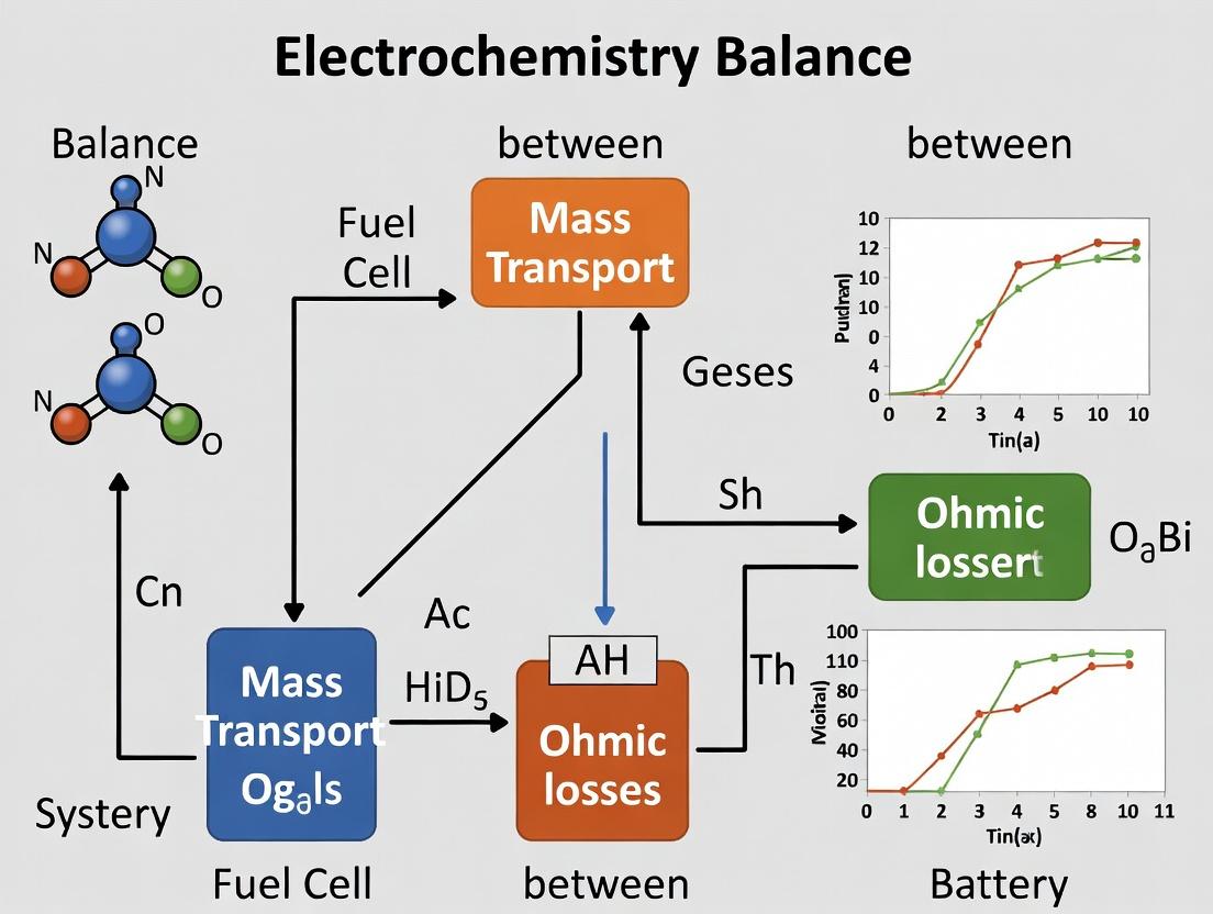
Mass Transport & Ohmic Balance in Biosensing: Visualizing the System
Diagram Title: Interplay of Mass Transport Mechanisms and Ohmic Losses
Diagram Title: Biosensor Mass Transport & Ohmic Loss Troubleshooting Workflow
Technical Support & Troubleshooting Center
Troubleshooting Guides
Issue 1: Unstable or Drifting Potential During Electrochemical Measurement
- Problem: The applied or measured electrode potential is not stable, making data interpretation difficult.
- Probable Cause: High and unstable ohmic drop due to improper electrode placement, low electrolyte conductivity, or a degraded reference electrode junction.
- Solution:
- Ensure the Luggin capillary (if used) is positioned correctly, typically ~2x its diameter from the working electrode.
- Increase supporting electrolyte concentration (e.g., 0.1 M to 0.5 M) to boost solution conductivity.
- Check and refresh reference electrode filling solution. Clean the porous frit.
- Use positive feedback iR compensation if available on your potentiostat, but only after verifying stability.
Issue 2: Inconsistent Results Between Duplicate Experiments in High-Resistance Media
- Problem: Replicate experiments show high variability, especially in low-ionic-strength solutions (e.g., some bio-electrolytes).
- Probable Cause: Small variations in electrode alignment or solution level significantly change the uncompensated resistance (Ru).
- Solution:
- Implement a rigid, reproducible cell and electrode fixture.
- Precisely measure and record the solution height.
- Routinely measure Ru via current interrupt or electrochemical impedance spectroscopy (EIS) before each experiment to ensure consistency.
- Consider using a more conductive buffer if chemically permissible for the system.
Issue 3: Distorted Voltammogram Shapes in Cyclic Voltammetry
- Problem: Peaks are drawn-out, asymmetric, or the scan rate dependence doesn't follow expected trends.
- Probable Cause: Significant iR drop distorting the effective potential at the working electrode, a critical concern when balancing mass transport and ohmic losses.
- Solution:
- Record a voltammogram of your system. Compare peak separation to theoretical expectations.
- Estimate Ru using EIS or current interrupt.
- Calculate the approximate iR drop (i * Ru) at the peak current. If it exceeds 10 mV, mitigation is needed.
- Apply iR compensation cautiously, ensuring no oscillation, or redesign the experiment to lower Ru (e.g., smaller electrode, closer reference).
Frequently Asked Questions (FAQs)
Q1: What is the simplest way to measure the uncompensated resistance (Ru) in my cell? A: Electrochemical Impedance Spectroscopy (EIS) is the most reliable method. Apply a small AC potential (e.g., 10 mV) at the open circuit potential over a high-frequency range (e.g., 100 kHz to 10 Hz). The high-frequency intercept on the real axis of the Nyquist plot gives Ru (primarily solution resistance). Alternatively, most modern potentiostats have an automated "Current Interrupt" or "Ru Test" function.
Q2: When should I NOT use positive feedback iR compensation? A: Avoid using it in the following scenarios: 1) With unstable or poorly conductive connections (leads to potentiostat oscillation). 2) In systems with rapidly changing current (e.g., during metal deposition), as it can over-compensate. 3) When Ru has not been measured accurately first. Incorrect compensation is worse than no compensation.
Q3: How does solution resistance directly impact studies focused on balancing mass transport and ohmic losses? A: In such research, you often vary parameters like electrolyte concentration, electrode spacing, or flow rate. Solution resistance (a key component of iR drop) changes with each. A high iR drop can mask the true kinetic and mass transport overpotentials, leading to incorrect conclusions about the dominant limiting factor. Precise iR measurement/compensation is essential to decouple these effects.
Q4: Can I reduce electrode resistance itself? A: Yes. For conductive substrates, ensure clean, solder-free connections. For porous or film-based electrodes (common in battery/drug sensor research), the electronic conductivity of the active material is key. Incorporate conductive additives (carbon, metals) and ensure good binder distribution. Use a more conductive current collector (e.g., gold vs. certain metal oxides).
Table 1: Contribution to Uncompensated Resistance (Ru)
| Component | Typical Range | Key Influencing Factors |
|---|---|---|
| Solution Resistance | 1 Ω - 10 kΩ | Electrolyte conductivity, distance between WE and RE, electrode geometry. |
| Working Electrode Resistance | 0.1 Ω - 1 kΩ | Substrate material, film thickness, conductivity of coated layers. |
| Counter Electrode Resistance | < 5 Ω | Electrode material, surface area, passivation. |
| Contact & Lead Resistance | < 1 Ω | Cable quality, connector cleanliness, clamp tightness. |
Table 2: iR Drop Impact on Common Techniques
| Technique | Primary Manifestation of iR Drop | Acceptable iR Drop (Rule of Thumb) |
|---|---|---|
| Cyclic Voltammetry | Increased ΔEp, peak broadening, shifted E1/2. | < 10 mV at peak current. |
| Chronoamperometry | Slow current transient, incorrect Cottrell slope. | < 5% of applied potential step. |
| Electrochemical Impedance | Distortion of high-frequency semicircle, inaccurate kinetic fitting. | Should be measured and subtracted during fitting. |
| Battery Charge/Discharge | Reduced usable voltage window, decreased energy efficiency. | Managed via cell design and conductive additives. |
Experimental Protocols
Protocol 1: Determining Uncompensated Resistance via EIS
Objective: Accurately measure Ru for iR compensation or cell diagnostics.
- Setup: Configure a standard 3-electrode cell. Ensure system is at steady-state (e.g., open circuit).
- Instrument Settings:
- Technique: Electrochemical Impedance Spectroscopy.
- DC Potential: Open Circuit Potential (OCP).
- AC Amplitude: 10 mV.
- Frequency Range: 200 kHz to 100 Hz (prioritize high-frequency data).
- Points per Decade: 10.
- Execution: Run the measurement.
- Analysis: Plot Nyquist plot (Z'' vs Z'). Identify the high-frequency intercept on the Z' (real) axis. This value is Ru.
Protocol 2: Systematic Evaluation of Ohmic Loss vs. Mass Transport Limitation
Objective: Decouple iR effects from diffusion control, core to balancing mass transport and ohmic losses.
- Baseline CV: Perform cyclic voltammetry of a reversible redox couple (e.g., 1 mM Ferrocenemethanol) in 0.1 M supporting electrolyte at 100 mV/s.
- Vary Conductivity: Repeat CV in supporting electrolytes of decreasing concentration (0.05 M, 0.01 M). Do not compensate.
- Measure Ru: Use Protocol 1 to measure Ru for each electrolyte.
- Apply Compensation: Repeat the CVs with 85-95% positive feedback iR compensation (based on measured Ru).
- Analysis: Compare peak separations (ΔEp) and shapes between uncompensated and compensated scans across different conductivities. Plot ΔEp vs. Ru to visualize the direct impact.
Diagrams
Diagram Title: Origins and Impact of iR Drop Components
Diagram Title: iR Drop Troubleshooting Decision Tree
The Scientist's Toolkit: Key Research Reagents & Materials
| Item | Primary Function | Role in Managing Ohmic Losses |
|---|---|---|
| Supporting Electrolyte (e.g., TBAPF6, KCl) | Carries current, minimizes migration of analyte. | Primary controller of solution resistance. Higher concentration lowers Ru. |
| Luggin Capillary | Houses reference electrode, allows close proximity to WE. | Minimizes solution resistance component by reducing WE-RE distance. |
| Conductive Carbon Additives (Vulcan XC-72, CNTs) | Mixed with active materials to form composite electrodes. | Reduces electrode resistance by creating percolating conductive networks in films. |
| Potentiostat with iR Compensation | Applies potential/current, measures electrochemical response. | Electronically corrects for iR drop via positive feedback or current interrupt methods. |
| Non-Aqueous Reference Electrode (Ag/Ag+) | Provides stable potential in organic electrolytes. | Enables accurate potential control in high-R solvents, a prerequisite for iR compensation. |
| Gold or Platinum Mesh Counter Electrode | Provides large surface area for current dissipation. | Prevents counter electrode polarization, which can indirectly increase measured cell resistance. |
Technical Support Center: Troubleshooting for Electrochemical Systems
Welcome to the technical support hub for researchers investigating the balance between mass transport and ohmic losses in electrochemical systems. This guide provides targeted solutions for common experimental challenges.
Frequently Asked Questions (FAQs)
Q1: During my flow cell experiment for drug precursor synthesis, my current efficiency dropped sharply after increasing the flow rate, even though reactant concentration was high. What is the primary cause? A1: This is a classic symptom of the inherent conflict. Enhanced flow rate improves convective mass transport, which can unmask or exacerbate ohmic losses. The increased current demand at higher reactant flux leads to a larger IR drop across the electrolyte, reducing the effective potential at the electrode and lowering efficiency. Check your inter-electrode gap and electrolyte conductivity.
Q2: My impedance spectra show a growing resistive loop at high flow velocities in a microfluidic electrolyzer. Is my electrode corroding? A2: Not necessarily. A growing low-frequency resistive loop often indicates that the system is becoming limited by ionic resistance (ohmic loss) rather than charge transfer. The improved mass transport shifts the limiting factor to electrolyte conductivity. Measure the solution resistance (R_s) directly from the high-frequency intercept on the Nyquist plot.
Q3: When I scaled up my paired electrosynthesis from an H-cell to a flow reactor, the product yield was lower despite identical charge passed. Why? A3: In scaling, the electrode area and current scale faster than the volume of electrolyte between electrodes, increasing current density. This intensifies the ohmic loss (V_loss = I * R). The increased resistance causes non-uniform potential distribution, leading to side reactions. Review your cell geometry and consider a segmented electrode to diagnose potential distribution.
Q4: How can I distinguish between mass transport limitation and ohmic loss limitation experimentally? A4: Perform a Current-Interrupt or Potentiostatic Electrochemical Impedance Spectroscopy (PEIS) measurement. A significant immediate voltage recovery upon current interrupt indicates dominant ohmic loss. In PEIS, a series resistance that is invariant with applied potential is ohmic, while one that changes with potential/flow rate often involves mass transport.
Troubleshooting Guides
Issue: Inconsistent Performance in High-Throughput Screening (HTS) Electrochemical Cells
- Symptoms: Poor reproducibility between cell channels, unexpected drop in Faradaic efficiency at high throughput rates.
- Diagnosis: This is frequently caused by uneven flow distribution leading to varied ohmic losses between parallel channels. A channel with slightly higher flow gets better mass transport, draws more current, and experiences a larger IR drop, altering its operational point.
- Solution:
- Verify Flow Uniformity: Use a tracer dye or flow sensor at each channel outlet.
- Implement Individual Reference Electrodes: Place a miniature reference electrode in each channel's outlet stream to monitor the actual working electrode potential.
- Independent Control: Consider using a multi-channel potentiostat for independent control of each channel's potential, compensating for variable IR drops.
Issue: Sudden Voltage Spike in a Continuous Electrosynthesis Flow Reactor
- Symptoms: Cell voltage increases dramatically during operation, potentially tripping safety limits.
- Diagnosis: This could be gas bubble formation (e.g., H2 or O2 from side reactions) trapped in the cell. Bubbles increase ohmic loss by blocking ionic current paths and reducing effective electrolyte conductivity.
- Solution:
- Immediate Action: Increase system pressure to dissolve bubbles or introduce a pulsed flow to dislodge them.
- Preventive Redesign: Orient the cell for vertical flow to facilitate bubble escape. Incorporate a gas-venting section or a porous membrane to separate gas.
- Operational Adjustment: Re-evaluate the applied potential/current to stay within the solvent's electrochemical window and minimize side reactions.
Experimental Protocols
Protocol 1: Quantifying the Mass Transport-Ohmic Loss Coupling
Objective: To measure the decrease in effective working electrode potential due to ohmic loss as a function of forced convection (flow rate). Methodology:
- Setup: Use a standard three-electrode flow cell (working, counter, reference). Place the reference electrode capillary tip (Luggin capillary) as close as permissible to the working electrode surface to minimize uncompensated resistance.
- Procedure: a. Circulate electrolyte at a baseline low flow rate (e.g., 10 mL/min). b. Apply a constant current relevant to your synthesis. c. Record the total cell voltage (Vcell) and the potential of the working electrode (Ewe) vs. the reference. d. Calculate the ohmic loss: Ohmic Loss (IR) = Vcell - |Ewe| (for a balanced reaction, adjust for counter electrode potential if needed). e. Incrementally increase the flow rate (e.g., to 50, 100, 200 mL/min) and repeat steps b-d at each step. f. For each flow rate, also perform linear sweep voltammetry to observe the limiting current plateau.
- Key Measurements: IR loss, Limiting current (i_lim).
Typical Data Summary:
| Flow Rate (mL/min) | Current Density (mA/cm²) | Measured E_we (V vs. Ref) | Total Cell Voltage (V) | Calculated IR Drop (V) | Observed Limiting Current (mA/cm²) |
|---|---|---|---|---|---|
| 10 | 5.0 | 0.51 | 1.85 | 1.34 | 6.2 |
| 50 | 7.5 | 0.49 | 2.91 | 2.42 | 9.8 |
| 100 | 9.2 | 0.48 | 3.65 | 3.17 | 12.1 |
| 200 | 10.5 | 0.47 | 4.72 | 4.25 | 14.5 |
Data shows IR drop increasing with flow rate as higher current is drawn, despite a more stable E_we.
Protocol 2: Mapping Potential Distribution in a Scalable Flow Electrode
Objective: To visualize how ohmic losses cause non-uniform reaction rates across a large electrode under flow. Methodology:
- Setup: Fabricate a segmented working electrode (e.g., 5 independent carbon felt strips in parallel aligned with flow). Use a common counter and reference. Connect each segment to a separate channel of a multi-potentiostat or a multiplexer.
- Procedure: a. Flow electrolyte with a redox probe (e.g., 1 mM ferrocyanide) at the target rate. b. Apply a constant total current to the entire assembly. c. Measure the current accepted by each individual segment. d. Alternatively, operate in potentiostatic mode and measure the current at each segment.
- Analysis: Calculate current density distribution. Segments near the inlet/current feeder will typically show higher current density, indicating uneven utilization due to resistive losses in the electrolyte path.
Visualizations
Diagram 1: The Core Conflict Workflow (96 chars)
Diagram 2: Feedback Loop Between Transport & Loss (99 chars)
The Scientist's Toolkit: Research Reagent & Material Solutions
| Item & Typical Example | Primary Function in Context |
|---|---|
| High Conductivity Supporting Electrolyte (e.g., TBAPF6 in MeCN, LiClO4 in non-aqueous systems) | Minimizes baseline solution resistance (Rs) to reduce the magnitude of IR drop (I*Rs). Choice is critical for organic electrosynthesis. |
| Luggin Capillary with Reference Electrode (e.g., Ag/AgCl, non-aqueous Ag/Ag+) | Allows accurate measurement of working electrode potential by positioning the reference probe close to the electrode surface, compensating for ohmic loss. |
| Conductivity Meter / Impedance Analyzer | Essential for directly measuring electrolyte conductivity and decomposing cell resistance into charge transfer and ohmic components via EIS. |
| Segmented or Interdigitated Electrode | Diagnostic tool for visualizing current and potential distribution across an electrode, directly identifying areas impacted by ohmic losses. |
| Peristaltic or HPLC Pump (Pulse-Free) | Provides precise, controllable convective flow to systematically vary mass transport rates independent of other variables. |
| Microfluidic Flow Cell with Small Electrode Gap | Hardware solution to reduce ohmic loss by minimizing the distance ions must travel between electrodes (R ∝ distance). |
| Redox Probe Molecules (e.g., Ferrocene, Potassium Ferricyanide) | Well-characterized, reversible redox couples used to benchmark mass transport and kinetic performance without complication from side reactions. |
Technical Support Center
Troubleshooting Guides & FAQs
Q1: My sensor's measured sensitivity (ΔSignal/ΔConcentration) has dropped significantly compared to the initial calibration. What could be the cause in the context of balancing mass transport and ohmic losses? A: A drop in sensitivity often points to an increased diffusion barrier or a rise in uncompensated resistance (ohmic loss).
- Primary Cause (Mass Transport): Fouling or passivation of the electrode surface. Protein adsorption or cellular debris forms a layer that impedes the diffusion of the analyte to the active surface, reducing the effective flux.
- Primary Cause (Ohmic Loss): Degradation of reference electrode stability or electrolyte depletion increases solution resistance. This leads to a larger iR drop, distorting the applied potential and reducing the faradaic efficiency for the redox reaction of interest.
- Troubleshooting Protocol:
- Surface Inspection: Perform cyclic voltammetry (CV) in a clean, known redox probe (e.g., 1 mM Ferro/ferricyanide). Compare the peak separation (ΔEp). An increase >59 mV indicates fouling (slowed kinetics) or increased resistance.
- Ohmic Loss Check: Use electrochemical impedance spectroscopy (EIS) to measure solution resistance (Rs) at high frequency. Compare to baseline.
- Action: If fouling is suspected, clean the electrode per manufacturer protocol (e.g., gentle polishing, electrochemical cleaning). If ohmic loss is high, replenish or change the electrolyte/buffer. Ensure reference electrode integrity.
Q2: How can I experimentally determine if my sensor's detection limit is being limited by mass transport constraints or by background noise/signal? A: The detection limit (DL) is defined as 3σ/S, where σ is the standard deviation of the blank and S is the sensitivity. The limiting factor depends on which term degrades.
- Experimental Protocol to Diagnose:
- Measure Blank Noise (σ): Record the signal (e.g., chronoamperometry or EIS phase angle) in analyte-free buffer for 10-20 minutes. Calculate the standard deviation.
- Measure Sensitivity (S) at Low [Analyte]: Perform a calibration with at least 3 low concentrations near the expected DL. Use a steady-state technique (e.g., rotating disk electrode) to ensure mass transport is not limiting the measurement itself. Plot signal vs. concentration; the slope is S.
- Analyze: Calculate DL = 3σ/S.
- Decouple: If you improve mass transport (e.g., increase stirring) and S increases significantly, your original DL was mass-transport-limited. If σ (noise) is high and dominates, investigate sources of electrical interference or stability issues (often related to interfacial resistance/ohmic losses).
Q3: My sensor's dynamic range seems to saturate at a lower concentration than theorized. How do I know if this is due to analyte depletion (mass transport) or electrode surface saturation (kinetics/ohmic effects)? A: Saturation occurs when the signal plateaus despite increasing analyte concentration.
- Diagnostic Experiment:
- Perform a Concentration vs. Rate Experiment: Use a technique sensitive to reaction rate, such as chronoamperometry with a step potential.
- Vary Mass Transport: Repeat the calibration curve under different stirring rates or flow conditions.
- Interpretation:
- If the saturation point (plateau concentration) increases with increased stirring/flow, saturation is due to analyte depletion (mass transport limitation) in the bulk-near surface region.
- If the saturation point remains unchanged despite faster stirring, saturation is due to surface site limitation (all binding sites occupied) or an ohmic loss effect where the potential at the electrode surface is so distorted that no further faradaic current can be driven.
Q4: When optimizing sensor geometry for my thesis research, what is the primary trade-off between improving mass transport and minimizing ohmic losses? A: This is a core design challenge. The table below summarizes the trade-offs.
| Sensor Geometry/Design Change | Impact on Mass Transport | Impact on Ohmic Losses (iR drop) | Net Effect on Key Metrics |
|---|---|---|---|
| Increasing electrode size | Increases total flux, but can decrease flux density. Can lead to larger diffusion layers. | Lowers overall current density, reducing iR drop. | May improve S/N, can lower DL, but may reduce sensitivity (S) if not uniformly active. Dynamic range may shift. |
| Using micro/nano-electrodes | Greatly enhances radial diffusion, increasing flux density. Redcess depletion. | Very low total current leads to negligible iR drop, even in low-conductivity media. | Dramatically improves sensitivity and lowers DL. Excellent for localized detection. |
| Adding a porous scaffold or nanostructure | Massively increases surface area and local analyte concentration, enhancing flux. | Can increase resistance if the structure is poorly conductive or traps ions, increasing iR drop within pores. | Sensitivity often increases, but electron transfer kinetics may slow (affecting S). Risk of higher background noise. |
| Reducing electrolyte concentration (for bio-relevance) | Minimal direct impact. | Sharply increases solution resistance (Rs), leading to large iR losses and potential distortion. | Can severely degrade all metrics: lowers S, raises DL, compresses dynamic range. Requires careful design (e.g., microelectrodes, supported electrolytes). |
| Implementing a redox mediator (shuttle) | Can enhance apparent transport via diffusion of the mediator. | Mediator can lower overpotential, effectively reducing the impact of iR loss on the driving potential. | Can improve sensitivity and extend dynamic range, but adds chemical complexity and potential instability. |
Essential Experimental Protocols
Protocol 1: Diagnosing Mass Transport vs. Kinetic (Ohmic) Limitation via Cyclic Voltammetry. Objective: Determine the rate-limiting step in your sensor's response. Method:
- Prepare your sensor in a standard electrolyte containing your target analyte or a redox probe.
- Record CV scans at multiple rates (e.g., 10, 25, 50, 100, 200 mV/s).
- Plot the peak current (Ip) vs. the square root of the scan rate (v^(1/2)).
- Interpretation: A linear relationship indicates a mass-transport-limited (diffusion-controlled) process. A linear plot of Ip vs. v indicates a surface-controlled (kinetically limited) process. A shift from linear Ip-v^(1/2) to non-linear at high rates indicates the onset of kinetic/ohmic limitations.
Protocol 2: Quantifying Ohmic Loss (iR Drop) with Electrochemical Impedance Spectroscopy (EIS). Objective: Measure the uncompensated resistance (Ru) in your sensor system. Method:
- Set up your sensor at its standard operating potential in your experimental buffer.
- Run an EIS spectrum from high frequency (e.g., 100 kHz) to low frequency (e.g., 0.1 Hz) with a small AC amplitude (e.g., 10 mV).
- Fit the resulting Nyquist plot to a suitable equivalent circuit model (e.g., Randles circuit). The high-frequency intercept on the real (Z') axis is the solution resistance (Rs), which is Ru.
- Application: Use this Ru value for post-measurement iR compensation in your potentiostat software or to assess buffer suitability.
The Scientist's Toolkit: Research Reagent Solutions
| Reagent/Material | Function in Context of Sensor Metrics |
|---|---|
| Potassium Ferri/Ferrocyanide Redox Probe | Standard for diagnosing electrode activity, fouling, and estimating electroactive area. Changes in its CV shape directly reflect mass transport and kinetic issues. |
| Ru(NH₃)₆³⁺/²⁺ Redox Probe | Outer-sphere, single-electron redox couple with fast kinetics. Ideal for isolating and studying mass transport and ohmic loss effects without complicating surface binding kinetics. |
| Nafion Perfluorinated Polymer | A common proton-conducting ionomer. Used to modify electrodes, it can enhance selectivity but may introduce additional mass transport resistance and affect local ohmic losses within the film. |
| Polyethylenimine (PEI) / Polydopamine | Scaffolding polymers for creating porous, high-surface-area films on sensors. Improve mass transport of analytes but require careful optimization to maintain electrical conductivity (minimize ohmic loss). |
| Supporting Electrolyte (e.g., KCl, PBS) | Provides high ionic strength to minimize solution resistance (ohmic loss). Its concentration and composition are critical variables when balancing bio-relevance with sensor performance. |
| Rotating Disk Electrode (RDE) System | Critical apparatus. Allows precise control of mass transport via rotation speed (Levich equation). The definitive tool for decoupling mass transport effects from reaction kinetics. |
| Microfluidic Flow Cell | Enables precise control of analyte delivery (convective mass transport) and allows study of sensors under dynamic flow, mimicking in vivo conditions relevant to drug development. |
Visualizations
Diagram 1: Factors Impacting Key Electrochemical Sensor Metrics
Diagram 2: Diagnostic Workflow for Sensor Performance Issues
Technical Support Center
FAQs & Troubleshooting Guides
Q1: In my 1D Nernst-Planck-Poisson (NPP) model for a charged species in a diffusion cell, the simulated concentration profile becomes unstable (wild oscillations) near the boundary. What is the cause and how can I fix this? A: This is typically a spatial discretization issue. The Poisson equation links concentration to potential, and coarse grids near steep boundary layers fail to resolve the coupling.
- Solution: Implement an adaptive mesh refinement (AMR) near the boundaries or switch to a higher-order discretization scheme (e.g., finite element method, FEM). Ensure your dimensionless potential
φ* = Fφ/RTis scaled correctly. A stability check is∆x < λ_D(Debye length) where gradients are large.
Q2: When adding migration to my diffusion model via the Nernst-Planck equation, my finite element simulation converges extremely slowly or not at all. A: Poor convergence often stems from a weak coupling treatment and ill-conditioned matrices.
- Solution:
- Use a coupled solver (Newton-Raphson) instead of a segregated one.
- Employ a robust preconditioner (e.g., Algebraic Multigrid - AMG) for the linear solver step.
- Verify your initial guess is physically plausible (e.g., from a simpler analytical model).
- Check that your applied current/voltage boundary conditions are consistent and do not create singularities.
Q3: How do I correctly implement concentration-dependent conductivity in a model balancing ion transport and ohmic loss?
A: Ohmic loss (η_ohm = j * L / σ) requires an accurate functional form for conductivity σ(c_i). A common error is using a constant value.
- Protocol: For a binary electrolyte, use the constitutive relation:
σ = F^2 * Σ (z_i^2 * u_i * c_i), whereu_iis the mobility. Implement this as a dependent variable in your FEM software (e.g., COMSOL's dependent variables, or as a user-defined function in FEniCS). The workflow is:
Title: Workflow for Coupled Conductivity Simulation
Q4: My simulated potential distribution in a porous electrode doesn't match experimental data. Which model complexity should I add first? A: Begin by incorporating a microstructure-aware effective conductivity.
- Protocol: Use Bruggeman's correction as a first step:
σ_eff = σ * ε^(1.5), whereεis porosity. If mismatch persists, move to a homogenized model using volume averaging, which introduces two coupled potential equations (solid and electrolyte phases). The logical escalation is:
Title: Model Complexity Escalation Path
Q5: What are the key benchmarks to validate a custom NPP-FEM code before applying it to novel drug transport problems? A: Always benchmark against analytical or canonical numerical solutions.
Table 1: Essential Benchmark Tests for Model Validation
| Test Case | Governing Equation(s) | Key Quantitative Output | Expected Result (Analytical/Numerical) |
|---|---|---|---|
| Cottrell Experiment | Fick's 2nd Law (Diffusion only) | Limiting current vs. time | I(t) = nFAc√(D/πt) |
| Poisson-Boltzmann | Poisson + Boltzmann distribution | Potential decay in stagnant layer | φ(x) = φ_0 exp(-x/λ_D) |
| Electroneutral Diffusion | NPP with Σ z_i c_i = 0 |
Concentration profile over time | Goldman-Hodgkin-Katz solution |
| Sand's Equation | NPP for supporting electrolyte | Transition time for current step | τ = πD (nFAc / 2j)^2 |
The Scientist's Toolkit: Research Reagent & Simulation Solutions
Table 2: Essential Resources for Multiphysics Transport Modeling
| Item / Solution | Function / Purpose | Example / Note |
|---|---|---|
| COMSOL Multiphysics | Commercial FEM platform with built-in "Transport of Diluted Species" and "Electrochemistry" modules. | Ideal for rapid prototyping of coupled NPP, fluid flow, and current distributions. |
| FEniCS / Firedrake | Open-source FEM platforms for full custom equation implementation. | Requires strong programming. Enables discretization-level control. |
| MPET (Multiphase Porous Electrode Theory) | Open-source numerical framework for porous electrode models. | Specialized for battery cells; adaptable to biological transport. |
| Bruggeman Correlation | Empirical relation for effective transport in porous media. | D_eff = D * ε / τ. Tortuosity τ is often ε^(-0.5). |
| Butler-Volmer Kinetics | Boundary condition for Faradaic reaction rates. | Links surface concentration to current density. Critical for drug redox assays. |
| Debye Length Calculator | Script/Formula to compute electrostatic screening length. | λ_D = √(ε_r ε_0 RT / (2 F^2 I)). Crucial for mesh sizing. |
| Micro-CT Data | 3D structural scans of porous materials or tissues. | Provides real geometry for the most accurate σ_eff and D_eff calculations. |
Advanced Design Strategies: Engineering Solutions to Balance Transport and Resistance
Troubleshooting Guides & FAQs
FAQ 1: I have fabricated a 3D nano-porous electrode, but my measured current density is lower than theoretical predictions, and the signal is unstable. What could be wrong?
- Answer: This is a classic symptom of a mass transport limitation overwhelming the benefits of increased surface area. While nano-structuring increases electroactive surface area (ESA), it can create diffusion bottlenecks if the pore network is too tortuous or deep. The instability often indicates a transition to a diffusion-limited current regime.
- Troubleshooting Steps:
- Characterize Pore Architecture: Use SEM to confirm pore interconnectivity. Perform Brunauer-Emmett-Teller (BET) analysis to measure pore size distribution. Mass transfer limitations are severe when pore diameters approach the analyte's diffusion layer thickness.
- Perform Electrochemical Diagnostics: Run Cyclic Voltammetry (CV) at multiple scan rates. A lack of linearity in the peak current (ip) vs. square root of scan rate (v^(1/2)) plot indicates restricted diffusion. Use Electrochemical Impedance Spectroscopy (EIS) to quantify the diffusional Warburg element.
- Protocol - Diagnostic CV: In a known redox couple (e.g., 1 mM Ferrocenemethanol in electrolyte), record CVs from 10 mV/s to 1000 mV/s. Plot the absolute anodic peak current vs. v^(1/2). A linear fit that does not pass through the origin suggests mixed kinetic-diffusion control.
- Solution: Consider hierarchical structuring: use larger micro-pores (1-10 µm) as transport channels feeding smaller nano-pores (<100 nm) for high surface area. This balances ESA with convective flow.
FAQ 2: After modifying my electrode with carbon nanotubes (CNTs), the ohmic drop (iR drop) has increased significantly, distorting my voltammograms. How can I mitigate this?
- Answer: Poor percolation in the CNT network or excessive polymer binder insulates the nanostructures, increasing sheet resistance and iR loss. This directly conflicts with the goal of balancing mass transport and ohmic losses.
- Troubleshooting Steps:
- Measure Sheet Resistance: Use a four-point probe on your modified electrode substrate. Compare to an unmodified control. A jump >10 Ω/sq is problematic for fast electron transfer.
- Check Binder Ratio: Excessive Nafion or chitosan (common for CNT immobilization) is highly resistive. Optimal ratios are often <0.1% wt for Nafion in CNT inks.
- Solution: Optimize the CNT ink formulation. Incorporate a conductive filler like graphene flakes (0D-2D) to improve inter-CNT connectivity. Use thermal or plasma annealing to reduce contact resistance between nanotubes. Always apply a potentiostatic iR compensation during experiments if your potentiostat allows.
FAQ 3: My flow-cell with a structured electrode shows excellent performance at low flow rates, but benefits diminish at high flow rates. Why?
- Answer: This indicates that external convective mass transfer is no longer the limiting factor. At high flow rates, the limitation shifts internally to within the electrode's architecture (intra-pore diffusion) or to the reaction kinetics itself.
- Troubleshooting Steps:
- Analyze Performance vs. Flow Rate: Plot limiting current or conversion efficiency vs. volumetric flow rate (or Reynolds number). The plateau point identifies the transition from external to internal diffusion control.
- Protocol - Flow Rate Sweep: Operate your electrochemical flow cell at a fixed overpotential in the mass-transport-limited region. Measure the steady-state current while incrementally increasing the flow rate. Record the current at each point after it stabilizes.
- Solution: Redesign the electrode's internal porosity. Implement a gradient porosity structure, where pore size increases from the current collector outward, to facilitate penetration of analyte at high flow rates.
FAQ 4: During the electrodeposition of a nano-structured metal foam, the growth is non-uniform across the substrate. What parameters should I control?
- Answer: Non-uniform growth stems from an uneven distribution of current density and metal ion concentration across the electrode surface during deposition.
- Troubleshooting Steps:
- Check Cell Geometry: Ensure a symmetric, parallel alignment between the working electrode and counter electrode. Distance should be optimized (typically 2-3x the electrode diameter).
- Monitor Deposition Potential/Current: Use a reference electrode placed close to the working electrode surface (via a Luggin capillary) to ensure the applied potential is consistent across the surface.
- Solution: Use pulsed electrodeposition instead of constant potential. The off-cycle allows for replenishment of metal ions in the diffusion layer, leading to more uniform growth. Add a leveling agent or surfactant to the plating bath.
Table 1: Impact of Electrode Architecture on Key Performance Parameters
| Architecture Type | Avg. Pore Size (nm) | Electroactive SA Increase (vs. Flat) | Typical Ohmic Increase (Ω) | Optimal Flow Regime (Reynolds No.) | Primary Limitation at High Current |
|---|---|---|---|---|---|
| Carbon Nanotube Forest | 5-50 (inter-tube) | 50-200x | Moderate (10-50) | Laminar (Re < 10) | Pore clogging, iR drop in deep layers |
| Dealloyed Nano-porous Metal | 20-100 | 100-500x | Low (5-20) | Stagnant / Low Flow | Intrapore diffusion, mechanical stability |
| 3D-Printed Lattice | 50,000+ (macro) | 10-30x | Very Low (<5) | Turbulent (Re > 2000) | Low intrinsic surface area |
| Hierarchical (Micro/Nano) | 50,000 & 50 | 200-1000x | Moderate-Low (10-30) | Broad (Re 10-1000) | Fabrication complexity |
Table 2: Diagnostic Electrochemical Signatures of Limiting Factors
| Observed Voltammetric Behavior | Likely Cause | Diagnostic Test | Characteristic EIS Feature |
|---|---|---|---|
| Peak current (ip) ∝ scan rate (v) | Surface-bound (thin-layer) redox | CV at varying v | Large capacitive loop |
| ip ∝ v^(1/2), but low magnitude | Semi-infinite planar diffusion | CV at varying v | 45° Warburg line at low frequency |
| Current plateau, independent of v & flow | Severe internal diffusion limitation | Flow rate sweep | Dominant, elongated Warburg line |
| Peak separation increases with scan rate | High ohmic loss (iR drop) | CV with/without iR compensation | High series resistance (Rs) intercept |
Experimental Protocols
Protocol 1: Fabrication of Hierarchical Nano/Micro-Porous Gold Electrode via Dealloying
- Objective: Create an electrode with bi-modal porosity for enhanced mass transfer and high surface area.
- Materials: Gold-silver leaf (Ag75/Au25 at%), concentrated nitric acid (70%), deionized water, furnace.
- Steps:
- Cut the Ag/Au leaf to desired substrate size.
- Anneal in air at 400°C for 2 hours to promote phase separation.
- Immerse in concentrated nitric acid at room temperature for 48 hours to selectively dissolve (dealloy) silver.
- Rinse thoroughly with copious amounts of DI water and ethanol.
- Dry under a nitrogen stream. Characterize with SEM and BET.
Protocol 2: Electrochemical Active Surface Area (ECSA) Determination via Underpotential Deposition (UPD)
- Objective: Quantify the true electroactive surface area of a nanostructured electrode.
- Materials: 0.1 M H2SO4, 5 mM Pb(NO3)2, N2 gas, standard 3-electrode cell.
- Steps:
- Purge electrolyte with N2 for 20 min.
- Perform CV in pure 0.1 M H2SO4 between -0.2 V and 1.2 V vs. Ag/AgCl (3 cycles) to clean.
- Add Pb(NO3)2 to a concentration of 5 mM.
- Record CV at 20 mV/s between -0.2 V and 0.6 V. The sharp deposition/stripping peaks correspond to Pb UPD.
- Integrate the charge (Q) under the Pb stripping peak. Use the conversion factor: ECSA = Q / (420 µC cm⁻² for Pb UPD on Au).
Visualizations
Diagram Title: The Optimization Cycle for Structured Electrodes
Diagram Title: Mass Transfer Pathways in a Hierarchical Electrode
The Scientist's Toolkit: Essential Research Reagents & Materials
| Item | Function/Explanation | Example Supplier/Product |
|---|---|---|
| Nafion Perfluorinated Resin | Binder for nanostructured layers; provides proton conductivity but can increase ohmic loss if overused. | Sigma-Aldrich, 5% wt solution in lower aliphatic alcohols. |
| Carbon Nanotubes (CNTs), MWCTs | Building block for high-surface-area, conductive 3D networks. Functionalized versions improve dispersion. | Cheap Tubes Inc., CVD-grown multi-walled tubes. |
| Ferrocenemethanol | Standard outer-sphere redox probe for diagnosing mass transfer limitations without complications from adsorption. | Sigma-Aldrich, 97% purity. |
| Potassium Ferricyanide K3[Fe(CN)6] | Common inner-sphere redox probe for testing electrochemical activity and surface fouling. | VWR Chemicals. |
| Chitosan | Natural, biocompatible polymer binder for enzyme/nanomaterial immobilization in biosensor architectures. | Sigma-Aldrich, low molecular weight. |
| Poly(diallyldimethylammonium chloride) PDDA | Polyelectrolyte for layer-by-layer assembly and surface charge modification of nanostructures. | Sigma-Aldrich, 20% wt in water. |
| Triton X-100 / PVP | Non-ionic surfactants used in electrodeposition baths to control nucleation and growth of nanostructures. | Fisher Scientific. |
| Holey Carbon Film Grids | TEM grids used as scaffolds for creating free-standing, electron-transparent nano-electrode samples for in-situ study. | Ted Pella Inc. |
Troubleshooting Guides & FAQs
This technical support center addresses common experimental challenges in electrochemical cell research, framed within the thesis context of Balancing Mass Transport and Ohmic Losses. Solutions aim to optimize the trade-off between efficient reactant delivery (mass transport) and minimizing resistive voltage drops (ohmic losses) across different cell designs.
Rotating Disk Electrode (RDE) Troubleshooting
Q1: My RDE experiment shows unstable limiting current, with values fluctuating over time. What could be the cause? A: This typically indicates an issue with the hydrodynamic boundary layer. Common causes and fixes:
- Cause 1: Incorrect alignment or a bent electrode shaft. This creates turbulent, non-laminar flow.
- Solution: Use a precision spirit level to ensure the cell is perfectly horizontal. Visually inspect the shaft for bends and replace if necessary.
- Cause 2: Formation of gas bubbles on the electrode surface (e.g., from side reactions).
- Solution: Degas the electrolyte solution thoroughly with an inert gas (N₂, Ar) for at least 20 minutes prior to experiments. Ensure rotation is started before applying potential.
- Cause 3: Loose electrode tip or damaged disk material.
- Solution: Tighten the electrode tip to the specified torque. Inspect the disk surface under a microscope for cracks or delamination and re-polish or replace.
Q2: How do I correct for ohmic losses (iR drop) in my RDE setup when testing poorly conductive electrolytes? A: Ohmic losses are critical in balancing cell performance. Use these methods:
- Method: Electronic iR Compensation. Most modern potentiostats offer positive feedback or current-interrupt iR compensation.
- Protocol: First, measure the uncompensated solution resistance (Rᵤ) using Electrochemical Impedance Spectroscopy (EIS) at high frequency (e.g., 100 kHz) or the current-interrupt method. Enable the potentiostat's iR compensation function and input the measured Rᵤ value. Caution: Over-compensation leads to potentiostat instability. Compensate typically 85-95% of Rᵤ.
- Alternative: Reduce Cell Resistance. Use a Luggin-Haber capillary to bring the reference electrode tip close to the RDE surface, minimizing the resistance path. Ensure a supporting electrolyte is used at sufficient concentration (e.g., 0.1 M).
Microfluidic Electrochemical Cell Troubleshooting
Q3: I observe inconsistent current responses between replicates in my microfluidic flow cell. What should I check? A: This often stems from mass transport variations or blockage.
- Check 1: Flow Profile Stability.
- Solution: Use a syringe pump with a high-precision, pulse-free motor. Incorporate a pulsation damper in-line. Verify flow rate calibration by measuring effluent volume over time.
- Check 2: Channel or Inlet Blockage.
- Solution: Flush the system with a strong solvent (e.g., 1M NaOH, isopropanol) compatible with your chip material. Filter all solutions and suspensions (using 0.2 µm filters) prior to loading into the syringe. Inspect the channel under a microscope.
- Check 3: Electrode Fouling.
- Solution: Implement an in-situ electrode cleaning protocol (e.g., cyclic voltammetry in clean supporting electrolyte between runs). Consider using anti-fouling coatings (e.g., PEGylated layers) for complex biofluids.
Q4: How can I quantify and minimize ohmic losses in a thin-layer microfluidic cell? A: Ohmic losses can be severe in microfluidic channels, especially with low ionic strength fluids like some biological buffers.
- Quantification Protocol: Use EIS across the two working/auxiliary electrodes. Measure the high-frequency real-axis intercept. Alternatively, measure the potential drop between two micro-reference electrodes placed upstream and downstream of the working electrode.
- Minimization Strategies:
- Integrate Closer Electrodes: Design cells with a counter electrode placed directly opposite the working electrode, separated only by the thin channel height.
- Increase Electrolyte Conductivity: Add a supporting electrolyte (e.g., KCl, NaClO₄) at a concentration that does not interfere with the reaction under study.
- Use Shorter Channels: For a given flow rate, pressure, and ohmic drop scale with channel length.
Quantitative Data Comparison: Cell Design Parameters
The following table summarizes key parameters affecting the mass transport-ohmic loss balance in different cell designs.
| Cell Design | Typical Mass Transport Coefficient (kₘ, cm/s) | Characteristic Length (mm) | Ohmic Drop Concern | Optimal Use Case |
|---|---|---|---|---|
| Static Cell | 0.0001 - 0.001 | 10 - 50 | High for bulk, low with Luggin capillary | Slow kinetics screening, high-concentration electrolytes. |
| RDE | 0.01 - 0.1 | Diffusion layer: 0.01 - 0.1 | Moderate; decreases with rotation. | Fundamental kinetics (Koutecký-Levich analysis), catalyst benchmarking. |
| Microfluidic (Laminar Flow) | 0.001 - 0.1 | Channel Height: 0.01 - 1 | High for low-conductivity streams, channel-length dependent. | Analysis of small volume samples, coupling with separation techniques, in-situ generation of reagents. |
| Gas Diffusion Electrode (GDE) | Very High (for gases) | Diffusion layer: < 0.001 | Low in thin catalyst layers, high in bulk electrolyte. | Fuel cells, CO₂ reduction, reactions involving gaseous reactants. |
Detailed Experimental Protocols
Protocol 1: Determining Kinetic Currents Using an RDE (Correcting for Mass Transport & Ohmic Losses) Objective: To extract the charge-transfer kinetic current (iₖ) for an O₂ reduction reaction, free from mass transport and iR drop influences. Reagents: Catalyst ink, 0.1 M KOH or HClO₄ electrolyte, O₂ or N₂ gas. Procedure:
- Electrode Preparation: Disperse catalyst, carbon, and Nafion binder in solvent (e.g., water/isopropanol). Deposit a thin, uniform film on a polished glassy carbon RDE tip to achieve a loading of 0.1-0.5 mg_cat/cm².
- Cell Setup: Use a standard 3-electrode cell with Pt wire counter and reversible hydrogen reference electrode (RHE). Position the Luggin capillary close to the RDE. Maintain electrolyte at 25°C.
- iR Drop Measurement: In O₂-saturated electrolyte, at a fixed rotation speed (e.g., 1600 rpm), perform a high-frequency EIS scan at open circuit potential (e.g., 100 kHz to 10 Hz) to determine uncompensated resistance (Rᵤ).
- Data Acquisition: With iR compensation set to 90% of Rᵤ, record linear sweep voltammograms (LSV) from 1.0 to 0.2 V vs. RHE at multiple rotation speeds (e.g., 400, 900, 1600, 2500 rpm).
- Data Analysis: Apply the Koutecký-Levich equation at each potential:
1/i = 1/iₖ + 1/(B*ω^(1/2))whereiis the measured current,ωis the rotation rate, andBis the Levich constant. Plot1/ivs.ω^(-1/2). The y-intercept equals1/iₖ. The kinetic currentiₖis now free from mass transport effects.
Protocol 2: Establishing a Steady-State Concentration Gradient in a Microfluidic Y-Channel Objective: To create and electrochemically probe a predictable laminar co-flow of two streams. Reagents: 1 mM K₃Fe(CN)₆ in 0.1 M KCl (Stream A), 0.1 M KCl only (Stream B). Procedure:
- Chip Priming: Place the PDMS/glass microfluidic chip (with integrated Au working electrode) under vacuum for 5 min. Fill both inlets with ethanol using a syringe to wet channels, then flush thoroughly with deionized water.
- Flow System Setup: Load Stream A and B into separate gas-tight glass syringes. Connect to chip inlets via tubing and nanoports. Mount syringes in a dual-syringe pump. Place a Ag/AgCl reference and Pt counter electrode in the chip's outlet reservoir.
- Flow Establishment: Start the syringe pump at a total flow rate of 10 µL/min (5 µL/min per stream). Allow flow to stabilize for 15 minutes to eliminate bubbles and establish a stable liquid-liquid interface.
- Electrochemical Mapping: Using a micromanipulator, position a microelectrode probe at the channel outlet to validate the diffusion interface. Alternatively, perform cyclic voltammetry at the integrated Au WE. The limiting current will be proportional to the concentration of Fe(CN)₆³⁻ delivered by Stream A, demonstrating controlled mass transport.
Visualizations
Title: RDE Current Instability Troubleshooting Flowchart
Title: Mass Transport vs Ohmic Loss Trade-offs
The Scientist's Toolkit: Research Reagent Solutions
| Item | Function & Relevance to Mass Transport/Ohmic Loss |
|---|---|
| High-Purity Supporting Electrolyte (e.g., NaClO₄, TBAPF₆) | Increases solution conductivity, minimizing ohmic losses. Must be electrochemically inert in the potential window of interest. |
| Luggin-Haber Capillary | Brings the reference electrode tip close to the working electrode, reducing the uncompensated resistance (Rᵤ) in the measurement circuit. Critical for accurate potential control. |
| Syringe Pump with Pulsation Damper | Provides precise, pulseless flow in microfluidics, ensuring stable and reproducible mass transport conditions (laminar flow profile). |
| Nafion Binder/Coating | Ionomer used in catalyst inks (RDE) or as a membrane coating (microfluidics). Facilitates proton transport to active sites, reducing local ohmic losses within the catalyst layer. |
| Microfluidic Chip with Integrated Electrodes | Embeds working, counter, and sometimes reference electrodes directly into the channel walls. Minimizes electrode spacing, dramatically reducing ohmic losses compared to external cells. |
| Rotational Speed Calibrator | Validates the rotation speed of an RDE, essential for accurate Levich analysis and reproducible mass transport conditions. |
Troubleshooting Guides & FAQs
FAQ 1: Why is my voltammogram distorted (peaked or drawn-out) despite using a supporting electrolyte?
- Answer: This indicates high uncompensated resistance (Ru). The supporting electrolyte concentration may be insufficient for the solvent/electrode geometry, or the chosen salt has low solubility/conductivity in your medium. Increase the concentration of your supporting electrolyte if solubility permits, or switch to a salt with a higher conductivity in your solvent (e.g., TBAPF6 over TBAClO4 in some organic solvents). Ensure your reference electrode is positioned correctly within the Luggin capillary.
FAQ 2: How do I choose between tetraalkylammonium salts and alkali metal salts?
- Answer: The choice balances minimizing resistance and avoiding interfering reactions.
- Tetraalkylammonium Salts (e.g., TBAPF6): Preferred for non-aqueous electrochemistry (acetonitrile, DMF) and wide negative potential windows. They minimize resistance and do not complex with reactants. However, they can adsorb on some electrodes at very negative potentials.
- Alkali Metal Salts (e.g., KCl, LiClO4): Standard for aqueous systems. They offer high conductivity but can participate in complexation or specific ion-pairing effects that may alter reaction mechanisms. Avoid if your analyte interacts with metal ions.
FAQ 3: My iR compensation causes oscillation or instability in the potentiostat feedback. What should I do?
- Answer: This occurs when the compensation level is set too high (>85-90%) or is incorrectly estimated. First, use positive feedback iR compensation cautiously. Manually determine Ru via current-interrupt or impedance methods. Apply compensation gradually, starting at 70-80%, and ensure your cell setup is stable (tight connections, no bubbles). For high-precision work, consider using a smaller working electrode or increasing electrolyte concentration instead of over-relying on electronic compensation.
FAQ 4: Can a supporting electrolyte interfere with my target analysis?
- Answer: Yes. Potential interferences include:
- Complexation: Salts like Li⁺ or Cl⁻ can complex with metalocene or organic species.
- Electrochemical Activity: Salts like TBABF4 may have BF4⁻ hydrolysis products that are electroactive. Perchlorate salts (ClO4⁻) require caution due to explosivity with organic materials.
- pH Buffering: In aqueous systems, the supporting electrolyte (e.g., phosphate buffer) can also act as a proton donor/acceptor, affecting reaction pathways.
- Always run a blank experiment with only supporting electrolyte to check its window and reactivity.
Experimental Protocol: Determining Optimal Supporting Electrote Concentration
Objective: To find the minimum concentration of supporting electrolyte required to minimize ohmic drop without introducing viscosity-related mass transport limitations or solubility issues.
Materials:
- Electrochemical cell (3-electrode: WE, CE, RE)
- Potentiostat with iR compensation capability
- Solvent of choice (e.g., Acetonitrile, dried)
- Supporting electrolyte salt (e.g., Tetrabutylammonium hexafluorophosphate - TBAPF6)
- Electroactive probe (e.g., 1.0 mM Ferrocene)
- Nitrogen gas for deaeration
Methodology:
- Prepare a 0.1 M stock solution of TBAPF6 in dry acetonitrile.
- Prepare a series of 10 mL solutions containing 1.0 mM Ferrocene and varying concentrations of TBAPF6 (e.g., 0.01 M, 0.05 M, 0.1 M, 0.2 M, 0.3 M).
- For each solution, assemble the cell, deaerate with N₂ for 10 minutes, and perform a cyclic voltammetry scan of the Ferrocene/Ferrocenium couple at a moderate scan rate (e.g., 100 mV/s).
- Record the peak-to-peak separation (ΔEp). Use the current-interrupt or AC impedance method (built into most potentiostats) to measure the uncompensated solution resistance (Ru) for each concentration.
- Plot Ru vs. [Electrolyte] and ΔEp vs. [Electrolyte]. The optimal range is typically where Ru and ΔEp plateau. A sharp increase in ΔEp at low concentration indicates significant ohmic distortion. A gradual increase in ΔEp at very high concentration may indicate increased viscosity slowing mass transport.
Data Presentation
Table 1: Effect of TBAPF6 Concentration on Resistance and Voltammetric Response in Acetonitrile (1 mM Ferrocene, 100 mV/s, 2 mm Pt disk electrode)
| [TBAPF6] (M) | Measured Ru (Ω) | ΔEp (mV) | Observed Peak Shape | Conductivity (mS/cm)* |
|---|---|---|---|---|
| 0.01 | 1250 | 95 | Drawn-out, asymmetric | ~0.8 |
| 0.05 | 185 | 72 | Slight fronting | ~5.5 |
| 0.10 | 85 | 65 | Near-Nernstian | ~12.0 |
| 0.20 | 42 | 64 | Sharp, symmetric | ~24.0 |
| 0.30 | 30 | 66 | Sharp, symmetric | ~32.0 |
Note: Example conductivity values for illustration; actual values vary by setup.
Table 2: Common Supporting Electrolytes and Their Properties
| Electrolyte | Common Solvent | Potential Window (Approx.) | Key Advantages | Potential Interferences |
|---|---|---|---|---|
| TBAPF6 | Acetonitrile, DCM | Wide (+2.5 to -3.0 V vs. Fc/Fc⁺) | High purity, good solubility, inert. | PF6⁻ hydrolysis in protic media. |
| LiClO4 | Acetonitrile, DMF | Wide | Very conductive, soluble. | Li⁺ complexation, Perchlorate = Explosive Hazard. |
| KCl | Water | Limited by H₂O electrolysis | High conductivity, inexpensive. | Cl⁻ can complex, not for non-aqueous use. |
| TBABF4 | Organic solvents | Wide | Good solubility in many organics. | BF4⁻ hydrolysis, can contain impurities. |
| Phosphate Buffer | Water | Moderate | Also provides pH control. | Can participate in proton-coupled reactions. |
The Scientist's Toolkit: Research Reagent Solutions
| Item | Function |
|---|---|
| Tetrabutylammonium Hexafluorophosphate (TBAPF6) | Gold-standard inert supporting electrolyte for non-aqueous electrochemistry. Minimizes resistance and ion pairing. |
| Ferrocene | Internal potential standard for non-aqueous experiments (E° is solvent-independent). Used to reference potentials and test cell resistance. |
| Luggin Capillary | A glass tube that positions the reference electrode tip close to the working electrode to minimize iR drop, without shielding. |
| Platinum Counter Electrode | Inert, high-surface-area electrode to complete the circuit without introducing contaminants. |
| Drying Column (Alumina) | For rigorous solvent purification to remove water and protic impurities that can react with electrolytes/analytes. |
| Conductivity Meter | To directly measure solution conductivity and optimize electrolyte concentration before electrochemical experiments. |
Workflow & Relationship Diagrams
Title: Optimization Workflow for Minimizing Uncompensated Resistance
Title: Core Thesis Conflict Between Mass Transport and Ohmic Loss
Technical Support Center: Troubleshooting & FAQs
This support center addresses common experimental challenges in integrating CNTs, graphene, and metallic nanostructures for research focused on balancing mass transport and ohmic losses in electrochemical and catalytic systems.
FAQ 1: My composite electrode (CNT/Metal NPs) shows high ohmic resistance despite using conductive materials. What could be wrong?
- Answer: This often stems from poor interfacial contact and excessive polymer binder insulating the conductive network.
- Troubleshooting Guide:
- Check Binder Ratio: High Nafion or PVDF content (>10 wt%) can block electron tunneling. Reduce to ≤5 wt% and ensure homogeneous dispersion.
- Verify Drying Conditions: Rapid drying can cause agglomeration, breaking percolation paths. Use controlled, slow drying (e.g., at room temperature for 12h).
- Assess Nanomaterial Functionalization: Overly aggressive acid treatment of CNTs can shorten them, reducing intrinsic conductivity. Use milder oxidation (e.g., 3M HNO₃ at 60°C for 4h instead of H₂SO₄/HNO₃ at 80°C) and confirm via Raman spectroscopy (ID/IG ratio <1.2).
- Test Contact: Use 4-point probe, not 2-point, to measure sheet resistance to exclude contact resistance.
- Troubleshooting Guide:
FAQ 2: How can I improve mass transport to active sites in a densely packed graphene foam electrode?
- Answer: Dense packing, while good for ohmic conduction, creates diffusion-limited conditions. Introduce hierarchical porosity.
- Troubleshooting Guide:
- Integrate Spacers: Incorporate non-aggregating spacers like silica nanoparticles (50-100 nm) during hydrogel formation. Etch them post-assembly (using 5% HF) to create macro-pores.
- Control Reduction: Rapid chemical reduction of graphene oxide (GO) collapses pores. Use a multi-stage, gentle reduction (e.g., thermal annealing at 200°C for 2h followed by 400°C in H₂/Ar for 1h).
- Design Flow Channels: For flow cells, pattern the foam into aligned channels (>100 µm) using 3D-printed molds to direct convective flow.
- Troubleshooting Guide:
FAQ 3: My deposited metallic nanostructures (e.g., Au NPs) on graphene agglomerate during electrochemical cycling. How to stabilize?
- Answer: Agglomeration indicates weak metal-support interaction and insufficient anchoring sites.
- Troubleshooting Guide:
- Pre-functionalize Support: Introduce oxygenated groups (via UV-ozone treatment for 10 min) or nitrogen doping (via ammonia annealing at 500°C) to provide nucleation anchors.
- Modify Deposition Protocol: Use pulse electrodeposition instead of constant potential. Example: Apply -0.8 V vs. Ag/AgCl for 0.1s, then 0 V for 1s, repeat 500 cycles in 1mM HAuCl₄/0.1M HClO₄.
- Apply Ultrathin Overcoats: Use atomic layer deposition (ALD) to apply 2-3 cycles of Al₂O₃ or TiO₂ to pin nanoparticles without fully encapsulating them.
- Troubleshooting Guide:
FAQ 4: How do I diagnose whether performance loss is due to mass transport or ohmic losses?
- Answer: Perform systematic electrochemical diagnostics.
- Troubleshooting Guide (Protocol):
- Record EIS: Measure electrochemical impedance spectra from 100 kHz to 0.1 Hz at open circuit. Fit to equivalent circuit. The high-frequency x-intercept is the ohmic resistance (RΩ). The low-frequency tail slope indicates diffusion limitations.
- Run CV at Different Scan Rates: For a capacitive system, plot peak current (ip) vs. scan rate (v). Linear relationship suggests capacitive (surface-limited) behavior. ip vs. v^(1/2) linearity suggests diffusion control.
- Rotating Disk Electrode (RDE) Studies: If using a slurry, coat on an RDE tip. Vary rotation rates (400-2500 rpm). Levich analysis distinguishes kinetic from mass transport current.
- Troubleshooting Guide (Protocol):
Table 1: Comparative Electrode Performance Metrics
| Material System | Ohmic Resistance (Ω cm²) | Limiting Current Density (mA cm⁻²) | Electrochemically Active Surface Area (m² g⁻¹) | Stability (Cycles @ 80% cap. retention) |
|---|---|---|---|---|
| Pristine Graphene Film | 5.2 | 1.5 (Diffusion-limited) | 50 | 100 |
| CNT Forest (Aligned) | 1.8 | 15.2 | 120 | 1000 |
| Au NPs / Graphene (Unstable) | 3.1 | 8.7 | 95 | 50 |
| Au NPs / N-doped Graphene (Stable) | 2.8 | 9.1 | 110 | 2000 |
| Hierarchical Graphene Foam w/ Macropores | 8.5 | 42.0 | 320 | 500 |
Key Experimental Protocol: Fabricating a Hierarchical CNT/Metal NP Composite
Objective: Create an integrated electrode with low ohmic loss and high mass transport for fuel cell catalysis. Procedure:
- CNT Functionalization: Sonicate 100 mg of multi-walled CNTs in a 3:1 mixture of H₂SO₄ (96%) and HNO₃ (65%) at 45°C for 3 hours. Dilute, filter, and wash to pH 7. Dry at 80°C for 6h.
- Electrophoretic Deposition (EPD): Prepare a suspension of 0.5 mg/mL functionalized CNTs in Mg(NO₃)₂ solution (10 mg/mL). Apply a constant voltage of 10 V between two conductive substrates (stainless steel, 2 cm apart) for 2 minutes. Dry in air.
- Pulse Electrodeposition of Pt NPs: Use the CNT-coated substrate as a working electrode in a 5mM H₂PtCl₆ + 0.5M H₂SO₄ solution. Apply a double-pulse: -0.7 V for 0.05s (nucleation), then +0.1 V for 1s (growth). Repeat for a total charge of 0.1 C cm⁻².
- Pore Former Leaching: Mix 20 wt% of PMMA microspheres (500 nm) with the EPD suspension in step 2. After deposition and metal growth, immerse the electrode in acetone for 24h to dissolve the PMMA, creating additional pores.
The Scientist's Toolkit: Research Reagent Solutions
Table 2: Essential Materials for Nanomaterial Integration
| Item | Function in Context of Mass Transport/Ohmic Loss |
|---|---|
| Nafion D520 Dispersion | Proton-conducting binder; must be used sparingly (<5 wt%) to avoid insulating active sites and blocking pores. |
| Chloroplatinic Acid (H₂PtCl₆) | Standard precursor for depositing Pt electrocatalyst nanoparticles directly onto conductive supports. |
| Poly(methyl methacrylate) PMMA Microspheres | Sacrificial template (50-1000 nm) to create tailored macro/mesopores in nanostructures, enhancing mass transport. |
| Nitrogen Gas (Ultra High Purity) | For creating inert atmospheres during sensitive annealing or reduction steps to prevent oxidation of nanostructures. |
| UV-Ozone Cleaner | For mild, controlled introduction of oxygenated anchoring sites on carbon surfaces to improve metal NP adhesion. |
| Triton X-100 Surfactant | Aids in dispersing hydrophobic nanomaterials (e.g., graphene) in aqueous solutions without permanent functionalization. |
| Potassium Ferricyanide K₃[Fe(CN)₆] | Redox probe for diagnostic CV to quantify active surface area and diffusion characteristics of modified electrodes. |
Visualizations
Diagram 1: Diagnosis of Performance Losses
Diagram 2: Fabrication Workflow for Stable NP-Carbon Composite
Troubleshooting & FAQs
Q1: In our amperometric glucose sensor, we observe a non-linear response at high glucose concentrations and signal saturation. What is the likely cause and how can we address it?
A: This is a classic mass transport limitation. The enzymatic reaction (e.g., with Glucose Oxidase) consumes glucose faster than it can diffuse to the electrode surface at high concentrations, creating a depletion layer. To balance transport with reaction kinetics:
- Solution 1: Optimize the enzyme loading in the immobilization matrix. Excessive enzyme can exacerbate local depletion.
- Solution 2: Introduce a permeable diffusion-limiting membrane (e.g., polyurethane, Nafion) to control the flux of glucose to the transducer. This extends the linear range.
- Solution 3: Enhance convective transport by implementing a stirred or flow-through system (e.g., in a continuous monitoring setup).
Q2: Our electrochemical immunosensor shows poor sensitivity and a high detection limit. We suspect poor antibody immobilization. What are the best practices for stable and oriented antibody binding on gold electrodes?
A: This issue directly impacts the binding efficiency (a kinetic parameter) and thus the overall sensor performance, which is governed by the balance between analyte capture and electron transfer.
- Protocol: Use a well-established self-assembled monolayer (SAM) protocol.
- Polish the gold electrode with 0.3 and 0.05 µm alumina slurry, sonicate in ethanol and water, and electrochemically clean in 0.5 M H₂SO₄.
- Immerse the electrode in a 1 mM solution of a heterobifunctional linker (e.g., 11-mercaptoundecanoic acid, 11-MUA) in ethanol for 12-24 hours to form the SAM.
- Activate the carboxyl groups by immersing in a solution containing 0.4 M EDC (1-ethyl-3-(3-dimethylaminopropyl)carbodiimide) and 0.1 M NHS (N-hydroxysuccinimide) in water for 30-60 minutes.
- Incubate with the antibody solution (10-100 µg/mL in a pH 7.4 buffer, e.g., PBS) for 2 hours. The amine groups on the antibody form stable amide bonds with the activated SAM, promoting oriented binding.
- Block remaining active sites with 1% BSA or ethanolamine.
Q3: During the development of a DNA hybridization sensor, we experience high background noise and non-specific adsorption. How can we minimize this?
A: Non-specific adsorption increases ohmic background and can swamp the faradaic signal from the target hybridization, disrupting the critical signal-to-noise balance.
- Solution 1: Incorporate a charged co-adsorbent (e.g., 6-mercapto-1-hexanol, MCH) alongside the thiolated DNA probe. MCH dilutes the probe layer, improves its orientation, and creates a negatively charged surface that repels non-specifically adsorbed nucleic acids.
- Solution 2: Optimize the stringency of the washing steps post-hybridization. Use buffers with controlled ionic strength and temperature.
- Solution 3: Apply a blocking agent (e.g., BSA, salmon sperm DNA) after probe immobilization to passivate uncoated electrode surfaces.
Q4: What causes significant iR drop (ohmic loss) in a low-conductivity buffer, and how does it affect our sensor's performance?
A: Ohmic loss (iR drop) occurs due to solution resistance (R) between working and reference electrodes when current (i) flows. It reduces the effective potential at the working electrode, slowing electron transfer kinetics, distorting voltammetric shapes, and lowering sensitivity.
- Mitigation Strategies:
- Increase Supporting Electrolyte Concentration: Use a phosphate buffer with at least 0.1 M ionic strength or add an inert salt (e.g., KCl, NaNO₃).
- Place Reference Electrode Close: Minimize the distance between the working and reference electrodes to reduce the resistance path.
- Use a Non-Faradaic Mode: Consider electrochemical impedance spectroscopy (EIS) which applies a small AC potential perturbation, minimizing iR drop.
- Employ Instrumentation with iR Compensation: Use a potentiostat with positive feedback iR compensation功能.
Table 1: Comparison of Biosensor Performance Metrics
| Biosensor Type | Typical Linear Range | Common Limit of Detection (LOD) | Key Transport/Loss Consideration |
|---|---|---|---|
| Enzymatic Glucose | 1-30 mM (blood range) | 5-50 µM | Mass transport limited by substrate diffusion; ohmic loss minimal in physiological buffers. |
| Electrochemical Immunosensor | pg/mL - ng/mL | 0.1-10 pg/mL | Kinetically limited by antibody-antigen binding; ohmic loss critical in low-conductivity, label-free buffers. |
| DNA Hybridization Sensor | fM - nM | 0.1-100 fM | Transport-limited for long DNA; kinetically limited for short DNA; ohmic loss significant in pure DNA solutions. |
Table 2: Impact of Experimental Parameters on Transport & Ohmic Effects
| Parameter | Effect on Mass Transport | Effect on Ohmic Loss (iR Drop) |
|---|---|---|
| Increased Stirring/Flow Rate | Enhances convective transport. | Negligible direct effect. |
| Higher Supporting Electrolyte Conc. | Minor effect on diffusion. | Dramatically Reduces solution resistance. |
| Smaller Electrode Size | Increases radial diffusion (enhances flux). | Increases current density but total i may be lower; impact varies. |
| Thicker Permeable Membrane | Decreases analyte flux (can linearize response). | Can Increase resistance if membrane is non-conductive. |
Experimental Protocol: Developing a Model DNA Hybridization Sensor
Objective: To demonstrate the balance between target DNA diffusion, surface hybridization kinetics, and electrochemical signal generation while managing ohmic losses.
Materials (The Scientist's Toolkit):
- Gold Disk Electrode (3 mm diameter): Transducer platform for thiol-based chemistry.
- Thiolated DNA Probe (e.g., 20-mer with 5' or 3' C6-SH): The capture element.
- 6-Mercapto-1-hexanol (MCH): A diluent and blocking agent to form a well-organized mixed SAM.
- Complementary & Non-Complementary DNA Target: For testing specificity.
- Methylene Blue (MB) or Hexaammineruthenium(III) chloride ([Ru(NH₃)₆]³⁺): Redox-active reporters that electrostatically interact with the DNA backbone.
- High Ionic Strength Buffer (e.g., 1M NaCl, 10 mM Tris, pH 7.4): Promotes DNA hybridization and reduces solution resistance.
- Low Ionic Strength Measurement Buffer (e.g., 10 mM Tris, pH 7.4): Used for [Ru(NH₃)₆]³⁺ measurement to enhance electrostatic sensitivity.
- Potentiostat/Galvanostat: For electrochemical measurements (CV, EIS, DPV).
Procedure:
- Electrode Preparation: Clean gold electrode as per Q2 protocol.
- Probe Immobilization: Co-immobilize thiolated DNA probe (1 µM) and MCH (1 mM) by incubating for 1 hour in the high ionic strength buffer. This forms a mixed SAM.
- Hybridization: Expose the functionalized electrode to solutions containing varying concentrations of the complementary DNA target in the high ionic strength buffer for 30-60 minutes at a controlled temperature (e.g., 37°C).
- Electrochemical Detection (Example using [Ru(NH₃)₆]³⁺):
- Rinse the electrode thoroughly with the low ionic strength measurement buffer.
- Transfer to a solution containing 50 µM [Ru(NH₃)₆]³⁺ in the low ionic strength measurement buffer. The cationic Ru complex electrostatically binds to the anionic phosphate backbone of the surface-confined DNA.
- Perform Square Wave Voltammetry (SWV) from -0.2 V to -0.5 V (vs. Ag/AgCl). The reduction current of [Ru(NH₃)₆]³⁺ is proportional to the amount of surface DNA (and thus the hybridized target).
- Control: Repeat with a non-complementary DNA sequence to assess non-specific adsorption.
Visualizations
Diagnosing and Solving Performance Issues: A Practical Guide for Researchers
Technical Support Center
Troubleshooting Guide
Issue 1: Poor Reproducibility in Current Density Measurements at High Overpotentials
- Symptoms: Current plateau is inconsistent between experimental runs; data scatter increases with applied potential.
- Likely Cause: Mass transport limitation becoming dominant. Uncontrolled convection from temperature gradients or vibration can alter diffusion layer thickness.
- Solution: Implement strict temperature control (e.g., use a water jacket). Use a Faraday cage and vibration isolation table. Ensure consistent electrode surface polishing between runs. Switch to a rotating disk electrode (RDE) for controlled mass transport.
Issue 2: Linear Sweep Voltammogram Shows Excessive Slope in the "Mass Transport Limited" Region
- Symptoms: The current continues to increase with potential instead of reaching a stable plateau.
- Likely Cause: Significant ohmic (iR) drop in the electrolyte, especially with low-conductivity solvents or large electrode spacings. The applied potential is not fully effective at the electrode surface.
- Solution: Increase electrolyte conductivity (e.g., use a supporting electrolyte at sufficient concentration). Employ iR compensation (positive feedback or current interruption). Move the reference electrode closer to the working electrode via a Luggin capillary.
Issue 3: Changing Electrode Area Does Not Scale Current Proportionally
- Symptoms: Doubling the geometric electrode area leads to less than double the current.
- Likely Cause: Non-uniform current distribution due to combined ohmic and kinetic/transport effects. At small areas, kinetics/transport may dominate; at large areas, ohmic effects become prominent.
- Solution: Perform experiments with a series of electrode areas. Use a well-designed cell geometry. Differentiate: mass transport scales with area, while ohmic effects are sensitive to geometry and electrode placement.
Issue 4: Unpredictable Response to Stirring or Rotation Rate Changes
- Symptoms: Current does not follow the Levich equation (current ∝ (rotation rate)^(1/2)) for an RDE.
- Likely Cause: The system is not under pure mass transport control. Mixed control involving slow kinetics or competing ohmic losses is present.
- Solution: Conduct a full rotation rate study. Plot inverse current (1/i) vs. inverse square root of rotation rate (1/ω^(1/2)) (Koutecký-Levich plot). The intercept reveals kinetic/ohmic contributions.
Frequently Asked Questions (FAQs)
Q1: How can I quickly diagnose if my system is limited by mass transport or ohmic losses? A: Perform a scan rate study in a stagnant solution. For a reversible system under mass transport control, peak current in cyclic voltammetry scales with v^(1/2). If the current scales linearly with v, the system is likely under ohmic or thin-layer control. Also, observe the peak separation; significant increase with scan rate indicates uncompensated resistance.
Q2: What is the most definitive experiment to confirm ohmic control? A: Measure the cell's electrochemical impedance spectrum (EIS) at the open-circuit potential. The high-frequency real-axis intercept in a Nyquist plot gives the uncompensated solution resistance (Ru). If the operating current (i) multiplied by Ru results in an iR drop comparable to your applied overpotential, ohmic losses are dominant.
Q3: Can both limitations be present simultaneously? How do I deconvolute them? A: Yes, most real systems operate under mixed control. Use the Koutecký-Levich equation for rotating disk experiments: 1/i = 1/(nFAkC) + 1/(0.62nFAD^(2/3)ν^(-1/6)Cω^(1/2)) Where the first term is the kinetic+ohmic contribution and the second is the mass transport term. Plotting 1/i vs. 1/ω^(1/2) yields an intercept that contains both the kinetic rate constant and ohmic contributions.
Q4: My iR compensation seems to cause oscillation. What should I do? A: Oscillation indicates over-compensation. This is a common pitfall. First, manually determine the solution resistance via EIS or current interrupt. Then, apply compensation gradually, starting at 70-80% of the measured R_u. Always verify compensation by checking the peak separation of a known outer-sphere redox couple (e.g., ferrocene) at different scan rates; it should remain constant and near 59 mV.
Table 1: Diagnostic Signatures of Limiting Factors
| Experimental Observation | Suggests Mass Transport Control | Suggests Ohmic Control |
|---|---|---|
| Current vs. Rotation Rate (RDE) | Linear Koutecký-Levich plot; follows Levich equation. | No dependence on rotation rate. |
| Current vs. Electrolyte Concentration | Current plateaus at high concentration. | Current increases linearly with concentration. |
| Peak Separation (CV, reversible couple) | Constant with increasing scan rate. | Increases linearly with scan rate. |
| Response to Stirring | Current increases significantly. | No change or minor change. |
| Potential Step Chronoamperometry | Current decays as i ∝ t^(-1/2) (Cottrell). | Current decays exponentially or remains stable. |
| Impedance Spectrum (Low Freq.) | Distinct Warburg (45°) diffusion tail. | Capacitive loop, no Warburg signature. |
Table 2: Typical Parameters for Model Systems
| System / Parameter | Value Range | Notes |
|---|---|---|
| Diffusion Coefficient (Aqueous ion) | 1 × 10^(-9) to 1 × 10^(-5) cm²/s | 1 × 10^(-5) cm²/s is typical for small molecules. |
| Uncompensated Resistance (1M electrolyte) | 1 - 50 Ω | Depends heavily on cell geometry and electrode distance. |
| Kinetic Rate Constant (k⁰, fast redox) | > 0.01 cm/s | Below ~0.001 cm/s, kinetics become limiting. |
| Diffusion Layer Thickness (stagnant) | 10 - 200 μm | Increases with time in unstirred solutions. |
Experimental Protocols
Protocol 1: Rotating Disk Electrode (RDE) for Diagnosis Objective: Distinguish kinetic, mass transport, and ohmic control.
- Setup: Use a standard 3-electrode cell with a Pt or glassy carbon RDE, Pt counter, and stable reference (e.g., Ag/AgCl). Use ≥0.1 M supporting electrolyte.
- Procedure: Record a series of linear sweep voltammograms (e.g., from 0 V to relevant reduction/oxidation potential) at different rotation rates (e.g., 400, 900, 1600, 2500 rpm).
- Analysis: Plot the limiting current (i_lim) at each rotation rate (ω) vs. ω^(1/2). A linear, zero-intercept plot indicates mass transport control. Perform a Koutecký-Levich analysis (1/i vs. 1/ω^(1/2)) to extract the kinetic intercept.
Protocol 2: Electrochemical Impedance Spectroscopy (EIS) for Ohmic Resistance Objective: Quantify uncompensated solution resistance (R_u).
- Setup: Same 3-electrode cell under open-circuit conditions or at a defined DC bias.
- Procedure: Apply a sinusoidal potential perturbation (10 mV amplitude) over a frequency range from 100 kHz to 0.1 Hz. Measure impedance.
- Analysis: Plot Nyquist plot (Z'' vs. Z'). The high-frequency intercept on the real (Z') axis is R_u. Use this value for accurate iR compensation.
Protocol 3: Scan Rate Dependence in Cyclic Voltammetry Objective: Diagnose control mechanism and detect ohmic distortion.
- Setup: Stagnant solution, standard 3-electrode cell with working electrode close to reference (Luggin capillary).
- Procedure: Record CVs of your system at increasing scan rates (e.g., 10, 50, 100, 500 mV/s).
- Analysis: For a reversible system: Plot peak current (ip) vs. v^(1/2). Linearity suggests mass transport control. Plot peak potential separation (ΔEp). ΔEp constant and ~59/n mV indicates negligible ohmic loss; increasing ΔEp indicates significant iR drop.
Visualization: Experimental Diagnostics
Title: Diagnostic Flowchart for Limiting Factors
Title: Components of Total Cell Overpotential
The Scientist's Toolkit: Research Reagent Solutions
Table 3: Essential Materials for Electrochemical Diagnostics
| Item | Function | Key Consideration |
|---|---|---|
| Rotating Disk Electrode (RDE) | Imposes controlled convection. Allows application of Levich/Koutecký-Levich analysis. | Material (Pt, GC, Au) must be inert. Surface polish is critical. |
| High Concentration Supporting Electrolyte (e.g., TBAPF6, KCl) | Minimizes ohmic resistance by increasing solution conductivity. Provides ionic strength. | Must be electrochemically inert in the potential window. Purify if necessary. |
| Potentiostat with iR Compensation & EIS | Applies potential, measures current. iR Comp. corrects for ohmic drop. EIS measures R_u. | Ensure positive feedback compensation is stable. Verify EIS calibration. |
| Luggin Capillary | Positions reference electrode close to working electrode to minimize uncompensated resistance. | Tip should be ~2x diameter from working electrode to avoid shielding. |
| Outer-Sphere Redox Probe (e.g., Ferrocene, Ru(NH3)6Cl3) | Provides a known, reversible reaction to diagnose cell setup, iR drop, and mass transport. | Kinetics should be fast; used to test uncompensated resistance. |
| Ultra-Pure Solvents & Salts | Reduces background current and interference from impurities. | Use HPLC-grade solvents. Dry and degas solvents for non-aqueous work. |
| Precision Polishing Kit (Alumina, diamond paste) | Ensines reproducible, clean electrode surface essential for quantifiable current densities. | Follow sequential grit polishing (e.g., 1.0, 0.3, 0.05 μm). |
Technical Support Center: Troubleshooting & FAQs
Q1: During cyclic voltammetry (CV) scan rate studies, my peak currents do not show a linear relationship with the square root of the scan rate. What could be causing this deviation from ideal Randles-Ševčík behavior?
A: This deviation indicates that the electrode process is not purely diffusion-controlled. Within the context of balancing mass transport and ohmic losses, consider these troubleshooting steps:
- Issue: Adsorption or surface-confined processes. This is common when the analyte binds to the electrode surface.
- Solution: Clean the electrode thoroughly (e.g., polish, sonicate). Test in a blank electrolyte to check for redox peaks. Consider using a different electrode material with lower adsorption potential.
- Issue: Significant uncompensated resistance (ohmic loss) distorting the voltammogram shape, especially at high scan rates.
- Solution: Increase electrolyte concentration (e.g., from 0.1 M to 0.5 M supporting electrolyte) to lower solution resistance. Use a potentiostat with positive feedback iR compensation, but apply cautiously to avoid oscillation.
- Issue: The electron transfer kinetics are slow relative to the scan rate, leading to a quasi-reversible or irreversible system.
- Solution: Reduce the scan rate to the range where linearity is observed. The deviation defines the kinetic window and is valuable data for calculating the electron transfer rate constant (k°).
Q2: How do I determine the optimal supporting electrolyte concentration for my redox system to minimize ohmic losses without introducing new problems?
A: The goal is to maximize conductivity while avoiding changes in speciation or double-layer effects.
- Baseline: Start with a standard concentration (e.g., 0.1 M KCl or TBAPF6).
- Perform a Concentration Series: Acquire CVs of your analyte at a medium scan rate (e.g., 100 mV/s) while incrementally increasing the supporting electrolyte concentration (e.g., 0.05 M, 0.1 M, 0.3 M, 0.5 M, 1.0 M).
- Analyze: Monitor the peak-to-peak separation (ΔEp). A significant decrease in ΔEp with increasing concentration indicates you are successfully reducing ohmic distortion. Optimal concentration is often where ΔEp stabilizes near the Nernstian value (59/n mV) for a reversible system.
- Warning: Very high concentrations (>1 M) can alter activity coefficients, increase viscosity (slowing mass transport), or cause solubility issues. Always check for precipitation or unexpected shifts in formal potential (E°').
Q3: When studying temperature effects, my electrochemical response becomes unstable. How can I design a robust variable-temperature experiment?
A: Temperature impacts both kinetic (Arrhenius) and mass transport (via diffusion coefficient) parameters.
- Stability Protocol:
- Equipment: Use a jacketed electrochemical cell connected to a precision circulating water or oil bath. Allow at least 15-20 minutes for thermal equilibration at each new temperature before measurement.
- Sealing: Ensure the cell is sealed to prevent solvent evaporation or condensation, which dramatically changes concentration and electrolyte strength.
- Reference Electrode: Use a reference electrode with temperature-stable junction (e.g., double-junction design) and confirm its potential is stable with temperature. Note that the potential of standard references like Ag/AgCl changes with temperature; this must be accounted for in data analysis.
- Order: Take measurements from low to high temperature to minimize condensation risks.
Data Presentation
Table 1: Effect of Electrolyte Concentration (KCl) on Ohmic Loss and Voltammetric Parameters for 1 mM Ferrocenemethanol (Hypothetical Data for a 1 mm diameter Pt disk electrode, 100 mV/s)
| [KCl] (M) | Solution Resistance, Ru (Ω) | Peak Separation, ΔEp (mV) | Peak Current, ipa (μA) | Observed Effect |
|---|---|---|---|---|
| 0.05 | 450 | 85 | 1.8 | High distortion |
| 0.10 | 225 | 70 | 2.0 | Moderate distortion |
| 0.30 | 75 | 65 | 2.1 | Near-optimal |
| 0.50 | 45 | 62 | 2.15 | Optimal (ΔEp ~ 59mV) |
| 1.00 | 22 | 61 | 2.14 | Minimal gain, risk of viscosity increase |
Table 2: Scan Rate Study Analysis for Diagnosing Control Mechanisms (Key Relationships from Cyclic Voltammetry)
| Observation (ip vs. v) | Diagnosis | Implication for Mass Transport/Kinetics |
|---|---|---|
| Linear with v1/2 | Diffusion-controlled current | Mass transport (diffusion) is rate-limiting. |
| Linear with v | Adsorption-controlled current | Surface process limits; no bulk diffusion. |
| Linear log(ip) vs. log(v) slope ~0.5 | Diffusion-controlled | Confirms Randles-Ševčík model. |
| Linear log(ip) vs. log(v) slope ~1.0 | Adsorption-controlled | Surface-confined species. |
| ΔEp increases significantly with v | Quasi-reversible or irreversible kinetics | Electron transfer kinetics (k°) becomes limiting. |
Experimental Protocols
Protocol 1: Systematic Scan Rate Study for Mechanism Elucidation
- Setup: Prepare a solution containing your analyte (0.5-2 mM) and sufficient supporting electrolyte (≥0.1 M) in deoxygenated solvent.
- Initial CV: Record a CV at 50 mV/s to identify redox peaks.
- Scan Rate Series: Record CVs across a wide range of scan rates (e.g., 10, 25, 50, 100, 200, 400, 600, 800, 1000 mV/s). Ensure the voltammogram shape remains stable.
- Data Analysis: Plot peak current (ip) for the oxidation or reduction peak vs. square root of scan rate (v1/2) and vs. scan rate (v). Use logarithmic plots (log ip vs. log v) to determine the slope.
Protocol 2: Optimizing Electrolyte Concentration to Balance Ohmic Loss
- Stock Solution: Prepare a concentrated stock solution of supporting electrolyte (e.g., 2.0 M KCl).
- Baseline Cell: In your electrochemical cell, add solvent, analyte, and a small volume of stock to achieve the lowest target concentration (e.g., 0.05 M). Mix thoroughly.
- Sequential Addition: Record a CV at a fixed, medium scan rate (e.g., 100 mV/s). Then, add a calculated aliquot of the electrolyte stock, mix, and record the next CV. Repeat until the final desired concentration is reached (e.g., 1.0 M).
- Measurement: Use your potentiostat's built-in function (if available) to measure uncompensated solution resistance (Ru) at each step via current interrupt or AC impedance.
- Determine Optimal Point: Identify the concentration where ΔEp no longer decreases meaningfully and Ru is acceptably low.
Mandatory Visualization
Title: Troubleshooting Scan Rate Data Workflow
Title: Key Factors Balancing Mass Transport & Ohmic Loss
The Scientist's Toolkit: Research Reagent Solutions
| Reagent / Material | Primary Function in Protocol Optimization |
|---|---|
| High-Purity Supporting Salt (e.g., TBAPF6, KCl) | Minimizes background current, provides known ionic strength, and is the primary tool for controlling ohmic loss. |
| Electrochemical Grade Solvent (e.g., Acetonitrile, DMF) | Low water content, wide potential window, and predictable viscosity for consistent mass transport studies. |
| Internal Redox Standard (e.g., Ferrocene, Decamethylferrocene) | Used for potential calibration and as a probe to diagnose ohmic loss and adsorption issues in-situ. |
| Polishing Kit (Alumina, Diamond Suspension) | Essential for reproducible electrode surfaces, removing adsorbed contaminants that skew scan rate data. |
| Thermostatic Circulator & Jacketed Cell | Enables precise temperature control studies to determine activation energies and separate kinetic from transport effects. |
| Purified Analyte Solution | Accurate knowledge of concentration is critical for quantitative comparison with Randles-Ševčík equation. |
Technical Support Center: Troubleshooting iR Drop in Electrochemical Experiments
Troubleshooting Guides & FAQs
Q1: During chronoamperometry in my drug degradation study, my measured current shows an abnormal, sharp initial drop, not the expected decay. What is happening and how can I fix it? A: This is a classic symptom of significant iR drop (ohmic loss). The uncompensated resistance (Ru) between your working and reference electrodes causes a voltage discrepancy, distorting the true potential at the working electrode surface (Eapplied - iRu = Etrue). This is critical in Balancing mass transport and ohmic losses research, as it can be mistaken for a mass transport limitation.
- Step 1: Measure your solution resistance using Electrochemical Impedance Spectroscopy (EIS) or current-interruption on your potentiostat.
- Step 2: If Ru is high (> 1 kΩ for non-aqueous solvents common in drug studies), implement Positive Feedback iR Compensation (see Protocol A). Start with a compensation level of 80% of measured Ru to avoid circuit oscillation.
- Step 3: Validate by running a fast cyclic voltammogram of a known redox couple (e.g., ferrocene). The peak separation (ΔEp) should approach 59 mV for a reversible system. iR drop will cause ΔEp to be larger.
Q2: I’ve enabled positive feedback iR compensation, but my potentiostat oscillates or the current reading is unstable. What should I do? A: Oscillation indicates over-compensation. The feedback loop is amplifying noise.
- Step 1: Immediately reduce the compensation percentage. Do not apply 100% compensation.
- Step 2: Ensure your reference electrode is placed correctly. Use a Luggin capillary to minimize Ru physically by bringing the reference electrode tip close to the working electrode.
- Step 3: Switch to the Current Interruption technique (see Protocol B) for more stable measurements, especially for transient techniques. This method is not continuous and avoids feedback loop instability.
Q3: How do I choose between Positive Feedback and Current Interruption for my experiment on transport losses? A: The choice depends on your technique and system stability.
| Technique | Recommended iR Mitigation Method | Rationale |
|---|---|---|
| Fast Cyclic Voltammetry, Chronoamperometry | Positive Feedback (with care) | Provides continuous compensation during the measurement, essential for fast techniques. |
| Potentiostatic Electrolysis (Bulk Drug Conversion) | Current Interruption | System is less sensitive to momentary interruptions; avoids long-term instability risk. |
| High-Precision Slow-Scan CV | Current Interruption | Prioritizes stability and accurate steady-state potential measurement. |
| Systems with rapidly changing resistance | Current Interruption | Positive feedback cannot track changing Ru accurately and will oscillate. |
Experimental Protocols
Protocol A: Implementing and Calibrating Positive Feedback iR Compensation Objective: To apply continuous iR drop compensation for dynamic electrochemical measurements.
- Cell Setup: Configure your standard 3-electrode cell (WE, CE, RE) for your experiment (e.g., drug oxidation).
- Measure Ru: Run a high-frequency impedance scan (e.g., 100 kHz) or use your potentiostat's current-interrupt function to determine the uncompensated solution resistance (Ru). Record this value.
- Enable Compensation: In your potentiostat software, enable "Positive Feedback iR Compensation" or "Ru Compensation."
- Set Initial Level: Input the measured Ru value, but set the initial % Compensation to 70-80%. Never start at 100%.
- Test and Iterate: Run a diagnostic experiment (e.g., fast CV of a standard). If stable, incrementally increase the compensation by 5% until the peak separation (ΔEp) is minimized without inducing oscillation.
Protocol B: Applying the Current Interruption Technique Objective: To measure and correct for iR drop by momentarily halting the current.
- Configure Technique: Select the "Current Interruption" or "iR Compensation (Interrupt)" module in your potentiostat software. This is often built into modern potentiostats.
- Set Interruption Parameters: Define a short interruption period (typically 10-100 µs). The current must drop to zero during this period for a valid measurement.
- Integrate with Experiment: Attach the interruption protocol to your main experiment (e.g., attach it to each potential step in a staircase voltammetry protocol).
- Data Processing: The instrument measures the potential difference immediately before and after the current interruption. The instantaneous drop in potential is equal to iRu. This value is either used to correct the displayed potential in real-time or is recorded for post-experiment correction.
Visualizations
Title: Decision Workflow for iR Drop Mitigation Technique
Title: Logical Impact of iR Drop on Transport Loss Research
The Scientist's Toolkit: Research Reagent Solutions
| Item | Function in iR Drop Mitigation Experiments |
|---|---|
| Potentiostat/Galvanostat with iR Compensation | Essential hardware/software capable of performing Positive Feedback and Current Interruption protocols. |
| Luggin Capillary | A glass capillary extension on the reference electrode to minimize physical distance (and thus Ru) to the working electrode. |
| Tetraalkylammonium Hexafluorophosphate Salts (e.g., TBAPF6) | Common supporting electrolyte in organic electrochemistry (drug studies). Provides high ionic conductivity to lower solution resistance. |
| Ferrocene | Internal redox standard (E1/2 ~ 0 V vs. Fc/Fc+) used to validate iR compensation efficacy by measuring ΔEp in CV. |
| Low-Resistance Reference Electrode (e.g., Ag/Ag+ in non-aq.) | Reference electrode with low impedance junction, chosen for compatibility with organic solvents in drug development. |
| Anhydrous, Electrochemical-Grade Solvent (Acetonitrile, DMF) | Pure solvent with minimal water to ensure predictable conductivity and avoid side reactions during drug molecule studies. |
Understanding the Core Principles
The choice between two-electrode (2E) and three-electrode (3E) configurations is fundamental in electrochemical experiments and is critical for research focused on balancing mass transport and ohmic losses. The decision hinges on the required accuracy of potential control versus the experimental simplicity and system constraints.
Key Comparison Table
| Feature | Two-Electrode Configuration | Three-Electrode Configuration |
|---|---|---|
| Electrodes | Working Electrode (WE), Counter Electrode (CE) | Working Electrode (WE), Counter Electrode (CE), Reference Electrode (RE) |
| Potential Control | Measures total cell voltage (WE vs. CE). Applied potential includes ohmic losses in solution. | Precisely controls potential at WE surface vs. RE. Minimizes impact of solution resistance. |
| Ohmic Loss (iR Drop) | Inherently uncompensated. Significant in low-conductivity solutions. | Can be measured and compensated (via positive feedback or current interrupt). |
| Primary Use Case | Systems where a true reference is impractical (e.g., sealed batteries, some in vivo bio-sensing). | Precise electrochemical kinetics studies (e.g., CV, EIS, corrosion), where accurate potential is paramount. |
| Impact on Mass Transport Studies | Cell voltage uncertainty complicates correlation of current with surface concentration. | Enables accurate correlation of current (flux) with applied surface potential for mass transport modeling. |
| Typical Setup Complexity | Simple. | More complex, requires stable RE placement and careful cell design. |
Troubleshooting Guides & FAQs
FAQ 1: My cyclic voltammogram is distorted and appears "tilted" or "slanted." Is this a sign of ohmic loss, and should I switch to a three-electrode setup? Answer: Yes, a slanted CV is a classic symptom of uncompensated solution resistance (iR drop). This is critical in studies balancing ohmic and mass transport effects. In a two-electrode setup, this is inherent. Action: Switch to a three-electrode configuration with a properly positioned Reference Electrode (RE). Use your potentiostat's iR compensation function (e.g., positive feedback) after measuring the uncompensated resistance. Note: Over-compensation can cause instability.
FAQ 2: I am testing a novel battery cell and only have access to two terminals (anode and cathode). Can I still obtain meaningful kinetic data? Answer: For a sealed battery system, a two-electrode configuration is your only option. Recognize that the measured voltage includes overpotentials at both electrodes. Action: To deconvolute contributions, pair your experiment with a separate three-electrode study of individual electrode materials in a controlled lab cell. This provides reference data to interpret the two-electrode battery results.
FAQ 3: My current response is noisy when I connect the Reference Electrode in my three-electrode system. What's wrong? Answer: This often indicates a high-impedance RE connection or a faulty RE. Troubleshooting Protocol: 1. Check the RE: Ensure the reference electrode (e.g., Ag/AgCl) is filled and not clogged. Verify its potential against a known standard. 2. Check Connections: Inspect cables and connectors for corrosion or looseness. 3. Shielding: Ensure the RE lead is properly shielded and kept away from power cords/CE lead to avoid capacitive coupling. 4. Electrolyte Conductivity: For low-conductivity media (e.g., organic solvents, pure water), iR drop may still be high even with 3E, causing instability. Consider adding a supporting electrolyte.
FAQ 4: When studying mass transport-limited currents, which configuration gives more reliable results? Answer: The three-electrode configuration is unequivocally superior. Reason: The mass transport-limited current (e.g., in a rotating disk experiment) is a function of the potential at the working electrode surface. A 3E setup directly controls this potential. In a 2E setup, the changing iR drop as current flows alters the effective potential at the WE, distorting the measured limiting current plateau.
Essential Experimental Protocols
Protocol 1: Measuring Uncompensated Resistance (Ru) for iR Compensation
Objective: Determine the solution resistance between WE and RE for accurate iR compensation in a 3E setup. Materials: Potentiostat, 3E cell, electrolyte. Steps:
- Set up a standard 3E configuration with WE, CE, and RE.
- Run Electrochemical Impedance Spectroscopy (EIS) over a high-frequency range (e.g., 100 kHz to 10 kHz).
- On the resulting Nyquist plot, identify the high-frequency intercept on the real (Z') axis. This value is Ru (Ω).
- In your potentiostat software, enter this Ru value to apply appropriate positive feedback or current interrupt compensation during subsequent experiments (e.g., CV).
Protocol 2: Validating Electrode Configuration for a Mixed Kinetics/Mass Transport Study
Objective: Decide whether a 2E or 3E configuration is necessary for a system where both reaction kinetics and mass transport are relevant. Steps:
- Perform a preliminary scan (CV or polarization) in a standard 3E configuration without iR compensation.
- Estimate the maximum current (imax) and Ru (from EIS, see Protocol 1).
- Calculate the maximum iR drop error: ΔE = imax * Ru.
- Decision Rule: If ΔE is > 1-2% of your applied potential window or significant relative to your reaction overpotential, you must use the 3E configuration with iR compensation. If ΔE is negligible and system constraints demand 2E, proceed with caution, clearly stating the inherent error.
Visualization: Decision Workflow for Electrode Configuration
Title: Decision Flowchart: Choosing 2E or 3E Configurations
The Scientist's Toolkit: Research Reagent Solutions
| Item | Function in Context |
|---|---|
| Potentiostat/Galvanostat | The core instrument for applying potential/current and measuring the electrochemical response. Must support 2E and 3E modes. |
| Ag/AgCl Reference Electrode (with KCl electrolyte) | A common, stable RE for aqueous 3E studies. Provides a constant potential reference point against which the WE is controlled. |
| Pseudoreference Electrode (e.g., Ag wire) | A simple metal wire used as a RE in non-aqueous or specialized cells. Must be calibrated vs. a known redox couple (e.g., Fc/Fc+) for each experiment. |
| Supporting Electrolyte (e.g., TBAPF6, KCl) | Added in high concentration (>0.1 M) to carry current and minimize ohmic losses. Crucial for balancing mass transport (dictated by analyte) and iR drop. |
| Luggin Capillary | A probe that positions the RE tip close to the WE to minimize iR drop in the uncompensated resistance, without shielding the WE. |
| Rotating Disk Electrode (RDE) | A key tool for studying mass transport. The rotation rate controls convective flux, allowing separation of kinetic and diffusion currents. Requires a stable 3E setup. |
| Ferrocene (Fc) | A reliable internal redox standard for non-aqueous electrochemistry. Used to calibrate the potential of a pseudoreference electrode, ensuring data is reported on a comparable scale. |
Common Pitfalls in Drug Transport Studies and In-Vitro Diagnostic Assays
Technical Support Center: Troubleshooting & FAQs
FAQs & Troubleshooting Guides
Q1: In our transwell assay for drug permeability, we observe inconsistent apparent permeability (Papp) values between replicates. What could be the cause? A: Inconsistent Papp values often stem from mass transport limitations not being controlled. Key culprits include:
- Unstirred Water Layers (UWL): Thick, static fluid layers above and below the cell monolayer create an additional diffusion barrier, distorting true membrane permeability. This is a classic pitfall where mass transport (diffusion through UWL) masks the actual cellular transport.
- Protocol: Implement a validated agitation method (e.g., orbital shaking at 50-100 rpm). Calculate the effective thickness of the UWL using a control compound with high permeability (e.g., propranolol) and adjust stirring to minimize it.
- Cell Monolayer Integrity: Variability in Tight Junction formation.
- Protocol: For every experiment, measure Transepithelial Electrical Resistance (TEER) before and after the assay. Accept only monolayers with TEER values above a validated threshold (e.g., >300 Ω·cm² for Caco-2). Use paracellular markers like Lucifer Yellow to confirm integrity.
Q2: Our electrochemical biosensor shows a weaker signal than expected, leading to poor diagnostic assay sensitivity. How can we improve it? A: This typically involves balancing ohmic losses (resistance in the system that reduces effective voltage) and mass transport of the analyte to the electrode.
- Ohmic Losses: High impedance in the solution or electrode leads to voltage drop, slowing electron transfer kinetics.
- Troubleshoot: Increase the electrolyte concentration (e.g., use 1x PBS instead of 0.1x) to improve conductivity. Ensure electrode surfaces are clean and properly functionalized to ensure low charge-transfer resistance.
- Mass Transport Limitation: Analyte cannot reach the active sensor surface fast enough.
- Troubleshoot: Incorporate active mixing or use a rotating disk electrode to enhance convective transport. Redesign the flow cell or microfluidic channel to reduce the diffusion boundary layer thickness.
Q3: When testing a low-solubility drug in transport assays, recovery is low. How can we address this? A: Low recovery invalidates permeability calculations and is a mass transport issue from precipitation.
- Solution: Use biorelevant media (e.g., FaSSIF/FeSSIF) that mimic intestinal fluids to enhance solubility. The concentration must not exceed the thermodynamic solubility in the chosen medium. Always measure donor and receiver concentrations to calculate mass balance; recovery should be 85-115%.
- Protocol: Pre-saturate the transport medium with the drug compound by stirring with excess solid drug for 24h, then filter (0.2 µm) to obtain a stable, saturated solution for the donor compartment.
Q4: Why is the calibration curve for our quantitative diagnostic assay nonlinear at high analyte concentrations? A: This is a direct sign of mass transport limitation overwhelming the assay kinetics. At high [analyte], the binding site saturation rate is faster than the analyte's diffusion rate to the immobilized capture probe (antibody, aptamer).
- Troubleshoot: Reduce the density of the capture probe on the solid surface (e.g., sensor chip, microplate well) to decrease the binding rate. Alternatively, increase mass transport via agitation or flow. Redesign the assay to operate in the mass transport-independent kinetic regime.
Table 1: Impact of Agitation on Apparent Permeability (Papp) of Model Compounds Demonstrates the mass transport pitfall of Unstirred Water Layers.
| Compound | Transport Mechanism | Papp (x10⁻⁶ cm/s) Static | Papp (x10⁻⁶ cm/s) Agitated (100 rpm) | % Increase with Agitation |
|---|---|---|---|---|
| Propranolol | Transcellular (High Perm) | 35.2 ± 5.1 | 48.7 ± 3.9 | 38% |
| Atenolol | Paracellular (Low Perm) | 0.8 ± 0.2 | 0.9 ± 0.1 | 13% |
| Ranitidine | P-gp Substrate | 1.5 ± 0.4 | 2.8 ± 0.5 | 87% |
Table 2: Effect of Electrolyte Concentration on Biosensor Performance Illustrates the mitigation of ohmic losses.
| Electrolyte (PBS) Concentration | Solution Resistance (kΩ) | Signal Current (nA) for 10 nM Target | Background Noise (nA) |
|---|---|---|---|
| 0.1x | 1.52 ± 0.15 | 15.2 ± 2.1 | 4.8 ± 0.7 |
| 1x (Standard) | 0.18 ± 0.02 | 22.5 ± 1.8 | 3.1 ± 0.4 |
| 5x | 0.05 ± 0.01 | 23.1 ± 2.0 | 5.5 ± 1.2 |
Experimental Protocols
Protocol 1: Validated Caco-2 Transwell Assay with UWL Control Objective: Determine intrinsic permeability of a drug candidate while minimizing mass transport artifacts.
- Culture Caco-2 cells on 12-well transwell inserts (1.12 cm², 0.4 µm pore) for 21-25 days.
- Pre-assay: Measure TEER. Wash with transport buffer (HBSS-HEPES, pH 7.4).
- Dosing: Add pre-warmed donor solution (typically apical for A-to-B). Include a high-permeability control (e.g., propranolol, 50 µM) and a paracellular marker (e.g., Lucifer Yellow, 100 µM). Add fresh buffer to the receiver compartment.
- Agitation: Place plate on an orbital shaker inside incubator (37°C, 5% CO₂). Set to 75 rpm.
- Sampling: At t=30, 60, 90, 120 min, sample 200 µL from receiver. Replace with fresh buffer. Sample donor at start and end.
- Analysis: Quantify drug concentrations via LC-MS/MS. Calculate Papp:
Papp = (dQ/dt) / (A * C₀), where dQ/dt is flux, A is membrane area, C₀ is initial donor concentration. - QC: Lucifer Yellow Papp < 1.0 x 10⁻⁶ cm/s; Propranolol Papp > 30 x 10⁻⁶ cm/s; Mass Balance Recovery: 85-115%.
Protocol 2: Optimizing Electrochemical Detection for an ELISA-like Assay Objective: Achieve sensitive, linear detection by balancing ohmic and mass transport effects.
- Electrode Preparation: Clean screen-printed carbon electrodes (SPCEs) via electrochemical cycling in sulfuric acid.
- Surface Functionalization: Immobilize capture antibody via EDC/NHS chemistry on SPCEs coated with carboxylated graphene.
- Assay Procedure: Perform standard sandwich immunoassay steps (block, incubate with sample, incubate with enzyme-labeled detection Ab) on the electrode surface.
- Electrochemical Measurement: Use a potentiostat. Add a stabilized electrochemical substrate (e.g., TMB/H₂O₂ for HRP enzyme).
- Key Step: Optimize electrolyte (1x PBS recommended). Implement chronoamperometry with gentle stirring (200 rpm magnetic stir bar) during measurement to ensure consistent mass transport.
- Data Analysis: Plot current vs. analyte concentration. A linear range over 2-3 logs indicates well-optimized transport and minimized ohmic loss.
Diagrams
The Scientist's Toolkit: Research Reagent Solutions
Table 3: Essential Materials for Transport & Diagnostic Assays
| Item | Function & Relevance to Thesis |
|---|---|
| Caco-2 Cell Line | Gold-standard in vitro model of intestinal permeability. Forms tight junctions. Critical for studying active transport vs. passive diffusion. |
| Corning or BD Falcon Transwell Inserts | Polycarbonate membranes (0.4 µm pores) for culturing cell monolayers. Standardized surface area is crucial for Papp calculations. |
| EVOM Voltohmmeter | For measuring TEER. Ensures monolayer integrity, preventing paracellular leakage from skewing mass transport data. |
| Lucifer Yellow CH | Fluorescent paracellular integrity marker. Its low Papp confirms tight junctions, isolating transcellular transport. |
| Biorelevant Media (FaSSIF/FeSSIF) | Simulates intestinal fluid for solubility. Prevents precipitation, a major mass transport pitfall, ensuring dissolved drug is available for transport. |
| Screen-Printed Carbon Electrodes (SPCEs) | Disposable, reproducible platforms for electrochemical biosensors. Low inherent resistance helps minimize ohmic losses. |
| Horseradish Peroxidase (HRP) Conjugates | Common enzyme label in immunoassays. Electrochemical detection of its product (e.g., from TMB) is sensitive but can be limited by substrate transport. |
| Magnetic Micro Stir Bars (1-2 mm) | For controlled agitation in small volumes (e.g., sensor cells, transwell plates). Reduces UWL thickness and standardizes convective mass transport. |
| Potentiostat/Galvanostat (e.g., PalmSens, CHI) | For electrochemical measurements. Applies precise potential while measuring current; essential for diagnosing ohmic losses (via EIS) and quantifying signal. |
Benchmarking Performance: Comparative Analysis of Techniques and Material Platforms
Technical Support Center: Troubleshooting & FAQs
This support center addresses common experimental challenges in research focused on balancing mass transport and ohmic losses, particularly in electrochemical systems for biosensing and drug development.
Troubleshooting Guide
FAQ 1: During sensor calibration, my sensitivity gains plateau despite increasing reaction kinetics. What could be the issue?
- Answer: This is a classic sign of emerging mass transport limitations. The reaction becomes limited by the rate at which analyte diffuses to the electrode surface, not by the electrode kinetics.
- Protocol for Diagnosis:
- Perform electrochemical impedance spectroscopy (EIS) across a range of frequencies (e.g., 100 kHz to 0.1 Hz) at your standard operating potential.
- Fit the Nyquist plot to a modified Randles circuit model.
- A significant increase in the Warburg diffusion element (σ) indicates strong mass transport control. Concurrently, monitor the solution resistance (R_s); a rise here signals increasing ohmic loss.
- Solution: Enhance convective mass transport (e.g., use a rotating disk electrode, implement flow-through cells) or redesign the electrode geometry/porosity to shorten diffusion paths.
FAQ 2: My system exhibits high background noise and unstable readings after modifying the electrode for higher surface area.
- Answer: Increasing electrode surface area (e.g., using nanomaterials) boosts capacitive charging currents and can increase effective solution resistance (ohmic loss), degrading the signal-to-noise ratio.
- Protocol for Diagnosis:
- Measure the double-layer capacitance (Cdl) via cyclic voltammetry in a non-Faradaic region. A large increase confirms higher capacitive background.
- Use EIS to measure the uncompensated solution resistance (Ru) in your specific electrolyte.
- Calculate the potential iR drop: ΔE = i * R_u, where 'i' is your operating current. A drop > 10 mV is problematic.
- Solution: Optimize the conductivity of your electrolyte buffer (within biocompatibility limits) and ensure proper placement of the reference electrode. Consider using positive feedback iR compensation if your potentiostat supports it.
FAQ 3: How do I quantitatively determine if the sensitivity gain from a new catalyst outweighs the ohmic penalty from its insulating support matrix?
- Answer: You must compare the gain in Faradaic current against the increase in iR loss and capacitive background.
- Comparative Protocol:
- Test Setup: Compare a bare electrode (Control) vs. a catalyst-modified electrode (Test) using the same geometric area.
- Measure Sensitivity: Record amperometric i-t curves or differential pulse voltammograms (DPV) for increasing analyte concentrations. Calculate sensitivity (Slope, in nA/μM).
- Measure Ohmic Penalty: Use high-frequency EIS (at 100 kHz) to obtain the series resistance (R_s). This approximates the ohmic resistance.
- Calculate Figure of Merit: Compute the ratio: Sensitivity Gain / Resistance Increase = (Stest / Scontrol) / (Rstest / Rscontrol). A ratio > 1 indicates a net benefit.
Table 1: Comparative Performance of Electrode Modifications
| Modification Type | Sensitivity Gain (vs. Planar Au) | Δ in Series Resistance (R_s) | Key Trade-off Observed | Optimal Use Case |
|---|---|---|---|---|
| Planar Gold Electrode | 1.0 (Baseline) | 0 Ω | N/A | Fast kinetics in high-conductivity buffers. |
| Porous Carbon Nanotube Layer | ~5.2 | + 180 Ω | Mass transport limitation at high [analyte]. | Detection of low-concentration, slow-diffusing molecules. |
| Insulating Polymer + Redox Mediator | ~12.7 | + 550 Ω | Significant iR drop at currents > 100 nA. | Low-current, potentiostatic operation. |
| Electrodeposited Nanostructured Pt | ~3.1 | + 25 Ω | Catalyst fouling over time. | Continuous monitoring in clean solutions. |
Table 2: Troubleshooting Metrics & Thresholds
| Parameter | Ideal Range | Warning Threshold | Diagnostic Method |
|---|---|---|---|
| Uncompensated Resistance (R_u) | < 100 Ω | > 500 Ω | Electrochemical Impedance Spectroscopy (EIS). |
| iR Drop (ΔE) | < 10 mV | > 25 mV | Calculated (i * R_u) or observed peak separation in CV. |
| Warburg Coefficient (σ) | Low, constant | Sharply increasing with [analyte] | Low-frequency EIS fitting. |
| Double Layer Capacitance (C_dl) | Application-specific | 10x increase over baseline | Cyclic Voltammetry in supporting electrolyte. |
Experimental Protocols
Protocol 1: Measuring Mass Transport & Ohmic Losses Simultaneously via Rotating Disk Electrode (RDE)
- Setup: Use a glassy carbon RDE in a standard 3-electrode cell with a Pt counter and Ag/AgCl reference electrode. Use a N₂-saturated PBS (pH 7.4) with 5 mM K₃[Fe(CN)₆] / K₄[Fe(CN)₆].
- Kinetics Scan: Perform cyclic voltammetry (CV) at a scan rate of 50 mV/s with the electrode static. Note the peak separation (ΔE_p).
- Mass Transport Modulation: Perform linear sweep voltammetry (LSV) from 0.6 V to -0.1 V at 10 mV/s at increasing rotation rates (e.g., 400, 900, 1600, 2500 rpm).
- Data Analysis: Plot the limiting current (ilim) vs. the square root of rotation rate (ω^(1/2)) – the Levich plot. Linearity confirms mass transport control. Extrapolate the Koutecký-Levich plot (1/i vs. 1/ω^(1/2)) to obtain the kinetic current (ik).
- Ohmic Check: The x-intercept of the high-frequency region on a Nyquist plot (from EIS) gives R_s. Monitor how iR drop affects the LSV shape at high currents.
Protocol 2: Protocol for Systematic Sensitivity-Ohmic Trade-off Analysis
- Fabrication: Create a series of working electrodes with incrementally increasing surface area/roughness (e.g., by varying electrodeposition time of a nanostructured material).
- Characterization (Step A - Ohmic Loss): For each electrode, in a pure supporting electrolyte, run EIS at the planned operating DC potential. Record R_s from the high-frequency intercept.
- Characterization (Step B - Sensitivity): For each electrode, using DPV or amperometry, record the signal response to a standard addition of your target analyte (e.g., 10 μM, 50 μM, 100 μM). Calculate sensitivity from the calibration curve slope.
- Correlation Plot: Create a 2Y-axis plot: Primary Y: Sensitivity (nA/μM) vs. Electrode Modification Level. Secondary Y: Series Resistance R_s (Ω) vs. Electrode Modification Level. The intersection point of the trend lines indicates the optimal modification level before ohmic penalties dominate.
Visualizations
Title: Optimization Workflow for Sensitivity vs. Ohmic Loss
Title: Key Factors in Sensitivity-Ohmic Balance
The Scientist's Toolkit: Research Reagent Solutions
| Item | Function & Relevance to Balance Research |
|---|---|
| Potentiostat/Galvanostat with EIS | Core instrument for applying potential/current and measuring electrochemical response. EIS capability is mandatory for quantifying ohmic resistance (R_s) and diffusion elements. |
| Rotating Disk Electrode (RDE) Setup | Allows precise control of convective mass transport. Critical for decoupling kinetic current (sensitivity) from mass-transport-limited current. |
| Redox Mediators (e.g., [Fe(CN)₆]³⁻/⁴⁻, Ru(NH₃)₆³⁺/²⁺) | Well-characterized benchmark probes for quantifying charge transfer kinetics and mass transport without surface-specific reactions. |
| Conductivity Meter / High-Purity Salts (KCl, PBS) | To accurately prepare and measure the conductivity of electrolyte solutions, the primary variable controlling ohmic loss. |
| Nanomaterial Inks (CNT, Graphene Oxide) | For fabricating high-surface-area working electrodes to experimentally probe the trade-off between increased sensitivity and increased resistance/capacitance. |
| Positive Feedback iR Compensation Module | Advanced potentiostat feature that can actively correct for ohmic drop during experiments, allowing study of kinetics under otherwise loss-prone conditions. |
| Microreference Electrode (e.g., mini Ag/AgCl) | To be placed close to the working electrode, minimizing the uncompensated resistance (R_u) in the cell setup. |
Technical Support Center
Troubleshooting Guide
Issue 1: Low Signal-to-Noise Ratio (SNR) with Microelectrode Arrays
- Problem: Recordings are noisy, obscuring neurotransmitter detection.
- Potential Causes & Solutions:
- Cause: Electrical interference from lab equipment.
- Solution: Use a Faraday cage, ground all equipment properly, and ensure all connections are shielded.
- Cause: High impedance at the microelectrode interface.
- Solution: Perform electrode conditioning (e.g., cyclic voltammetry in PBS) and apply conductive polymer coatings (e.g., PEDOT:PSS) to improve charge transfer.
- Cause: Non-faradaic capacitive currents dominating the signal.
- Solution: Use a low-pass filter appropriate for your analyte's kinetics and employ background subtraction techniques (e.g., fast-scan cyclic voltammetry).
Issue 2: Inconsistent Recordings Between Array Channels
- Problem: Signal amplitude varies significantly across electrodes on the same array.
- Potential Causes & Solutions:
- Cause: Variability in electrode surface area or coating integrity.
- Solution: Perform electrochemical impedance spectroscopy (EIS) on each channel to check consistency. Re-coat electrodes if necessary.
- Cause: Poor alignment of array with tissue sample.
- Solution: Use precision micromanipulators and microscopic guidance during placement. Ensure tissue is securely immobilized.
- Cause: Localized tissue damage or glial scarring on specific electrodes.
- Solution: Optimize insertion speed and use biocompatible coatings like laminin.
Issue 3: Rapid Signal Attenuation with Macroelectrodes in Dense Tissue
- Problem: Signal decreases over time or with tissue depth during chronic implantation.
- Potential Causes & Solutions:
- Cause: Ohmic losses (iR drop) due to high current density and tissue resistance.
- Solution: For voltammetry, switch to a slower scan rate to reduce current. Consider using potentiometry or amperometry with lower applied potentials.
- Cause: Biofouling and protein adsorption on the electrode surface.
- Solution: Use anti-fouling membranes (e.g., Nafion, PEG-based hydrogels) and regular calibration in situ.
- Cause: Local depletion of analyte due to poor mass transport.
- Solution: Increase time between sampling pulses to allow analyte replenishment. This directly addresses the balance between mass transport (replenishment) and ohmic losses (signal drop).
Frequently Asked Questions (FAQs)
Q1: For my thesis on balancing mass transport and ohmic losses, when should I choose a microelectrode array over a single macroelectrode? A: Choose microelectrode arrays when you need high spatial resolution to map neurotransmitter release across a region (e.g., brain slice) or when operating in high-resistance media where ohmic losses are crippling. Their small size minimizes iR drop and reduces diffusion layer overlap, improving mass transport. Use macroelectrodes for bulk detection in well-stirred, low-resistance solutions or when signal averaging over a larger area is acceptable.
Q2: What is the optimal scan rate for Fast-Scan Cyclic Voltammetry (FSCV) to minimize ohmic loss without sacrificing temporal resolution? A: There is a trade-off. Higher scan rates (>400 V/s) increase temporal resolution and current, but exacerbate ohmic losses (iR drop). Lower scan rates (<100 V/s) reduce iR drop but blur fast neurotransmission events. For a balanced approach in brain tissue, 300-400 V/s is often used. You must empirically determine this for your specific system by measuring signal distortion and background charging current.
Q3: How do I calibrate my electrode response in situ, and how does this relate to mass transport limitations? A: Perform post-experimal calibration by flowing known concentrations of your target analyte in artificial cerebrospinal fluid (aCSF) over the implanted electrode. The slope of the calibration curve (sensitivity, nA/µM) will be lower in tissue than in free solution due to restricted mass transport (tortuosity, uptake). This measured in-situ sensitivity is critical for accurate quantification and directly reflects the mass transport conditions of your experiment.
Q4: What coating materials best improve selectivity while managing the trade-offs of increased electrode size/insulation? A: Nafion (perfluorinated polymer) repels anions like ascorbate and DOPAC. Chitosan or overoxidized polypyrrole can enhance cation selectivity. While coatings improve selectivity, they add a diffusion barrier, potentially slowing mass transport and increasing response time. The thickness must be optimized: too thin offers no selectivity, too thick impedes transport and increases impedance.
Table 1: Key Performance Characteristics Comparison
| Feature | Microelectrode Array (MEA) | Macroelectrode (Single) |
|---|---|---|
| Typical Radius | 1 - 50 µm | 50 - 500 µm |
| Spatial Resolution | High (µm scale) | Low (mm scale) |
| Ohmic Loss (iR Drop) | Very Low | Can be Significant |
| Mass Transport Rate | High (Spherical Diffusion) | Lower (Planar Diffusion) |
| Signal-to-Noise Ratio | Lower per electrode, higher with averaging | Generally Higher (larger area) |
| Primary Use Case | Multiplexed mapping, localized detection | Bulk solution analysis, deep brain stimulation/recording |
| Impact on Mass Transport vs. Ohmic Loss Thesis | Minimizes ohmic loss, enabling study of pure mass transport limits. | Ohmic losses are significant; system performance is a complex balance of both factors. |
Table 2: Recent Experimental Findings from Literature (2023-2024)
| Study Focus | Key Quantitative Result (MEA) | Key Quantitative Result (Macroelectrode) | Implication for Mass Transport/Ohmic Balance |
|---|---|---|---|
| Dopamine Detection in Mouse Striatum | Limit of Detection (LOD): 6.2 nM; Temporal Resolution: 10 ms. | LOD: 25 nM; Temporal Resolution: 100 ms. | MEAs achieve lower LOD due to reduced capacitive currents, revealing faster mass transport kinetics. |
| Chronic Implant Stability | Signal衰减 by ~40% after 7 days (due to gliosis). | Signal衰减 by ~70% after 7 days (due to biofouling & iR drop). | Macroelectrode performance decays faster due to combined mass transport (biofouling) and ohmic issues. |
| Serotonin Detection Sensitivity | Sensitivity: 0.05 nA/µM (PEDOT coated). | Sensitivity: 0.5 nA/µM (Carbon fiber). | Macroelectrode has higher total current, but MEA has higher current density, favoring mass transport-limited measurements. |
Detailed Experimental Protocols
Protocol 1: Coating a Microelectrode Array with PEDOT:PSS for Enhanced Performance
- Clean Electrodes: Sonicate MEA in isopropyl alcohol for 5 minutes, then rinse with deionized water.
- Electrochemical Deposition: Prepare a solution of 0.1M EDOT and 0.1M PSS in deionized water. Place the MEA and a Pt counter electrode in the solution. Using a potentiostat, apply a constant potential of 1.0 V vs. Ag/AgCl reference for 20-30 seconds per electrode site.
- Rinse and Test: Rinse thoroughly with PBS. Characterize using Cyclic Voltammetry (CV) in 1 mM potassium ferricyanide from -0.5 V to +0.8 V at 50 mV/s. A successful coating shows a significant increase in redox peak currents.
Protocol 2: In-Vivo Calibration of a Carbon-Fiber Macroelectrode for Dopamine
- Post-Recording Placement: After the in-vivo recording, carefully retract the electrode and place it in a flowing stream of degassed, warm aCSF.
- Flow Injection Analysis: Use a calibration apparatus to inject boluses of known dopamine concentrations (e.g., 0.5, 1.0, 2.0 µM) into the aCSF stream flowing over the electrode.
- Signal Recording: Record the amperometric (at +0.6 V vs. Ag/AgCl) or FSCV response for each bolus. Measure peak current.
- Calculate Sensitivity: Plot peak current vs. concentration. The slope of the linear fit is the in-situ sensitivity (nA/µM). This accounts for in-vivo mass transport limitations.
Visualizations
Diagram Title: Experimental Design Decision Flow for Neurotransmitter Detection
Diagram Title: Key Pathways in Electrochemical Neurotransmitter Detection
The Scientist's Toolkit: Research Reagent Solutions
Table 3: Essential Materials for Neurotransmitter Detection Experiments
| Item | Function & Relevance to Research Thesis |
|---|---|
| Poly(3,4-ethylenedioxythiophene):Poly(styrene sulfonate) (PEDOT:PSS) | Conductive polymer coating. Decreases electrode impedance, reducing ohmic losses and improving charge transfer efficiency for sensitive detection. |
| Nafion Perfluorinated Resin Solution | Cation-exchange polymer coating. Enhances selectivity for cationic neurotransmitters (e.g., dopamine) over anions. Modifies mass transport by adding a selective diffusion layer. |
| Artificial Cerebrospinal Fluid (aCSF) | Physiological buffer for in-vitro and in-vivo calibration. Provides a controlled ionic medium to study mass transport without biological variables. |
| Potassium Ferricyanide | Redox standard for electrode characterization. Used in Cyclic Voltammetry to measure electroactive area and kinetics, fundamental for quantifying mass transport rates. |
| Phosphate Buffered Saline (PBS) | Standard electrolyte for basic electrochemical testing and cleaning. Provides consistent ionic strength to assess baseline ohmic losses. |
| Laminin or Poly-L-Lysine | Biocompatible adhesion coatings for MEAs. Promote cell/tissue adhesion, reducing micromotion that can cause signal drift and complicate mass transport analysis. |
| Agarose or Hydrogel Scaffolds | Used for in-vitro brain slice models or to simulate tissue density. Directly controls mass transport properties (tortuosity) for systematic study. |
Troubleshooting Guides & FAQs
Q1: Why do I see a depressed semicircle in my Nyquist plot, and how does it affect the deconvolution of charge transfer resistance (Rct) and solution resistance (Rs)? A1: A depressed, non-ideal semicircle often indicates surface heterogeneity, roughness, or a distribution of relaxation times. It complicates deconvolution by making the high-frequency intercept with the real axis (Rs) ambiguous and can lead to overestimation of Rct if a simple Randles circuit is used.
- Troubleshooting: Use a constant phase element (CPE) instead of a pure capacitor in your equivalent circuit model. The ZView or EC-Lab software packages offer CPE fitting. Ensure electrode surface preparation is consistent and clean.
Q2: During a titration experiment, my solution resistance (Rs) changes unexpectedly. How can I validate if this is due to bulk property changes or electrode fouling? A2: Sudden Rs shifts can stem from bulk conductivity changes (expected) or from a passivation layer on the electrode (fouling), which introduces an erroneous series resistance.
- Troubleshooting Protocol:
- Pause the experiment.
- Replace the analyte solution with a fresh sample of the initial, known-conductivity electrolyte (e.g., 1x PBS).
- Re-measure the impedance spectrum.
- Compare the new Rs value to the initial measurement. A return to the baseline Rs indicates the change was bulk-related. A persistent deviation indicates electrode fouling, requiring cleaning/re-preparation.
Q3: How can I minimize the impact of stray capacitance and inductance in high-frequency measurements (>100 kHz) when trying to accurately determine Rs? A3: At high frequencies, parasitic effects can distort the impedance, skewing the Rs intercept.
- Troubleshooting:
- Use shielded cables and keep them as short as possible.
- Use a Faraday cage.
- Employ a two-electrode configuration with careful geometry for high-frequency measurements if only R_s is of interest.
- In your equivalent circuit model, include a series inductance (L) element to account for wire inductance.
Q4: My fitting software returns a negative value for a circuit component. What does this mean in the context of separating Rs and Rct? A4: A negative resistance is physically impossible and indicates an incorrect model or poor initial parameter estimates leading to a local minimum in the fitting algorithm.
- Troubleshooting: Constrain the values of Rs and Rct to be positive during fitting. Start with a simpler model (e.g., pure Randles) and use the "Simplex" fitting method first to find good initial estimates before switching to a more complex model or the "Levenberg-Marquardt" algorithm.
Q5: For a system with fast redox kinetics, the semicircle is very small and the plot is dominated by the Warburg diffusion tail. How can I reliably extract R_ct? A5: This is common in systems with excellent charge transfer. The key is to ensure high data point density in the high-frequency region.
- Troubleshooting Protocol: Increase the number of measurement points per frequency decade (e.g., from 10 to 20). Use a potentiostat with a high current range resolution. Verify the stability of your reference electrode potential. Fit the data using a model that includes the Randles circuit (Rs, Rct, CPE) in series with a Warburg (W) element.
Research Reagent Solutions & Essential Materials
| Item | Function & Relevance to Thesis |
|---|---|
| Potassium Ferri/Ferrocyanide Redox Couple ([Fe(CN)₆]³⁻/⁴⁻) | A well-understood, reversible redox probe for validating electrode kinetics and calibrating R_ct measurements. Essential for baseline studies on ohmic losses. |
| Phosphate Buffered Saline (PBS) | A standard, physiologically relevant electrolyte with stable ionic strength and pH. Critical for studying mass transport limitations and R_s under controlled conditions. |
| Nafion Perfluorinated Membrane | A proton-exchange membrane used to modify electrode surfaces or in cell designs. Can help study the interplay between charge transfer, ion transport, and ohmic drop. |
| Electrolyte with Varying Supporting Salt Concentration (e.g., KCl from 0.1 M to 1.0 M) | Systematically varies solution conductivity (R_s) independently of redox concentration, allowing for the experimental decoupling of ohmic and mass transport overpotentials. |
| Ultra-Microelectrode (UME) | Electrode with a radius in micrometers. Minimizes iR drop (ohmic loss) and increases mass transport rates, serving as a key tool to isolate and study charge transfer kinetics. |
Table 1: Typical Impedance Parameters for a Model System (1 mM [Fe(CN)₆]³⁻/⁴⁻ in 0.1 M KCl)
| Parameter | Symbol | Typical Value (Glassy Carbon Electrode) | Notes |
|---|---|---|---|
| Solution Resistance | R_s | 100 - 300 Ω | Highly dependent on cell geometry and electrode distance. |
| Charge Transfer Resistance | R_ct | 500 - 2000 Ω | Varies with overpotential and electrode pretreatment. |
| Double Layer Capacitance | C_dl | 20 - 40 µF | Modeled as a CPE with n ~ 0.9 for rough surfaces. |
| Warburg Coefficient | σ | 700 - 1500 Ω⋅s⁻⁰·⁵ | Indicates diffusion control at low frequencies. |
Table 2: Impact of Supporting Electrolyte Concentration on Deconvoluted Parameters
| [KCl] (M) | Measured R_s (Ω) | Fitted R_ct (Ω) | R_s Contribution to Total Overpotential at 1 µA (%) |
|---|---|---|---|
| 0.05 | 450 ± 25 | 1200 ± 150 | 27.3 |
| 0.10 | 220 ± 15 | 1150 ± 120 | 16.1 |
| 0.50 | 45 ± 5 | 1100 ± 100 | 3.9 |
| 1.00 | 22 ± 3 | 1080 ± 90 | 2.0 |
Experimental Protocol: Systematic Deconvolution of Rs and Rct
Objective: To measure the impedance spectrum of a redox couple and fit the data to an equivalent circuit to deconvolute Rs and Rct.
Materials: Potentiostat with EIS capability, 3-electrode cell (WE: glassy carbon, CE: Pt wire, RE: Ag/AgCl), 5 mM K₃[Fe(CN)₆] / K₄[Fe(CN)₆] (1:1) in 0.1 M KCl, pH 7.4 PBS.
Method:
- Electrode Preparation: Polish the working electrode sequentially with 1.0 µm, 0.3 µm, and 0.05 µm alumina slurry on a microcloth. Rinse thoroughly with deionized water and sonicate for 1 minute.
- Cell Setup: Fill the electrochemical cell with 10 mL of the prepared redox solution. Insert the electrodes, ensuring consistent spacing between WE and RE.
- Open Circuit Potential (OCP) Measurement: Allow the system to stabilize for 300 seconds and record the OCP.
- EIS Measurement Setup: In the potentiostat software, set the following parameters:
- DC Bias Potential: The measured OCP.
- AC Amplitude: 10 mV (rms).
- Frequency Range: 100 kHz to 100 mHz (or 10 mHz for clearer Warburg tail).
- Points per Decade: 10.
- Run Experiment: Initiate the EIS scan. Ensure the system is quiet and free from vibrations.
- Data Fitting:
- Export the data (Zreal, Zimag, Frequency).
- Import into fitting software (e.g., ZView).
- Select the Randles Circuit with CPE: Rs + (Rct || CPE) + W (if diffusion tail is present).
- Input sensible initial guesses (from Table 1).
- Run the "Simplex" fit, then refine with "Complex Non-linear Least Squares."
- Record the fitted values for Rs, Rct, CPE (Y₀, n), and W.
Visualizations
EIS Deconvolution Workflow
Equivalent Circuit for Parameter Deconvolution
EIS Role in Thesis on Loss Balancing
Troubleshooting Guides & FAQs
Q1: My electrochemical point-of-care (POC) sensor shows consistently low signal output. What could be the cause? A: Low signal often stems from mass transport limitations or high ohmic losses. First, verify the buffer conductivity to minimize ohmic drop. Ensure adequate mixing or use a stirred system if your protocol allows. Check for passivation or fouling of the electrode surface, which impedes analyte transport. Re-calibrate using a standard solution with known concentration to isolate the issue to the sensor or the sample matrix.
Q2: How can I distinguish between an issue with mass transport versus electrode kinetics in my sensor data? A: Perform a scan rate study using cyclic voltammetry on a benchmark redox couple (e.g., ferricyanide). Plot peak current (Ip) vs. square root of scan rate (v^(1/2)). A linear relationship indicates mass transport-limited (diffusive) behavior. A linear plot of Ip vs. v suggests a kinetic-limited (adsorptive) process. Deviations at higher scan rates may indicate significant uncompensated resistance (ohmic loss).
Q3: My sensor reproducibility has degraded across multiple tests. What are the systematic checks? A: 1. Reagent Stability: Check expiration dates of immobilized enzymes/antibodies. 2. Fluidics: Inspect for clogging or inconsistent sample volume delivery in cartridge-based systems. 3. Surface Regeneration: If reusable, ensure the regeneration protocol is strictly followed. 4. Electrical Contact: Ensure contacts between strip/device are clean and secure. 5. Calibration Drift: Re-calibrate the instrument with fresh standards.
Q4: What are best practices for storing commercial POC sensor strips/cartridges to maintain performance? A: Always follow manufacturer specifications. Generally, store in a sealed, desiccated environment at 4°C unless otherwise stated. Avoid freeze-thaw cycles. Allow packages to equilibrate to room temperature before opening to prevent condensation. For protein-based sensors, avoid prolonged exposure to light.
Q5: How do I optimize assay time without sacrificing sensitivity, considering the trade-off with mass transport? A: To balance speed and sensitivity: 1. Increase Convection: Implement gentle shaking or stirring during incubation if the platform permits. 2. Reduce Diffusion Distance: If modifying cartridges, explore thinner membranes or smaller fluidic channels. 3. Increase Capture Surface Area: Use nanostructured or porous electrodes. 4. Temperature: Slightly elevated temperature (e.g., 37°C) can enhance diffusion and reaction kinetics, but validate with your analyte stability.
Experimental Protocols
Protocol 1: Evaluating Ohmic Losses in a Lateral Flow Electrochemical Cell Objective: Quantify the effective resistance of a commercial POC sensor strip under operating conditions. Materials: Potentiostat, commercial electrochemical sensor strips, 1X PBS (Phosphate Buffered Saline), 1 mM Potassium Ferricyanide in 1X PBS. Method:
- Connect the sensor strip to the potentiostat as per the device manual.
- Pipette 50 µL of 1 mM ferricyanide solution onto the sensor's detection zone.
- Run Electrochemical Impedance Spectroscopy (EIS) from 100 kHz to 0.1 Hz at the open circuit potential with a 10 mV amplitude.
- Fit the high-frequency semicircle in the Nyquist plot to a simple equivalent circuit (Solution Resistance (Rs) in series with a Constant Phase Element (CPE)). The Rs value represents the ohmic losses.
- Repeat with samples of varying ionic strength (e.g., DI water vs. 10X PBS) to observe the impact on Rs.
Protocol 2: Characterizing Mass Transport Limitations via Chronoamperometry Objective: Determine if the sensor operates under diffusion-limited conditions. Materials: Potentiostat, commercial amperometric sensor strips, analyte standard at known concentration (e.g., glucose), stirring plate. Method:
- Apply the working potential specified for the sensor.
- Upon baseline stabilization, introduce the standard solution without stirring.
- Record the current transient until a steady-state is reached (typically 60-120s). This is the diffusion-limited current (i_lim, static).
- Repeat the experiment with constant, gentle stirring.
- Record the new steady-state current (i_lim, stirred).
- Calculate the enhancement factor: E = (ilim, stirred) / (ilim, static). An E >> 1 indicates significant mass transport limitation in the static design.
Data Presentation
Table 1: Comparison of Key Performance Parameters in Commercial POC Sensor Platforms
| Platform Type | Typical Assay Time (min) | Reported Sensitivity (varies by analyte) | Sample Volume (µL) | Dominating Limitation (Mass Transport / Ohmic) | Common Clinical Use Case |
|---|---|---|---|---|---|
| Lateral Flow (Optical) | 10-20 | ~nM-µM | 50-100 | Mass Transport | hCG (pregnancy), Infectious Ag (e.g., COVID-19) |
| Lateral Flow (Electrochemical) | 2-5 | ~nM | 10-50 | Ohmic (in low ionic strength samples) | Glucose, Lactate |
| Microfluidic Cartridge | 15-30 | ~pM-nM | 10-100 | Mass Transport | Cardiac Troponin, PCR-based detection |
| Handheld SPR | 5-15 | ~ng/mL | ~20-50 | Kinetic (Binding) | Biomolecular Interaction (Antibody affinity) |
Table 2: Impact of Buffer Ionic Strength on Sensor Performance Metrics
| Buffer Conductivity (mS/cm) | Measured Ohmic Drop (mV)* | Signal Response (nA)* | Signal-to-Noise Ratio | Recommended Use Case |
|---|---|---|---|---|
| 1.5 (Low, e.g., diluted sample) | 85 | 120 | 5:1 | Not recommended - high error. |
| 12.5 (Standard, e.g., 1X PBS) | 15 | 250 | 25:1 | Ideal for most biofluids (serum, buffer). |
| >50 (High, e.g., 10X PBS) | <5 | 255 | 24:1 | Can be used, risk of protein precipitation. |
*Data simulated for a model amperometric sensor with 1 µA baseline current and 500 Ω uncompensated resistance.
Visualizations
Title: Sensor Signal Failure Diagnosis Tree
Title: Optimization Workflow for Sensor Limitations
The Scientist's Toolkit: Research Reagent Solutions
| Item | Function in Context of Mass Transport/Ohmic Research |
|---|---|
| Potassium Ferri/Ferrocyanide Redox Couple | A well-characterized, reversible redox probe for fundamental electrochemical characterization of sensor platforms (kinetics, conductivity). |
| Polymer Electrolyte Membranes (e.g., Nafion) | Used to modify electrode surfaces; can enhance selectivity but also introduce additional mass transport resistance and ohmic drop. |
| Supporting Electrolytes (e.g., KCl, PBS) | High-concentration inert salts used to increase solution conductivity, thereby minimizing ohmic losses and isolating mass transport effects. |
| Micro/Nano-particle Labels (e.g., Au nanoparticles, Enzymes) | Common signal amplifiers in POC sensors. Their diffusion and binding kinetics are central to mass transport considerations. |
| Blocking Agents (e.g., BSA, Casein) | Used to prevent non-specific binding on sensor surfaces. Overuse can create a thick layer that hinders mass transport of the analyte. |
| Viscogens (e.g., Glycerol, PEG) | Used to modulate solution viscosity in controlled experiments to study the direct impact of diffusion coefficient on sensor response. |
Technical Support Center: Troubleshooting & FAQs
Q1: Our in-vitro electrochemical assay shows excellent catalyst activity, but in-vivo performance in a mouse model is drastically lower. What are the primary mass transport culprits? A: This common discrepancy often stems from unaccounted in-vivo mass transport limitations. Key culprits include:
- Protein Fouling: Immediate formation of a protein corona on the implant surface, creating a diffusion barrier not present in clean buffer solutions.
- Limited Convective Flow: Unlike stirred or rotated in-vitro setups, transport in tissue relies on slower interstitial fluid exchange and capillary perfusion.
- Local pH and Metabolite Shifts: The tissue microenvironment can have vastly different pH and competing redox-active species (e.g., ascorbate, urate) compared to your controlled electrolyte.
- Fibrous Encapsulation: Over longer terms (>1 week), a foreign body response creates a fibrous, avascular capsule, severely limiting reactant supply.
Protocol for Simulating Protein Fouling In-Vitro:
- Prepare Solution: Spike your standard PBS or electrochemical cell culture medium with 40-50 mg/mL of Bovine Serum Albumin (BSA) or 10% fetal bovine serum (FBS).
- Pre-incubation: Incubate your electrode/device in this protein-rich solution for a minimum of 1 hour at 37°C prior to electrochemical testing.
- Testing: Perform your standard cyclic voltammetry or amperometry protocol without removing the adsorbed protein layer. Compare current density and overpotential to results from a clean buffer.
Q2: How do I differentiate between an ohmic loss (iR drop) problem and a genuine mass transport limitation when translating from a 3-electrode benchtop cell to a miniaturized in-vivo system? A: Use a combination of electrochemical techniques to deconvolute these losses.
Diagnostic Electrochemical Protocol:
- Electrochemical Impedance Spectroscopy (EIS): Measure the uncompensated solution resistance (Ru) at high frequency (e.g., 100 kHz) in your in-vivo setup. This is your primary iR drop contributor.
- iR Compensation: Repeat your voltammetry with post-experiment iR compensation (applied via your potentiostat software) set to the measured Ru.
- Analysis: If the compensated voltammogram still shows severe current limitation (plateauing) and a shift in half-wave potential under increased scan rates, mass transport is the dominant issue. If the curve shape normalizes after compensation, ohmic loss was the key problem.
Q3: Our drug-release implant functions perfectly in a well-stirred phosphate buffer (in-vitro) but shows burst release and premature depletion in subcutaneous tissue. How can we better model the in-vivo environment? A: The issue is the lack of a realistic diffusion boundary layer. Moving from perfect sink conditions to a stagnant, tissue-like environment is critical.
Protocol for a Diffusion-Limited Release Test:
- Construct a Diffusion Cell: Use a Franz diffusion cell or a custom two-chamber setup separated by a porous membrane (e.g., polycarbonate, 0.1-0.4 μm pores).
- Add a Diffusion Barrier: On the receptor chamber side of the membrane, add a layer of agarose gel (1-2%) or Matrigel to simulate tissue density and diffusional resistance.
- Measure Release: Place your implant in the donor chamber filled with a small volume of buffer. Sample from the receptor chamber over time and compare the release profile to that from a standard sink condition (directly in a large, stirred buffer volume).
Data Presentation
Table 1: Comparison of Key Parameters in In-Vitro vs. In-Vivo Electrochemical Environments
| Parameter | Typical In-Vitro (Bench) Setting | Typical In-Vivo (Subcutaneous/ Tissue) Setting | Impact on Performance |
|---|---|---|---|
| Convective Flow | High (stirred/rotated) | Very Low (interstitial diffusion) | Major reduction in current/delivery rate in-vivo. |
| Uncompensated Resistance (Ru) | Low (5-100 Ω, high ion strength) | High (500-5000 Ω, lower/fluctuating ion strength) | Significant iR drop, distorting voltammetry & efficiency. |
| Relevant [O2] | 0.2-1.0 mM (air-saturated buffer) | 0.02-0.07 mM (hypoxic tissue) | ~10x lower flux for O2-reduction based devices. |
| pH | Controlled (7.4) | Fluctuating (6.5-7.4, inflammatory sites lower) | Can degrade catalyst/electrode stability and kinetics. |
| Fouling Agents | Minimal (clean buffers) | Immediate (proteins, cells, extracellular matrix) | Creates diffusion barrier and can deactivate surfaces. |
Table 2: Troubleshooting Matrix: Symptom vs. Likely Cause & Test
| Observed Symptom (In-Vivo) | Primary Suspected Cause | Recommended Diagnostic Experiment |
|---|---|---|
| Drastically lower current/output than in-vitro | Mass Transport Limitation | Perform CV at increasing scan rates in-vitro with a gel barrier (see Protocol Q3). |
| Distorted, drawn-out voltammogram shape | High Ohmic Loss (iR Drop) | Perform EIS to measure Ru, then repeat CV with iR compensation. |
| Rapid performance decay over hours | Surface Fouling (Biofouling) | Pre-foul electrode in serum protein (Protocol Q1) and re-test kinetics. |
| Erratic or noisy potentiostat readings | Poor Reference Electrode Stability/ Junction Potential | Use a ruggedized reference (e.g., Ag/AgCl with enhanced junction) and check stability in PBS. |
The Scientist's Toolkit: Research Reagent Solutions
| Item | Function & Rationale |
|---|---|
| Agarose Gel (1-3%) | Simulates the diffusional resistance of tissue extracellular matrix for in-vitro release or transport studies. |
| Bovine Serum Albumin (BSA), 40 mg/mL | Standard protein for simulating the initial biofouling layer that forms immediately upon implantation. |
| Matrigel / Synthetic Hydrogels | More physiologically relevant 3D matrices for cell culture and modeling tissue penetration barriers. |
| Electrochemical Impedance Spectroscope (EIS) | Critical tool for measuring uncompensated resistance (Ru) and tracking electrode fouling/interface changes in real time. |
| Ruggedized Reference Electrode (e.g., leak-free Ag/AgCl) | Essential for stable potential measurement in fluctuating, low-ionic-strength in-vivo environments. |
| Potentiostat with Current Ranging <1nA | Necessary for measuring the low faradaic currents often encountered in tissue due to mass transport limits. |
| Fluorescent or Radioactive Tracers (e.g., Fluorescein, 3H-sucrose) | Used to directly measure and image diffusion coefficients and concentration gradients in tissue explants or in-vivo. |
Experimental Workflow & Pathway Diagrams
Title: Iterative Workflow for Translational Device Optimization
Title: In-Vivo Mass Transport Barriers to an Implanted Device
Conclusion
The optimization of electrochemical biosensors necessitates a deliberate and informed balancing of mass transport and ohmic losses. As detailed, foundational understanding of these phenomena informs the selection of advanced materials (e.g., nanostructured electrodes) and methodologies (e.g., microfluidics, iR compensation). A systematic troubleshooting approach allows researchers to diagnose the dominant limitation in their specific system. Ultimately, validation through comparative analysis confirms that no universal solution exists; the optimal balance is application-dependent, dictated by the target analyte, sample matrix, and required performance metrics. Future directions point toward the intelligent, adaptive design of sensors using machine learning and the development of novel composite materials that intrinsically decouple transport from resistance, promising transformative advances for real-time biomarker monitoring and high-throughput drug screening in clinical and research settings.