Cyclic Voltammetry Parameter Calculation: A Comparative Guide for Accurate Electrochemical Analysis in Drug Development
This article provides a comprehensive comparison of methods for calculating essential cyclic voltammetry (CV) parameters—transfer coefficient (α), diffusion coefficient (D₀), and heterogeneous electron transfer rate constant (k₀)—tailored for researchers and...
Cyclic Voltammetry Parameter Calculation: A Comparative Guide for Accurate Electrochemical Analysis in Drug Development
Abstract
This article provides a comprehensive comparison of methods for calculating essential cyclic voltammetry (CV) parameters—transfer coefficient (α), diffusion coefficient (D₀), and heterogeneous electron transfer rate constant (k₀)—tailored for researchers and professionals in drug development. It establishes foundational CV principles for interpreting voltammograms, details specific calculation methodologies with a case study on paracetamol, addresses common troubleshooting and optimization challenges, and presents rigorous validation and comparative frameworks. By synthesizing current research and practical applications, this guide aims to enhance the reliability and accuracy of electrochemical data, supporting its critical role in pharmaceutical analysis and DNA-interaction studies.
Decoding the Cyclic Voltammogram: Essential Parameters and Their Meaning
Cyclic voltammetry (CV) is a cornerstone electrochemical technique, serving as a primary tool for investigating redox processes in fields ranging from drug development to materials science. The power of CV lies in its ability to provide both qualitative and quantitative information about an electrochemical reaction through the analysis of the resulting voltammogram. This guide focuses on the critical parameters—peak currents, peak potentials, and peak separation—that researchers extract from voltammograms to determine reaction reversibility, calculate kinetic rates, and understand underlying mechanisms. The accurate interpretation of these parameters is fundamental to drawing meaningful scientific conclusions; however, recent research has revealed that some long-established calculation methods may be flawed, necessitating a fresh comparison of available methodologies [1] [2].
The cyclic voltammogram itself is a plot of current (i) against the applied potential (E), typically producing a "duck-shaped" profile for a simple, reversible redox couple. The forward scan generates an oxidative peak (anodic peak current, ipa, at potential Epa) as species are oxidized at the electrode surface. Upon reversing the scan direction, a reductive peak (cathodic peak current, ipc, at potential Epc) appears as the generated products are reduced back to their original form [3] [4]. The differences and relationships between these peaks form the basis of our analysis. This article will objectively compare the established and emerging methods for calculating key parameters, supported by experimental data and clear protocols, to serve as a reliable resource for scientists navigating the complexities of voltammetric data analysis.
A Practical Guide to Voltammogram Features
Fundamental Parameters and Their Significance
The first step in interpreting a cyclic voltammogram is to correctly identify its fundamental features. The diagram below illustrates these key parameters and their interrelationships.
The following table details the core parameters extracted from a cyclic voltammogram and their standard interpretations for a reversible system.
Table 1: Fundamental Parameters of a Cyclic Voltammogram
| Parameter | Symbol | Description | Diagnostic Significance for Reversible Reactions | ||
|---|---|---|---|---|---|
| Anodic Peak Current | ( i_{pa} ) | Maximum current during the forward (oxidative) potential sweep. | Ratio ( \left | i{pc}/i{pa}\right | \approx 1 ) [5] |
| Cathodic Peak Current | ( i_{pc} ) | Maximum current during the reverse (reductive) potential sweep. | Ratio ( \left | i{pc}/i{pa}\right | \approx 1 ) [5] |
| Anodic Peak Potential | ( E_{pa} ) | Potential at which ( i_{pa} ) occurs. | ( \Delta Ep = E{pa} - E_{pc} \approx \frac{59}{n} \, \text{mV} ) at 25°C [5] | ||
| Cathodic Peak Potential | ( E_{pc} ) | Potential at which ( i_{pc} ) occurs. | ( \Delta Ep = E{pa} - E_{pc} \approx \frac{59}{n} \, \text{mV} ) at 25°C [5] | ||
| Peak Separation | ( \Delta E_p ) | Difference between anodic and cathodic peak potentials (( E{pa} - E{pc} )). | Direct indicator of electron transfer kinetics. | ||
| Formal Potential | ( E^{0'} ) or ( E_{1/2} ) | Midpoint potential ( \frac{E{pa} + E{pc}}{2} ). | Approximates the standard redox potential of the couple [3] [5] |
Determining Reaction Reversibility
The parameters in Table 1 are primarily used to diagnose the reversibility of an electrochemical reaction, which is split into two concepts:
- Chemical Reversibility: The electron transfer can be reversed without side reactions consuming the generated species. This is indicated by a peak current ratio ( \left|i{pc}/i{pa}\right| ) close to 1 [5] [4].
- Thermodynamic (Nernstian) Reversibility: The electron transfer kinetics are fast enough to maintain equilibrium at the electrode surface, as defined by the Nernst equation, during the potential scan. This is indicated by a peak separation ( \Delta E_p ) close to ( 59/n ) mV [5].
A system must exhibit both to be considered "electrochemically reversible." An increase in ( \Delta Ep ) beyond the theoretical value or a decrease in the ( i{pc}/i_{pa} ) ratio indicates quasi-reversible or irreversible behavior, often due to slow electron transfer kinetics or coupled chemical reactions [5].
Comparison of Parameter Calculation Methodologies
A critical choice researchers face is the selection of an appropriate method to calculate kinetic and thermodynamic parameters from cyclic voltammetry data. Different methods are applicable under different conditions and can yield varying results for the same electrochemical system.
Table 2: Comparison of Electrochemical Parameter Calculation Methods
| Method | Key Equation/Principle | Applicability / Required Conditions | Reported Advantages | Reported Limitations / Discrepancies |
|---|---|---|---|---|
| Nicholson & Shain [1] [2] | ( k^0 = \Psi \left( \frac{\pi n D F \nu}{RT} \right)^{1/2} ) | Quasi-reversible couples; ( \Delta E_p < 200 ) mV [1] | Popular standard for quasireversible systems; largely unaffected by α variation (0.3-0.7) [1] | Can overestimate ( k^0 ) values [2]. Not suitable for large ( \Delta E_p ) [1]. |
| Klingler-Kochi (Conventional) [1] | ( k^0 = 2.18 \left( \frac{n \alpha D F \nu}{RT} \right)^{1/2} \exp\left[ -\frac{\alpha^2 n F}{RT}(E{pa}-E{pc}) \right] ) | Quasi-/irreversible couples; ( \Delta E_p \geq 150 ) mV; 0 < α < 1 [1] | Designed for systems with large peak separation. | Recently identified as flawed. Yields values markedly different from other methods and simulations [1]. |
| Klingler-Kochi (Corrected) [1] | Revised equations (see Section 3.1) | Quasi-/irreversible couples; ( \Delta E_p \geq 150 ) mV. | Proposed correction to the conventional method. Theoretically and experimentally validated with simulations [1]. | Newer method requiring broader adoption and validation. |
| Kochi & Gileadi [2] | Not specified in results. | Quasi-reversible reactions. | Cited as a reliable alternative for ( k^0 ) calculation [2]. | - |
| Digital Simulation (DigiSim, DigiElch) [1] [6] | Fitting experimental CVs by numerically solving differential equations for mass transport and kinetics. | All reaction types, including complex mechanisms. | High accuracy; can deconvolute multiple processes; "gold standard" for validation [1] [6]. | Requires commercial software/license; more time-consuming than purely analytical methods. |
Critical Analysis and Case Study: The Klingler-Kochi Method
A 2025 study directly challenges the long-standing conventional Klingler-Kochi (K-K) method, which has been used for over four decades to assess kinetics of quasireversible couples [1]. The research demonstrates that the original equations for formal potential (( E_f^0 )), standard rate constant (( k^0 )), and the dimensionless kinetic parameter (( \Psi )) are erroneous.
The corrected Klingler-Kochi method derives new equations using a similar analytical approach but was validated against digital simulations, a more reliable numerical technique [1]. The study strongly advises against using the conventional K-K equations and recommends the corrected version or simulation-based methods for accurate determination of ( k^0 ) and ( Ef^0 ), especially for systems with ( \Delta Ep \geq 150 ) mV [1].
Experimental Evidence: The invalidity of the conventional K-K method was demonstrated using voltammetric data from several redox couples, including ( [UO2(CO3)3]^{4-}/[UO2(CO3)3]^{5-} ), ( [PuO2(CO3)3]^{4-}/[PuO2(CO3)3]^{5-} ), ( Fe^{3+}/Fe^{2+} ), and ( Eu^{3+}/Eu^{2+} ) [1]. The parameters calculated via the conventional method deviated significantly from those obtained via the corrected method and digital simulations.
Case Study: Paracetamol Electroanalysis
A comparative study on the electro-oxidation of paracetamol provides a practical example of method selection [2]. The research aimed to determine the transfer coefficient (α), diffusion coefficient (D₀), and heterogeneous electron transfer rate constant (k⁰).
- Experimental Protocol: Cyclic voltammograms of a 1 × 10⁻⁶ M paracetamol solution (with 0.1 M LiClO₄ supporting electrolyte) were recorded at scan rates from 0.025 V/s to 0.300 V/s using a glassy carbon working electrode [2].
- Findings on k⁰ Calculation: The study concluded that the Kochi and Gileadi methods were reliable for calculating k⁰. In contrast, the Nicholson and Shain method using a single scan rate was found to overestimate k⁰ values [2]. However, they noted that a plot of ( \nu^{-1/2} ) versus ( \Psi ) (from the Nicholson equation) agreed well with the other reliable methods, highlighting the importance of multi-scan rate analysis.
Best Practices and Essential Methodologies
Detailed Experimental Protocol for Method Comparison
To ensure reproducible and accurate CV measurements for parameter calculation, the following protocol, synthesized from the search results, is recommended.
Step-by-Step Workflow for Reliable CV Analysis:
The Scientist's Toolkit: Essential Research Reagents and Materials
Table 3: Key Research Reagent Solutions and Materials
| Item | Function / Purpose | Example from Literature |
|---|---|---|
| Supporting Electrolyte | Minimizes solution resistance; ensures current is limited by analyte diffusion, not ion migration. | 0.1 M Lithium Perchlorate (LiClO₄) [2] |
| Potentiostat | Applies controlled potential and measures resulting current. | CHI 760D Electrochemical Workstation [2] |
| Glassy Carbon (GC) Electrode | An inert working electrode with a wide potential window for many organic and inorganic analytes. | Used for paracetamol study [2] |
| Ag/AgCl Reference Electrode | Provides a stable, known reference potential for the working electrode. | Saturated Calomel Electrode (SCE) used [2] |
| Platinum Counter Electrode | Completes the electrical circuit by carrying the current flowing from the working electrode. | Used as counter electrode [2] |
| Digital Simulation Software | Models the entire CV system to fit experimental data and extract accurate kinetic parameters. | DigiSim, DigiElch, or custom Python/Matlab programs [1] [6] |
This comparison guide underscores that the choice of calculation method significantly impacts the determined electrochemical kinetic parameters. The key takeaways for researchers are:
- Method Selection is Critical: The Nicholson method is suitable for quasireversible systems with low peak separation, while the (corrected) Klingler-Kochi method should be used for systems with large ( \Delta E_p ) [1] [2].
- Question Established Conventions: The recent finding that the conventional Klingler-Kochi method is flawed [1] highlights the need for critical evaluation of even long-established protocols.
- Validation is Paramount: Regardless of the analytical method chosen, parameters should be confirmed by comparing simulated voltammograms with experimental data—a step that is often neglected but is crucial for reliable kinetics [1].
- Embrace a Multi-Method Approach: Using more than one technique (e.g., Kochi & Gileadi and validated Nicholson plots) and cross-referencing results provides a more robust analysis, as no single method is universal for all reaction types [2].
Future advancements in voltammetric analysis will likely rely increasingly on digital simulation and modeling to deconvolute complex reaction mechanisms and provide a more accurate, fundamental understanding of charge transfer processes [7]. By applying these compared methodologies and best practices, researchers and drug development professionals can ensure the accurate interpretation of voltammograms, thereby strengthening the scientific conclusions drawn from their electrochemical data.
In the field of electrochemistry, particularly in techniques like cyclic voltammetry, the accurate determination of key parameters is essential for understanding reaction mechanisms and kinetics. Three fundamental parameters—the transfer coefficient (α), diffusion coefficient (D₀), and heterogeneous electron transfer rate constant (k₀)—provide critical insights into electrochemical processes. The transfer coefficient (α) represents the symmetry factor affecting activation energy at the electrode surface, thereby influencing reaction direction. The diffusion coefficient (D₀) is a transport parameter governing how species move toward and away from the electrode surface. The heterogeneous electron transfer rate constant (k₀) indicates the facility of electron transfer between the electrode and redox species, determining whether a reaction is reversible, quasi-reversible, or irreversible [2] [8].
Electrochemical reactions are systematically categorized based on the value of k₀: reversible (k₀ > 2×10⁻² cm/s), quasi-reversible (k₀ between 2×10⁻² cm/s and 3×10⁻⁵ cm/s), and irreversible (k₀ < 3×10⁻⁵ cm/s) [2]. The determination of these parameters requires careful methodological selection, as no universal approach works optimally for all reaction types. This guide provides a comparative analysis of established methodologies for calculating α, D₀, and k₀, supported by experimental data and protocol details to assist researchers in selecting the most appropriate techniques for their specific electrochemical systems.
Comparative Analysis of Calculation Methodologies
Methodologies for Determining the Transfer Coefficient (α)
The transfer coefficient (α) is a dimensionless parameter that quantifies the symmetry of the energy barrier for electron transfer. A value of 0.5 indicates a perfectly symmetric barrier [9] [8]. Different electrochemical methods offer distinct approaches for determining α.
Table 1: Comparison of Methods for Determining the Transfer Coefficient (α)
| Method | Key Equation/Principle | Application Context | Advantages | Limitations |
|---|---|---|---|---|
| Ep − Ep/2 Equation [2] | α = (1 − n)(RT/F) / (Ep − Ep/2) | Quasi-reversible reactions | Direct calculation from cyclic voltammetry data; Effective for quasi-reversible systems | Requires prior knowledge of number of electrons (n) |
| Tafel Plot Analysis [9] | αc = –(RT/F)(dln⎮jc⎮/dE) | Single elementary step reactions | Direct measurement from current-potential relationship; Independent of mechanistic considerations | Only applicable to single-step electron transfers |
The Ep − Ep/2 method has been demonstrated as particularly effective for calculating α in quasi-reversible systems, as evidenced in paracetamol electrochemical studies where it provided reliable values [2]. For systems involving single elementary electron transfer steps, the Tafel plot analysis offers a robust approach based on the fundamental definition of the transfer coefficient, though its application becomes more complex for multi-step electrode processes [9].
Methodologies for Determining the Diffusion Coefficient (D₀)
The diffusion coefficient represents the magnitude of the molar flux through a surface per unit concentration gradient, with typical values around 10⁻⁵ to 10⁻⁶ m²/s for gases and 10⁻⁹ to 10⁻¹⁰ m²/s for liquids [10] [11]. Accurate determination of D₀ is crucial for understanding mass transport limitations in electrochemical systems.
Table 2: Comparison of Methods for Determining the Diffusion Coefficient (D₀)
| Method | Key Equation/Principle | Application Context | Advantages | Limitations |
|---|---|---|---|---|
| Modified Randles-Ševčík Equation [2] | ip = 2.69×10⁵n³/²ACD₀¹/²ν¹/² | Diffusion-controlled reversible reactions | Widely used; Direct relationship with peak current | Assumes reversible system; Requires known concentration |
| Stokes-Einstein Equation [11] [12] | D = kT/(6πηr) | Particles in viscous liquids | Theoretical prediction; Relates D to viscosity and particle size | Limited to spherical particles in continuum; Accuracy varies |
| Fick's Laws of Diffusion [11] [12] | J = -D(dc/dx) and ∂c/∂t = D(∂²c/∂x²) | General diffusion processes | Fundamental principles; Applicable to various systems | Requires concentration gradient measurements |
For electrochemical applications, the modified Randles-Ševčík equation has proven particularly effective when applied to diffusion-controlled systems, as confirmed through validation studies comparing calculated and simulated values [2]. The Stokes-Einstein equation provides a valuable theoretical foundation, especially for understanding the temperature dependence of diffusion through its relationship with solvent viscosity [11].
Methodologies for Determining the Rate Constant (k₀)
The heterogeneous electron transfer rate constant (k₀) defines the kinetic facility of a redox couple, with higher values indicating faster electron transfer kinetics [8]. Different computational approaches offer varying levels of accuracy and applicability.
Table 3: Comparison of Methods for Determining the Rate Constant (k₀)
| Method | Key Equation/Principle | Application Context | Advantages | Limitations |
|---|---|---|---|---|
| Kochi and Gileadi Method [2] | Not specified in source | Quasi-reversible reactions | Reliable alternative; Good agreement with simulations | Limited documentation in literature |
| Nicholson and Shain Method [2] | k₀ = Ψ(πnD₀Fν/RT)¹/² | Heterogeneous electron transfer | Established theoretical foundation | Can overestimate k₀ values |
| Nicholson and Shain Plot Method [2] | Plot of ν⁻¹/² versus Ψ | Quasi-reversible reactions | Good agreement with other reliable methods | Requires multiple measurements at different scan rates |
Comparative studies have demonstrated that the Kochi and Gileadi method provides reliable k₀ values that agree well with digital simulations [2]. While the standard Nicholson and Shain equation may overestimate k₀, the alternative approach using a plot of ν⁻¹/² versus Ψ yields values consistent with other established methods, making it a valuable tool for analyzing quasi-reversible systems [2].
Experimental Protocols for Parameter Determination
Cyclic Voltammetry Experimental Setup
The accurate determination of α, D₀, and k₀ requires careful experimental design and execution. The following protocol outlines a standardized approach for acquiring high-quality electrochemical data:
- Instrumentation: Utilize a potentiostat (e.g., CHI 760D Electrochemical Workstation) with a conventional three-electrode cell configuration [2].
- Electrode System: Employ a glassy carbon working electrode (surface area: 0.0706 cm²), platinum counter electrode, and saturated calomel reference electrode (SCE) [2].
- Electrode Preparation: Polish the working electrode with 0.2 µm aluminum powder before each experiment to ensure reproducible surface conditions [2].
- Solution Preparation: Prepare analyte solutions (e.g., 1×10⁻⁶ M paracetamol) with supporting electrolyte (0.1 M LiClO₄) in deionized water. Purge solutions with nitrogen gas for 15 minutes prior to measurements to remove dissolved oxygen [2].
- Data Acquisition: Perform cyclic voltammetry scans across a range of scan rates (typically 0.025 V/s to 0.300 V/s with 0.025 V/s increments) to enable kinetic analysis [2].
- Parameter Extraction: Obtain peak potentials (Epa, Epc) and peak currents (Ipa, Ipc) directly from cyclic voltammograms for subsequent calculations [2] [5].
Step-by-Step Parameter Calculation Protocol
- Step 1: Determine Reaction Reversibility: Calculate peak separation (ΔEp = |Epc - Epa|) and peak current ratio (Ipc/Ipa). For reversible reactions, ΔEp ≈ 59/n mV and Ipc/Ipa ≈ 1 at all scan rates [5].
- Step 2: Calculate Transfer Coefficient (α): Use the Ep - Ep/2 method for quasi-reversible systems: α = (1 - n)(RT/F)/(Ep - Ep/2), where n is the number of electrons, R is the gas constant, T is temperature, and F is Faraday's constant [2].
- Step 3: Determine Diffusion Coefficient (D₀): Apply the modified Randles-Ševčík equation using peak current data: ip = 2.69×10⁵n³/²ACD₀¹/²ν¹/², where A is electrode area and C is concentration [2] [5].
- Step 4: Calculate Rate Constant (k₀): Utilize the Kochi and Gileadi method or the Nicholson and Shain plot approach (ν⁻¹/² versus Ψ) for most accurate results with quasi-reversible systems [2].
- Step 5: Validation: Verify calculated parameters through digital simulation of cyclic voltammograms using software such as DigiSim [2].
Signaling Pathways and Methodological Relationships
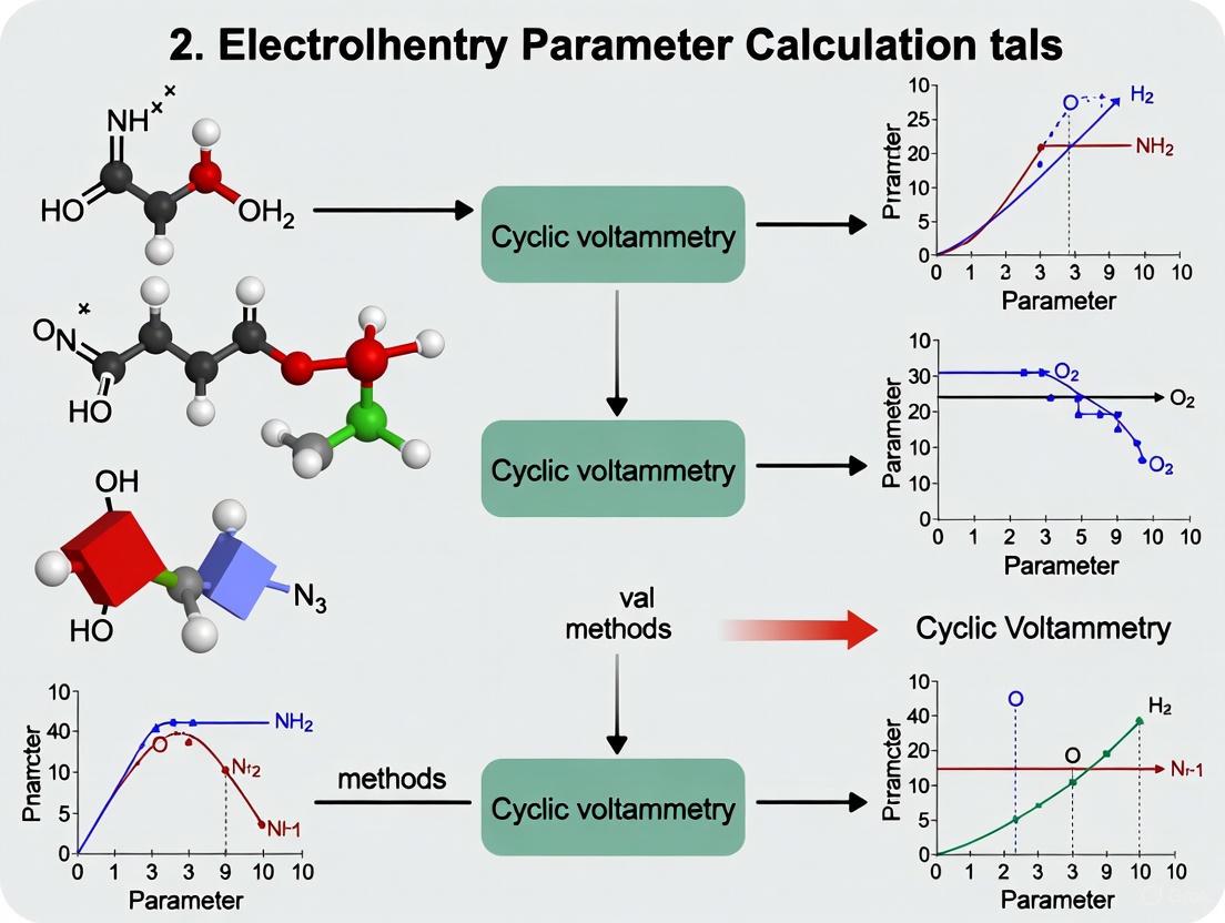
The relationship between experimental parameters and calculation methodologies follows a logical pathway that ensures accurate parameter determination. The diagram below illustrates this methodological framework:
Methodological Framework for Parameter Calculation
Essential Research Reagent Solutions
Successful electrochemical parameter determination requires specific materials and reagents optimized for reliable performance. The following table details essential research reagent solutions and their functions in electrochemical experiments:
Table 4: Essential Research Reagent Solutions for Electrochemical Parameter Determination
| Reagent/Material | Function | Specifications | Application Notes |
|---|---|---|---|
| Glassy Carbon Electrode | Working electrode surface for electron transfer | Standard surface area: 0.0706 cm² | Polish with 0.2 µm aluminum powder before each use for reproducible results [2] |
| Supporting Electrolyte (LiClO₄) | Maintains constant ionic strength; minimizes migration effects | 0.1 M concentration in aqueous solutions | Electrochemically inert in studied potential window; prevents ohmic resistance effects [2] |
| Paracetamol Standard | Model electroactive compound for method validation | 1×10⁻⁶ M concentration in deionized water | Exhibits quasi-reversible electron transfer with coupled chemical reactions [2] |
| Nitrogen Gas | Removal of dissolved oxygen from solutions | High purity (>99.9%) | Purge for 15 minutes before measurements to prevent oxygen interference [2] |
| Reference Electrode (Saturated Calomel) | Provides stable reference potential | SCE with constant KCl saturation | All reported potentials referenced to SCE potential [2] |
| Polishing Material (Aluminum Powder) | Electrode surface renewal | 0.2 µm particle size | Essential for reproducible electrode surface conditions [2] |
The comparative analysis presented in this guide demonstrates that method selection critically impacts the accuracy of α, D₀, and k₀ determination in electrochemical studies. For quasi-reversible systems, the Ep - Ep/2 equation for α calculation and the modified Randles-Ševčík equation for D₀ determination emerge as optimal choices, while the Kochi and Gileadi method provides reliable k₀ values. The experimental protocols and reagent specifications outlined herein provide researchers with a standardized framework for electrochemical parameter determination, facilitating more accurate and reproducible characterization of electrode processes across diverse applications in electrocatalysis, sensor development, and energy storage systems.
In electrochemical research, the classification of reactions as reversible, quasi-reversible, or irreversible forms a cornerstone for understanding electron transfer kinetics and designing experimental methodologies. This classification profoundly impacts parameter calculation, analytical sensitivity, and mechanistic interpretation across diverse fields including pharmaceutical development, sensor design, and energy storage systems. The terminology of "reversibility" in electrochemistry often causes confusion, as it encompasses both chemical reversibility (the stability of redox products) and electrochemical reversibility (the kinetics of electron transfer relative to mass transport) [13] [14].
The distinction between these reaction types is not merely academic; it determines the appropriate mathematical models for calculating essential parameters such as the heterogeneous electron transfer rate constant (k₀), transfer coefficient (α), and diffusion coefficient (D₀) [2] [14]. Misclassification can lead to significant errors in parameter estimation, potentially compromising pharmaceutical quantification accuracy or battery material assessments. This guide systematically compares these reaction classes using experimental data and standardized protocols to empower researchers in making accurate electrochemical classifications.
Fundamental Definitions and Theoretical Framework
Electrochemical versus Chemical Reversibility
A critical conceptual foundation lies in distinguishing between electrochemical and chemical reversibility:
Electrochemical Reversibility concerns the kinetics of electron transfer between the electrode and solution species. A system is electrochemically reversible when electron transfer occurs rapidly compared to mass transport, establishing Nernstian equilibrium at the electrode surface throughout the potential scan [13] [15]. This is quantified by the standard heterogeneous rate constant, k₀.
Chemical Reversibility refers to the stability of the electrogenerated product. A system is chemically reversible when the product of the electron transfer remains stable long enough to be re-converted to the original species during the reverse potential scan [13] [14]. When the product undergoes a subsequent irreversible chemical reaction (denoted in EC mechanisms), chemical irreversibility occurs [14].
This distinction explains why a system can be electrochemically reversible yet chemically irreversible, or vice versa, necessitating careful diagnostic approaches.
The Butler-Volmer Framework and Key Parameters
Electron transfer kinetics are described by the Butler-Volmer equations [14]: [ kf = k^0 \exp\left({-\alpha f (E - E^{0'})}\right) ] [ kb = k^0 \exp\left({(1-\alpha) f (E - E^{0'})}\right) ]
Where ( kf ) and ( kb ) are the forward and backward rate constants, ( k^0 ) is the standard heterogeneous rate constant (cm/s), ( \alpha ) is the charge transfer coefficient, ( E ) is the applied potential, and ( E^{0'} ) is the formal potential [14].
The parameter ( k^0 ) indicates the kinetic facility of a redox couple—its intrinsic ability to exchange electrons [14]. However, the observed electrochemical reversibility depends on the ratio of charge transfer to mass transfer rates, formalized in the reversibility parameter, ( \Lambda ) [14] [15]: [ \Lambda = \frac{k^0}{(D f \nu)^{0.5}} ]
Where ( D ) is the diffusion coefficient, ( f = F/RT ), and ( \nu ) is the scan rate. This relationship highlights that reversibility is not defined by ( k^0 ) alone but by its value relative to experimental conditions, particularly scan rate.
Table 1: Key Parameters Defining Electrochemical Reaction Types
| Parameter | Reversible | Quasi-Reversible | Irreversible |
|---|---|---|---|
| Heterogeneous Rate Constant (k⁰) | k⁰ > 2 × 10⁻² cm/s [2] | 2 × 10⁻² > k⁰ > 3 × 10⁻⁵ cm/s [2] | k⁰ < 3 × 10⁻⁵ cm/s [2] |
| Reversibility Parameter (Λ) | Λ ≥ 15 [14] | 15 ≥ Λ ≥ 10⁻²⁽¹⁺ᵅ⁾ [14] | Λ ≤ 10⁻²⁽¹⁺ᵅ⁾ [14] |
| Peak Separation (ΔEₚ) | ΔEₚ ≈ 59/n mV at 25°C [16] | ΔEₚ > 59/n mV, increases with ν [2] | ΔEₚ > 59/n mV, large and ν-dependent [2] |
| Electron Transfer Kinetics | Fast relative to mass transport [13] [14] | Intermediate [17] | Slow relative to mass transport [13] [14] |
| Current Function | ( I_p \propto \nu^{1/2} ) [16] | ( I_p \propto \nu^{1/2} ) (with deviations) [2] | ( I_p \propto \nu^{1/2} ) [2] |
Experimental Protocols for Reaction Classification
Standard Cyclic Voltammetry Methodology
Cyclic Voltammetry (CV) is the primary experimental technique for classifying electrochemical reactions [16]. The protocol involves applying a linear potential sweep between defined limits while measuring the resulting current, then reversing the scan direction to complete the cycle [18] [15].
Recommended Experimental Protocol [2] [18]:
- Electrode Preparation: Polish the working electrode (typically glassy carbon) with 0.2 µm alumina powder to ensure a reproducible surface [2]. Clean and dry before use.
- Solution Preparation: Prepare analyte solution with supporting electrolyte (e.g., 0.1 M LiClO₄ or KNO₃) to minimize ohmic drop [2] [19]. Degas with inert gas (N₂ or Ar) for 15 minutes to remove oxygen [2].
- Instrument Parameters: Use a standard three-electrode system (working, reference, counter) [2]. Set initial potential positive of the expected redox event. Apply scan rates typically from 0.025 V/s to 0.300 V/s (or broader: 10 mV/s to 1000 V/s) [2] [15].
- Data Collection: Record cyclic voltammograms at multiple scan rates. Measure peak potentials (Epa, Epc), peak currents (Ipa, Ipc), and calculate peak separation (ΔEp = |Epc - Epa|) and formal potential (E₁/₂ = |Epc - Epa|/2) for each scan rate [2].
Diagnostic Data Analysis Workflow
The classification of reaction type relies on specific diagnostic relationships observable in CV data:
Diagram 1: Diagnostic workflow for classifying electrochemical systems using cyclic voltammetry data.
Comparative Analysis of Reaction Types
Reversible Systems
Definition and Characteristics: Reversible systems exhibit fast electron transfer kinetics where Nernstian equilibrium is maintained at the electrode surface throughout the potential scan [13]. The redox couple rapidly interconverts with the applied potential, creating the characteristic "duck-shaped" voltammogram with symmetrical forward and reverse peaks [14].
Diagnostic Features [2] [16] [15]:
- Peak separation (ΔEₚ) remains constant at approximately 59/n mV (at 25°C) regardless of scan rate
- Ratio of reverse to forward peak currents (Ipc/Ipa) is close to unity
- Peak current (Iₚ) is proportional to the square root of scan rate (ν¹/²)
- Formal potential (E₁/₂) is located midway between the anodic and cathodic peaks
- Peak potentials are independent of scan rate
Quantitative Example: For a reversible one-electron transfer process, the Randles-Ševčík equation describes the peak current [19] [15]: [ I_p = 0.446 \times n F A C \sqrt{\frac{n F \nu D}{R T}} ] Where n is electron number, F is Faraday's constant, A is electrode area, C is concentration, ν is scan rate, D is diffusion coefficient, R is gas constant, and T is temperature.
Quasi-Reversible Systems
Definition and Characteristics: Quasi-reversible systems represent the intermediate case where electron transfer kinetics are comparable to the mass transfer rate, causing deviation from ideal Nernstian behavior while still displaying both forward and reverse peaks [2] [17]. Most experimentally studied systems fall into this category [2].
Diagnostic Features [2]:
- Peak separation (ΔEₚ) exceeds 59/n mV and increases with scan rate
- Both peak potentials shift with changing scan rate
- Peak current remains proportional to ν¹/², but with a smaller proportionality constant than reversible systems
- The reverse peak decreases relative to the forward peak but remains observable
Experimental Case Study - Paracetamol: Research on paracetamol oxidation demonstrated quasi-reversible behavior with ΔEₚ increasing from 0.128 V to 0.186 V as scan rate increased from 0.025 V/s to 0.300 V/s [2]. The Ipc/Ipa ratio remained constant at 0.59 ± 0.03, indicating coupled chemical reactions consuming the oxidized product [2]. This example highlights how quasi-reversible behavior often accompanies complex reaction mechanisms.
Irreversible Systems
Definition and Characteristics: Irreversible systems exhibit slow electron transfer kinetics where the reverse peak is absent or significantly diminished because the electrogenerated species undergoes chemical transformation before the reverse scan can occur [13] [20].
- No reverse peak is observed, or it is greatly diminished relative to the forward peak
- Large peak separation that increases significantly with scan rate
- Peak current remains proportional to ν¹/²
- Peak potential shifts with scan rate according to: Eₚ = E⁰ - (RT/αnF)[0.78 - ln(k⁰/D¹/²) + ln(αnFν/RT)¹/²]
Mechanistic Insight: True electrochemical irreversibility arises from slow electron transfer kinetics, while apparent irreversibility may result from coupled chemical reactions (EC mechanisms) where the product is chemically consumed [20] [14]. Distinguishing these requires additional experiments varying time scales (scan rates) or complementary techniques.
Table 2: Comparative Diagnostic Features from Cyclic Voltammetry
| Diagnostic Feature | Reversible | Quasi-Reversible | Irreversible |
|---|---|---|---|
| Peak Separation (ΔEₚ) | ~59/n mV, constant [2] [16] | >59/n mV, increases with ν [2] | Large, strongly ν-dependent [2] |
| Iₚc/Iₚa Ratio | ~1 [2] | ≤1, may decrease with ν [2] | <<1 or no reverse peak [20] |
| Peak Potential (Eₚ) | Independent of ν [2] | Shifts with ν [2] | Shifts significantly with ν [2] |
| Scan Rate Dependence (Iₚ vs ν¹/²) | Linear [16] | Linear [2] | Linear [2] |
| Key Identifying Feature | Ideal "duck shape", constant ΔEₚ [14] | Observable but separated peaks, ν-dependent ΔEₚ [2] | Missing or small reverse peak [20] |
Parameter Calculation Methods Compared
Determining Key Electrochemical Parameters
Accurate parameter calculation requires method selection based on the classified reaction type. A comparative study using paracetamol as a model system evaluated different methodologies [2]:
Transfer Coefficient (α) Calculation:
- The Eₚ - Eₚ/₂ method proved most effective for quasi-reversible reactions [2]
- This method utilizes the potential difference between the peak potential and the potential at half peak current
Diffusion Coefficient (D₀) Determination:
- The modified Randles-Ševčík equation provided optimal results [2]
- Requires accurate values for n, α, and concentration
Heterogeneous Electron Transfer Rate Constant (k₀) Estimation:
- Kochi and Gileadi methods provided reliable calculations for quasi-reversible systems [2]
- The Nicholson and Shain method (k₀ = Ψ(πnD₀Fν/RT)¹/²) tended to overestimate k₀ values [2]
- However, the plot of ν⁻¹/² versus Ψ (from the Nicholson and Shain equation) agreed well with Kochi and Gileadi methods [2]
Impact of Reaction Classification on Parameter Accuracy
Misclassifying reaction type introduces significant errors in calculated parameters. For example, applying reversible models to quasi-reversible systems overestimates k₀ values, while using irreversible models for quasi-reversible systems underestimates kinetic parameters [2]. The transfer coefficient (α) calculation is particularly sensitive to proper classification, as different mathematical relationships apply to each reaction type [2].
Table 3: Recommended Calculation Methods by Reaction Type
| Parameter | Reversible | Quasi-Reversible | Irreversible |
|---|---|---|---|
| Transfer Coefficient (α) | From Tafel plot [19] | Eₚ - Eₚ/₂ method [2] | Tafel analysis [19] |
| Diffusion Coefficient (D₀) | Randles-Ševčík equation [2] [19] | Modified Randles-Ševčík equation [2] | Randles-Ševčík equation (irreversible form) [19] |
| Rate Constant (k⁰) | From ΔEₚ and ν dependence [2] | Kochi and Gileadi methods [2] | Nicholson and Shain method (with correction) [2] |
| Electron Count (n) | From ΔEₚ [2] | From digital simulation [2] | From Tafel slope [19] |
The Scientist's Toolkit: Essential Research Reagents and Materials
Table 4: Essential Materials for Electrochemical Classification Studies
| Material/Reagent | Function/Application | Example Specifications |
|---|---|---|
| Glassy Carbon Electrode | Working electrode providing inert surface for electron transfer [2] | 0.0706 cm² surface area, polished with 0.2 µm alumina [2] |
| Supporting Electrolyte | Minimizes ohmic drop, carries current without participating in reaction [2] | 0.1 M LiClO₄ or KNO₃ in deionized water [2] [19] |
| Paracetamol Standard | Model compound for quasi-reversible reaction studies [2] | 1 × 10⁻⁶ M in supporting electrolyte [2] |
| Potentiostat | Instrument for applying potential and measuring current [2] [18] | CHI 760D or equivalent with three-electrode capability [2] |
| Reference Electrode | Provides stable potential reference [2] [19] | Saturated Calomel Electrode (SCE) or Ag/AgCl [2] |
| Counter Electrode | Completes electrical circuit without contamination [2] | Platinum wire or graphite rod [2] [19] |
| Digital Simulation Software | Verifies parameters and reaction mechanisms [2] | DigiSim software or EC-Lab CV Sim/CV Fit [2] [15] |
Implications for Pharmaceutical Research and Development
Proper classification of electrochemical reactions carries particular significance in pharmaceutical applications where redox properties influence drug stability, metabolic pathways, and analytical detection [16]. Paracetamol serves as an exemplary case where its quasi-reversible oxidation with coupled chemical reactions mirrors metabolic oxidation pathways [2]. The Ipc/Ipa ratio <1 observed in paracetamol voltammetry indicates chemical consumption of the oxidized product, providing insight into decomposition pathways relevant to pharmaceutical formulation stability [2].
Understanding these electrochemical classifications enables researchers to:
- Select appropriate quantification methods for electroactive pharmaceuticals
- Predict stability issues related to redox processes
- Design sensors with optimized potential windows
- Interpret mechanistic pathways for drug metabolism
- Develop accelerated stability testing protocols based on electrochemical screening
The systematic approach to classification presented in this guide provides pharmaceutical scientists with a framework for incorporating electrochemical characterization into drug development pipelines, potentially identifying stability issues or metabolic activation pathways early in the development process.
The Critical Role of Accurate Parameter Calculation in Pharmaceutical and Bioanalytical Applications
In pharmaceutical and bioanalytical sciences, understanding the electrochemical behavior of molecules is not merely an academic exercise—it is a critical component of drug stability profiling, metabolite prediction, and biosensor development. Cyclic voltammetry (CV) stands as a frontline technique for investigating redox processes of pharmacologically relevant compounds, providing invaluable insights into their electron transfer mechanisms and coupled chemical reactions [2] [21]. The accurate determination of electrochemical parameters—including the heterogeneous electron transfer rate constant (k⁰), charge transfer coefficient (α), and diffusion coefficient (D₀)—directly impacts the reliability of data used to make critical decisions in drug development pipelines.
Despite the widespread application of CV in pharmaceutical research, a significant challenge persists: the selection of appropriate mathematical formalisms for parameter calculation can lead to substantially different results, potentially compromising data integrity and reproducibility [1] [2]. Recent investigations have revealed that conventional methods long trusted by researchers may contain fundamental flaws, necessitating a systematic comparison of available approaches and their applicability domains for specific pharmaceutical scenarios [1]. This guide provides an objective comparison of contemporary CV parameter calculation methodologies, supported by experimental data, to empower researchers in selecting the most reliable approaches for their specific bioanalytical applications.
Comparative Analysis of Parameter Calculation Methods
Key Electrochemical Parameters and Their Pharmaceutical Relevance
In pharmaceutical electroanalysis, three fundamental parameters provide critical insights into drug compound behavior. The heterogeneous electron transfer rate constant (k⁰) quantifies the kinetics of electron transfer between a drug molecule and electrode surface, with values categorizing reactions as reversible (k⁰ > 2 × 10⁻² cm/s), quasi-reversible (k⁰ = 2 × 10⁻² to 3 × 10⁻⁵ cm/s), or irreversible (k⁰ < 3 × 10⁻⁵ cm/s) [2]. This parameter directly impacts drug reactivity predictions and sensor design. The charge transfer coefficient (α) describes the symmetry of the energy barrier for electron transfer, affecting how reaction rates change with applied potential and providing mechanistic insights for metabolically relevant redox processes [2]. The diffusion coefficient (D₀) characterizes the transport of analyte molecules to the electrode surface, influencing response times in analytical systems and providing information about molecular size and aggregation behavior in physiological environments [2] [21].
Methodologies for Parameter Calculation: A Comparative Assessment
Table 1: Comparison of Methods for Calculating Charge Transfer Coefficient (α)
| Method | Theoretical Basis | Applicability | Limitations | Reported Values for Paracetamol |
|---|---|---|---|---|
| Ep − Ep/2 Equation | Based on potential difference between peak and half-peak potential [1] | Irreversible systems with ΔEp ≥ 443 mV | Requires specific peak separation; less reliable for quasi-reversible systems | α = 0.52 (most effective) [2] |
| Tafel Plot Analysis | Plot of log|Ired| vs. E; slope = -2.303RT/αnF [1] | Systems with dominant faradaic process | Sensitive to background current; requires careful baseline correction | Comparable results when properly implemented [2] |
Table 2: Comparison of Methods for Calculating Heterogeneous Electron Transfer Rate Constant (k⁰)
| Method | Theoretical Basis | Applicability | Limitations | Reported k⁰ for Paracetamol (cm/s) |
|---|---|---|---|---|
| Nicholson and Shain | k⁰ = Ψ(πnD₀Fν/RT)¹/² [1] [2] | Quasi-reversible systems | Can overestimate k⁰ values; limited to ΔEp < 200 mV | 6.72 × 10⁻³ (overestimated) [2] |
| Kochi and Gileadi | Based on peak potential separation analysis [2] | Quasi-reversible and irreversible systems | Requires validation across multiple scan rates | 2.15 × 10⁻³ (reliable alternative) [2] |
| Conventional Klingler-Kochi | k⁰ = 2.18(nαcD₀Fν/RT)¹/²exp[-αc²nF/RT(Epa-Epc)] [1] | Quasi-reversible couples with ΔEp ≥ 150 mV | Recently identified as flawed; provides erroneous values | Not recommended due to inaccuracies [1] |
| Corrected Klingler-Kochi | Revised methodology validated by digital simulation [1] | Quasi-reversible and irreversible couples | Requires specialized fitting procedures | Accurate for validated redox couples [1] |
Table 3: Comparison of Methods for Calculating Diffusion Coefficient (D₀)
| Method | Theoretical Basis | Applicability | Limitations | Implementation Considerations |
|---|---|---|---|---|
| Modified Randles-Ševčík | Ip = 2.69×10⁵n³/²AD₀¹/²Cν¹/² [2] [5] [22] | Reversible and quasi-reversible systems | Assumes reversible electron transfer; requires independent n determination | Most effective when electron transfer is sufficiently fast [2] |
| Chronoamperometry | It = nFAD₀¹/²C/(πt)¹/² (Cottrell equation) [21] | Systems with stable redox products | Sensitive to charging current at short times | Useful for validation of CV-based results [21] |
The recent investigation by Agarwal (2025) has fundamentally challenged the long-standing conventional Klingler-Kochi method, demonstrating through digital simulations and experimental studies with uranium and plutonium complexes that the traditional expressions yield erroneous kinetic parameters [1]. This finding is particularly relevant for pharmaceutical researchers studying compounds with similar quasi-reversible redox behavior. The paracetamol case study further reinforces that method selection significantly impacts results, with the Kochi and Gileadi approach providing more reliable k⁰ values compared to the overestimation observed with the Nicholson and Shain method [2].
Experimental Protocols for Method Validation
Case Study: Comprehensive Electrochemical Analysis of Paracetamol
Materials and Experimental Conditions:
- Analyte: Paracetamol (1 × 10⁻⁶ M) in deionized water [2]
- Supporting Electrolyte: LiClO₄ (0.1 M) to maintain ionic strength [2]
- Working Electrode: Glassy carbon (surface area: 0.0706 cm²) polished with 0.2 µm aluminum powder [2]
- Reference Electrode: Saturated calomel electrode (SCE) [2]
- Counter Electrode: Platinum wire [2]
- Instrumentation: CHI 760D Electrochemical Workstation [2]
Experimental Procedure:
- Solution Preparation: Dissolve paracetamol in deionized water with 0.1 M LiClO₄ as supporting electrolyte [2]
- Deaeration: Purge solution with nitrogen gas for 15 minutes to remove dissolved oxygen [2]
- Electrode Preparation: Polish glassy carbon working electrode with 0.2 µm aluminum powder to ensure reproducible surface [2]
- Voltammetric Data Collection:
- Data Analysis:
Validation Approach:
- Compare parameter values obtained from different calculation methods [2]
- Validate results through digital simulation of cyclic voltammograms using DigiSim software [2]
- Confirm reaction mechanism through diagnostic criteria including peak separation dependence on scan rate and peak current ratios [2]
Workflow for Method Selection and Validation
Advanced Validation Using Digital Simulation
The most robust approach for validating electrochemical parameters involves digital simulation of experimental voltammograms [1] [2]. This process involves:
- Initial Parameter Estimation: Using analytically determined values of k⁰, α, and D₀ as initial inputs for simulation software [1]
- Theoretical CV Generation: Simulating cyclic voltammograms incorporating all relevant experimental parameters (electrode geometry, scan rates, uncompensated resistance) [15]
- Iterative Refinement: Adjusting kinetic parameters until optimal fit between simulated and experimental voltammograms is achieved [2]
- Goodness-of-Fit Assessment: Quantitatively comparing simulated and experimental data across all scan rates [1]
Commercial software packages such as DigiSim and DigiElch provide robust platforms for this validation approach, while custom simulations can be developed in Python or Matlab for more specialized applications [1].
The Scientist's Toolkit: Essential Research Reagent Solutions
Table 4: Essential Materials and Reagents for Pharmaceutical Cyclic Voltammetry
| Item | Function | Pharmaceutical Application Notes |
|---|---|---|
| Glassy Carbon Electrode | Working electrode platform | Biocompatible surface; minimal catalytic activity; suitable for drug compounds [2] |
| Platinum Counter Electrode | Current conduction | Inert surface prevents contamination; essential for clean baseline [2] |
| Saturated Calomel Reference | Potential reference | Stable reference potential; compatible with aqueous pharmaceutical systems [2] |
| Lithium Perchlorate | Supporting electrolyte | Minimal specific adsorption; wide potential window [2] |
| DigiSim Software | CV simulation | Experimental validation; mechanism verification [1] [2] |
| Nitrogen Gas | Solution deaeration | Removes dissolved oxygen that interferes with drug redox signals [2] |
Method Selection Guidelines for Pharmaceutical Applications
The selection of appropriate parameter calculation methods must be guided by the specific electrochemical behavior of the pharmaceutical compound under investigation and the required reliability of the resulting data. Based on comparative experimental evidence:
- For charge transfer coefficient (α) determination, the Ep - Ep/2 method provides the most reliable results for quasi-reversible pharmaceutical compounds like paracetamol [2]
- For heterogeneous electron transfer rate constant (k⁰), the Kochi and Gileadi method offers superior reliability compared to conventional approaches, while the corrected Klingler-Kochi method should be employed for systems with ΔEp ≥ 150 mV [1] [2]
- For diffusion coefficient (D₀) calculation, the modified Randles-Ševčík equation provides accurate results when applied to appropriate reversible systems [2]
- Digital simulation validation should be incorporated as a standard procedure for high-stakes pharmaceutical applications to verify accuracy of analytically determined parameters [1] [2]
The convergence of evidence from multiple studies indicates that the uncritical application of conventional electrochemical parameter calculation methods can lead to significantly inaccurate results, potentially compromising pharmaceutical development decisions based on these data. Implementation of the validated approaches outlined in this guide will enhance the reliability and reproducibility of electrochemical data in pharmaceutical and bioanalytical applications.
A Practical Guide to Calculating CV Parameters: Methods, Equations, and a Paracetamol Case Study
In electrochemical research, the transfer coefficient (α) is a fundamental kinetic parameter that signifies the symmetry of the activation energy barrier for an electron transfer reaction, profoundly influencing the reaction rate and mechanism [2]. Accurate determination of α is essential for a deep understanding of processes in catalysis, sensors, batteries, and drug development [2]. Cyclic voltammetry (CV) is a frontline technique for investigating these parameters, but the selection of a feasible calculation method requires careful consideration due to the complex nature of electrode reactions [2]. This guide provides a comparative analysis of methodological approaches for determining the transfer coefficient, focusing on the Eₚ - Eₚ/₂ equation and contrasting it with alternative methods to guide researchers in selecting the most appropriate protocol for their specific electrochemical system.
The determination of the transfer coefficient often relies on analyzing the shape and position of peaks in a cyclic voltammogram. The following sections detail the prominent methods, with a focus on the Eₚ - Eₚ/₂ technique.
The Eₚ - Eₚ/₂ Equation
This method is applied to irreversible or quasi-reversible systems. It utilizes the potential difference between the peak potential (Eₚ) and the potential at half the peak current (Eₚ/₂) from a cyclic voltammogram. The value of the transfer coefficient (α) for a reduction reaction is calculated using the following equation:
α = (47.7 / n) mV / (Eₚ - Eₚ/₂) (at 25 °C)
The number of electrons transferred in the reaction is denoted by 'n' [2]. The strength of this method lies in its direct extraction of information from a single voltammogram, requiring no variation of scan rate.
Alternative Methods for Determining (α)
Other established methods provide alternative pathways for determining the transfer coefficient, often relying on scan rate-dependent data.
Tafel Plot Analysis: This method involves plotting the overpotential (η) against the logarithm of the current (log |i|) in a region where the reverse reaction and mass transport effects are negligible. The slope of the linear Tafel region is equal to ±(2.3RT/αnF), from which α can be extracted. This method is typically suitable for fully irreversible systems.
Variation of Peak Potential with Scan Rate: For an irreversible system, the peak potential (Eₚ) shifts with the logarithm of the scan rate (log ν). The transfer coefficient can be determined from the slope of a plot of Eₚ versus log ν, as described by the equation: slope = (2.3RT / 2αnF). This method requires collecting voltammograms at multiple scan rates.
Analysis of the Full Voltammetric Shape using Digital Simulation: This is considered the most rigorous approach. It involves simulating the entire cyclic voltammogram using specialized software (e.g., DigiSim, DigiElch) by varying kinetic parameters until the simulated curve matches the experimental data [6]. The value of α used to achieve the best fit is then reported. This method is powerful for complex mechanisms involving coupled chemical reactions.
Table 1: Comparative Analysis of Methods for Determining the Transfer Coefficient (α)
| Method | Underlying Principle | Experimental Data Required | Applicable System | Key Advantages | Key Limitations |
|---|---|---|---|---|---|
| Eₚ - Eₚ/₂ Equation | Peak shape analysis (Eₚ - Eₚ/₂) | One CV at a single scan rate | Irreversible, Quasi-Reversible | Simple, fast, requires only one CV | Accuracy can be affected by baseline current and signal-to-noise. |
| Tafel Plot | Linear region of log |i| vs. η | Current-potential data from a single CV | Irreversible | Direct kinetic information from a single scan | Requires a well-defined Tafel region unaffected by mass transport. |
| Eₚ vs. log ν | Scan rate dependence of Eₚ | Multiple CVs at different scan rates | Irreversible | Confirms irreversibility; provides kinetic info | Time-consuming; requires multiple experiments. |
| Digital Simulation | Whole curve fitting | One or multiple CVs | All system types | Most accurate for complex mechanisms; provides full kinetic picture | Requires specialized software and expertise; computationally intensive. |
Experimental Case Study: Paracetamol Electro-oxidation
A recent study directly compared different electrochemical methodologies for calculating α, D₀, and k₀, using the electro-oxidation of paracetamol as a case study [2]. The experimental workflow and key findings are summarized below.
Figure 1: Experimental workflow for the comparative determination of electrochemical parameters using paracetamol as a model compound [2].
Detailed Experimental Protocol
- Reagents and Solution Preparation: A 10 mL solution of 1 x 10⁻³ M paracetamol was prepared using deionized water, with 0.1 M lithium perchlorate (LiClO₄) as the supporting electrolyte [2].
- Instrumentation and Electrodes: Cyclic voltammetry was performed using a CHI 760D Electrochemical Workstation. A conventional three-electrode cell was employed, consisting of a glassy carbon (GC) working electrode (surface area: 0.0706 cm²), a platinum counter electrode, and a saturated calomel electrode (SCE) as the reference [2].
- Electrode Preparation and Measurement: Before measurements, the glassy carbon working electrode was polished with 0.2 µm aluminum powder to ensure a clean and reproducible surface. The solution was purged with nitrogen gas for 15 minutes to remove dissolved oxygen. Cyclic voltammograms were recorded at scan rates ranging from 0.025 V/s to 0.300 V/s, with an incremental change of 0.025 V/s [2].
- Data Analysis: From the collected voltammograms, key parameters like anodic peak potential (Epa), cathodic peak potential (Epc), anodic peak current (Ipa), and cathodic peak current (Ipc) were directly measured. The transfer coefficient (α) was then calculated using the Eₚ - Eₚ/₂ method [2].
Key Research Reagent Solutions
Table 2: Essential Materials and Reagents for Electrochemical Parameter Determination
| Item | Function / Role in the Experiment |
|---|---|
| Potentiostat/Galvanostat | Instrument for applying potential and measuring current (e.g., CHI 760D, WaveDriver 200) [2] [6]. |
| Glassy Carbon Electrode | Common working electrode material providing a reproducible and inert surface for electron transfer [2]. |
| Saturated Calomel Electrode (SCE) | Reference electrode providing a stable and known potential against which the working electrode is measured [2]. |
| Platinum Counter Electrode | Completes the electrical circuit in the three-electrode cell, allowing current to flow [2]. |
| Supporting Electrolyte (e.g., LiClO₄) | Minimizes solution resistance and governs ionic strength, ensuring the current is due to the analyte's faradaic process [2]. |
| Aluminum Powder (0.2 µm) | Abrasive material for polishing the working electrode to achieve a fresh, clean, and reproducible surface before experiments [2]. |
| Digital Simulation Software | Software (e.g., DigiSim, DigiElch) used for modeling and fitting entire voltammograms to extract kinetic parameters [6]. |
Results and Discussion
The comparative study on paracetamol provided critical insights into the performance of the Eₚ - Eₚ/₂ method relative to other techniques for parameter calculation.
The findings demonstrated that for the studied quasi-reversible system, the Eₚ - Eₚ/₂ equation for α and the modified Randles–Ševčík equation for the diffusion coefficient (D₀) were particularly effective [2]. The calculated kinetic parameters were further validated through digital simulation of the cyclic voltammograms, confirming the reliability of this combined approach [2].
It is crucial to note that the accuracy of the calculated heterogeneous electron transfer rate constant (k₀) is highly dependent on the precise determination of both α and D₀ [2]. The study also highlighted that the widely used Nicholson and Shain method (k₀ = Ψ(πnD₀Fν/RT)¹/²) can lead to overestimated k₀ values. In contrast, the Kochi and Gileadi methods, as well as an alternative plot of ν⁻¹/² versus Ψ, were identified as reliable alternatives for calculating k₀ [2].
The selection of a methodology for determining the transfer coefficient (α) is not a one-size-fits-all process. Based on the comparative data, the Eₚ - Eₚ/₂ method stands out for its simplicity and effectiveness, especially for quasi-reversible systems like the paracetamol model presented. It provides a direct calculation from a single voltammogram, making it an excellent first choice for initial kinetic assessment.
For systems with more complex mechanisms or when the highest accuracy is required, digital simulation of the entire voltammogram, though more demanding, provides the most comprehensive and reliable results [6]. Researchers should therefore base their choice on the nature of their electrochemical reaction, the required precision, and the available experimental resources. This guide underscores that a critical evaluation of methodological strengths and limitations is fundamental to advancing accurate electrochemical research in fields ranging from drug development to energy storage.
In electrochemical research, the diffusion coefficient (D₀) is a fundamental physical parameter that quantifies the rate at which an electroactive species travels through a solution under a concentration gradient [23]. Accurate determination of D₀ is essential for understanding reaction mechanisms, optimizing electrochemical systems, and developing applications in fields ranging from battery research to sensor design [16] [24]. Cyclic voltammetry (CV), a widely used electroanalytical technique, serves as a primary tool for extracting this parameter by studying the current response of a system to a linearly cycled potential [16].
The Randles-Ševčík equation represents the classical theoretical relationship for determining diffusion coefficients from cyclic voltammetry data, specifically for reversible, diffusion-controlled systems with soluble reactants and products [25] [26]. Despite its long-standing utility, the application of this equation has specific limitations, leading to the development of modified approaches and alternative methodologies for more complex scenarios, including quasi-reversible and irreversible systems, or those involving adsorbed species [1] [2] [23]. This guide provides a comparative analysis of these methods, presenting experimental data and protocols to help researchers select the optimal approach for their specific electrochemical system.
Theoretical Framework of the Randles-Ševčík Equation
The Fundamental Equation and Its Components
The Randles-Ševčík equation describes the effect of scan rate on the peak current (ip) in a cyclic voltammetry experiment. For a simple, electrochemically reversible redox reaction where both reactants and products are soluble, the peak current is given by:
ip = 0.4463 n F A C (n F v D₀ / R T)^(1/2) [25]
Where:
- ip = peak current (Amperes, A)
- n = number of electrons transferred in the redox event
- F = Faraday constant (96485 C mol⁻¹)
- A = electrode surface area (cm²)
- C = concentration of the redox species (mol cm⁻³)
- v = scan rate (V s⁻¹)
- D₀ = diffusion coefficient of the oxidized analyte (cm² s⁻¹)
- R = universal gas constant (8.314 J mol⁻¹ K⁻¹)
- T = absolute temperature (K) [25]
For practical use at a standard temperature of 25 °C (298.15 K), the constants can be combined, simplifying the equation to:
ip = (2.69 × 10⁵) n³⁄² A C D₀¹⁄² v¹⁄² [25] [27] [5]
The theoretical foundation of this equation relies on solving Fick's laws of diffusion under specific initial and boundary conditions that assume electrochemical reversibility and semi-infinite linear diffusion [25] [26].
The Randles-Ševčík Plot: A Practical Tool for Analysis
In experimental practice, the Randles-Ševčík equation is typically applied by measuring peak currents across a range of scan rates. The results are presented in a Randles-Ševčík plot, where the peak current (ip) is graphed against the square root of the scan rate (v¹⁄²) [28].
Table 1: Interpretation of Randles-Ševčík Plot Characteristics
| Plot Characteristic | Interpretation | System Behavior |
|---|---|---|
| Linear Trend | The system is diffusion-controlled and likely reversible [28] [5]. | The redox species is freely diffusing, and electron transfer kinetics are fast. |
| Symmetry between Anodic and Cathodic Lines | The redox reaction is reversible [28]. | Equal numbers of molecules are oxidized and reduced each cycle. |
| Deviation from Linearity | The system may be quasi-reversible, irreversible, or involve surface-adsorbed species [28] [26]. | Electron transfer kinetics are slow, or the mechanism is not purely diffusion-limited. |
Comparative Analysis of Methodologies for Determining D₀
While the Randles-Ševčík equation is ideal for reversible systems, several other methods have been developed to handle more complex electrochemical reactions.
Method 1: The Standard Randles-Ševčík Equation
This method is the benchmark for electrochemically reversible systems. Its major application is determining the diffusion coefficient when other parameters are known, but it can also be used to find the electrochemical active area (EAA) of an electrode or the concentration of a redox species [28].
Limitations: The equation is strictly valid only for reversible processes with freely diffusing species and fast electron transfer kinetics. Applying it to quasi-reversible or irreversible systems, or those with adsorbed species, yields inaccurate results [26]. Furthermore, the peak current must be corrected for any significant background charging current [5].
Method 2: The Nicholson and Shain Approach for Quasi-Reversible Systems
For systems that are not fully reversible, the Nicholson and Shain method provides a more general framework. It utilizes the dimensionless kinetic parameter (Ψ), which is related to the standard heterogeneous electron transfer rate constant (k⁰) [1] [2]:
Ψ = k⁰ / [π D₀ n F v / (R T)]^(1/2) [1]
The parameter Ψ can be obtained from experimentally determined peak potential differences (ΔEp) [1]. A plot of Ψ against (n π D₀ F v / R T)^(-1/2) yields a straight line, and the value of k⁰ can be determined from the slope [1]. However, a recent comparative study on the paracetamol redox system found that this method can sometimes overestimate the value of k⁰ [2].
Limitations: The traditional Nicholson method is primarily applicable when the peak potential difference (ΔEp) is less than 200 mV [1]. Its accuracy can be compromised for systems with complex coupled chemical reactions.
Method 3: The Klingler-Kochi Method and Its Recent Correction
The Klingler-Kochi (K-K) method was developed for assessing electrochemical kinetic parameters, including k⁰, for quasi- and irreversible couples with a ΔEp exceeding 150 mV [1]. For years, conventional K-K equations were widely used. However, a 2025 study by Agarwal identified these conventional expressions as flawed, leading to significant errors [1].
The study, which involved digital simulations and experiments with actinide and lanthanide complexes, introduced a corrected Klingler-Kochi method. This revised methodology was validated theoretically and experimentally, showing strong agreement with simulated data for redox couples like [UO₂(CO₃)₃]⁴⁻/⁵⁻ and [PuO₂(CO₃)₃]⁴⁻/⁵⁻, unlike the conventional expressions [1]. The authors advise against using the conventional K-K method and recommend the corrected version for accurate parameter assessment [1].
Method 4: Advanced Regression and Simulation Techniques
Modern computational approaches offer powerful alternatives to traditional analytical methods.
- Digital Simulation (DigiSim, DigiElch): This involves fitting experimental cyclic voltammograms with theoretically generated curves by varying parameters like D₀ and k⁰ [1] [2]. This method is highly versatile and can model complex mechanisms with coupled chemical reactions (EC, EC₂, etc.) [23]. While computationally intensive, it is considered a robust validation tool [1].
- Machine Learning Regression (SVR, GPR): Advanced techniques like Support Vector Regression (SVR) and Gaussian Process Regression (GPR) can be trained on simulated voltammograms to directly estimate diffusion coefficients from experimental data [23]. These methods can achieve high accuracy that is largely independent of the rate constants governing the reaction and can handle various reaction mechanisms more effectively than the standard Nicholson-Shain approach [23].
Table 2: Comparison of Methods for Determining the Diffusion Coefficient D₀
| Method | Best For | Key Formula/Technique | Pros | Cons |
|---|---|---|---|---|
| Randles-Ševčík | Reversible, diffusion-controlled systems [25] [26]. | ip = (2.69 × 10⁵) n³⁄² A C D₀¹⁄² v¹⁄² | Simple, straightforward, widely understood [28]. | Only valid for reversible processes [26]. |
| Nicholson & Shain | Quasi-reversible systems (ΔEp < 200 mV) [1] [2]. | Uses the Ψ parameter and ΔEp [1]. | Well-established for its applicable range. | Can overestimate k⁰; limited to ΔEp < 200 mV [1] [2]. |
| Corrected Klingler-Kochi | Quasi- and irreversible systems (ΔEp ≥ 150 mV) [1]. | Revised equations for Ef⁰ and k⁰ [1]. | Addresses flaws in the conventional method; validated for actinides [1]. | Newer method, requires validation for diverse systems. |
| Digital Simulation | Complex systems with coupled chemical reactions [23]. | Computer-based fitting of full CV curves. | Highly accurate; models complex mechanisms [1]. | Time-consuming; requires expertise and software [23]. |
| Machine Learning (SVR/GPR) | High-throughput analysis of complex data [23]. | Regression models trained on simulated data. | Fast, accurate, and mechanism-flexible after training [23]. | Requires large training datasets and computational resources. |
Experimental Protocols for Key Methods
Protocol 1: Determining D₀ via the Randles-Ševčík Plot
This is a foundational experiment for characterizing a new electroactive species or electrode material [28] [24].
Workflow Overview:
Materials and Reagents:
- Three-Electrode Cell: Consisting of a working electrode (e.g., Glassy Carbon, Pt), a reference electrode (e.g., Saturated Calomel Electrode, Ag/AgCl), and a counter electrode (e.g., Pt wire) [2].
- Potentiostat: An instrument capable of performing cyclic voltammetry and controlling the applied potential.
- Electroactive Species Solution: A solution of known concentration (typically mM range) of the molecule of interest (e.g., paracetamol, ferrocene) [2].
- Supporting Electrolyte: A high-concentration electrolyte (e.g., LiClO₄, KNO₃) to ensure sufficient conductivity and minimize ohmic resistance (IR drop) [1] [2].
Step-by-Step Procedure:
- Solution Preparation: Prepare a solution with a known, precise concentration (C) of the redox species in a suitable solvent, containing a high concentration (e.g., 0.1 M) of an inert supporting electrolyte [2].
- Electrode Preparation: Clean and polish the working electrode according to standard protocols to ensure a reproducible surface area (A) [2].
- Data Collection: Deoxygenate the solution by purging with an inert gas (e.g., N₂ or Ar) for ~15 minutes. Run cyclic voltammetry experiments at a fixed temperature, cycling the potential through the redox event of interest. Repeat this process for a wide range of scan rates (v), for example, from 0.025 V/s to 0.300 V/s [2].
- Peak Current Measurement: For each voltammogram, measure the absolute value of the peak current (iₚ) for either the oxidation or reduction peak.
- Plotting and Calculation: Create a plot of iₚ versus the square root of the scan rate (v¹⁄²). For a diffusion-controlled system, this will yield a straight line. The slope of this line is equal to (2.69 × 10⁵) n³⁄² A C D₀¹⁄². With n, A, and C known, D₀ can be calculated [25] [28].
Protocol 2: Validating Kinetic Parameters Using the Corrected Klingler-Kochi Method
This protocol is recommended for systems suspected to be quasi- or irreversible, especially those with larger peak separations (ΔEp ≥ 150 mV) [1].
Workflow Overview:
Step-by-Step Procedure:
- Data Collection: Follow steps 1-4 from Protocol 1 to obtain cyclic voltammograms at multiple scan rates.
- Peak Potential Separation: For each scan rate, measure the anodic (Epa) and cathodic (Epc) peak potentials and calculate the peak potential difference, ΔEp = |Epa - Epc| [1] [2].
- Determine Charge Transfer Coefficient (α): The cathodic charge transfer coefficient (αc) is a required parameter. It can be determined from a Tafel plot (log|Ired| vs. E) or, for fully irreversible couples (ΔEp ≥ 443 mV), using the equation: Epc - Epc/2 = -1.857 RT/(αc n F) [1].
- Apply Corrected K-K Equations: Use the experimentally determined ΔEp and αc values in the corrected Klingler-Kochi equations to calculate the formal potential (Ef⁰) and the dimensionless kinetic parameter (ψ), which in turn allows for the calculation of the standard rate constant (k⁰) [1].
- Validation via Simulation: The study by Agarwal strongly recommends validating the calculated kinetic parameters by simulating cyclic voltammograms using software like DigiSim or DigiElch with the obtained parameters and comparing them directly with the experimental CVs [1].
The Scientist's Toolkit: Essential Research Reagent Solutions
The following reagents and materials are critical for obtaining reliable electrochemical data for diffusion coefficient calculation.
Table 3: Essential Reagents and Materials for CV Experiments to Determine D₀
| Item | Function/Role | Examples & Notes |
|---|---|---|
| Supporting Electrolyte | To provide ionic conductivity, minimize ohmic resistance (IR drop), and control the ionic strength and double-layer structure [2] [5]. | LiClO₄, KNO₃, TBAPF₆. Must be electrochemically inert in the potential window of study and of high purity [1] [2]. |
| Redox Probe / Analyte | The electroactive species under investigation. Its concentration must be known precisely. | Ferrocene, Paracetamol, [Fe(CN)₆]³⁻/⁴⁻. Often used as internal standards or model systems [25] [2]. |
| Working Electrode | The surface where the redox reaction of interest occurs. Its area (A) must be known or determinable. | Glassy Carbon (GC), Platinum (Pt), Gold (Au). Requires meticulous cleaning and polishing before experiments [2]. |
| Reference Electrode | Provides a stable, known potential against which the working electrode potential is measured. | Saturated Calomel Electrode (SCE), Ag/AgCl. Choice depends on solvent compatibility [2]. |
| Counter Electrode | Completes the electrical circuit by carrying the current flowing from the working electrode. | Platinum wire or coil. Typically made from an inert material [2]. |
| Solvent | The medium in which the electrochemical reaction takes place. | Water, Acetonitrile (ACN), Dichloromethane (DCM). Must be dry and pure, with an appropriate potential window [2]. |
The accurate determination of the diffusion coefficient D₀ hinges on selecting a methodology appropriate for the electrochemical reversibility and mechanism of the system under study.
For simple, reversible systems with fast electron transfer kinetics, the standard Randles-Ševčík equation remains the most efficient and direct method. Its requirement is a linear Randles-Ševčík plot with symmetric anodic and cathodic peaks [28].
For quasi-reversible systems, the choice is more nuanced. The Nicholson and Shain method is applicable for smaller peak separations (ΔEp < 200 mV), though its accuracy should be verified [2]. For systems with larger peak separations (ΔEp ≥ 150 mV), the recently published corrected Klingler-Kochi method presents a robust analytical alternative, provided the charge transfer coefficient can be accurately determined [1].
For the most complex systems, particularly those involving coupled chemical reactions (EC, EC₂, etc.) or when the highest accuracy is required, digital simulation represents the gold standard, despite its computational cost [1] [23]. Emerging machine learning techniques like SVR and GPR offer a promising path for high-throughput, accurate analysis of complex voltammetric data [23].
In practice, a combined approach is often most effective: using an analytical method (e.g., corrected K-K) for an initial estimate and then validating the resulting parameters through digital simulation of the experimental voltammograms [1]. This workflow ensures reliability and deepens the mechanistic understanding of the electrochemical process.
The heterogeneous electron transfer rate constant (k₀) is a fundamental parameter in electrochemistry, providing critical insights into the kinetics of redox reactions at electrode interfaces. Accurate determination of k₀ is essential for advancing applications in electrocatalysis, sensor development, and energy storage systems. Cyclic voltammetry (CV) stands as the predominant experimental technique for probing these kinetics, with several analytical methods developed to extract k₀ from voltammetric data.
Among the most established methodologies are the Nicholson-Shain, Kochi, and Gileadi approaches, each with distinct theoretical foundations, applicability ranges, and practical limitations. This guide provides a systematic comparison of these three methods, supported by experimental case studies and clear protocols to assist researchers in selecting and applying the most appropriate methodology for their electrochemical systems. The evaluation is framed within the critical context of recent research highlighting necessary corrections to conventional practices, ensuring the accurate determination of this crucial kinetic parameter.
Methodological Principles and Theoretical Foundations
The three methods for determining k₀ are based on the analysis of cyclic voltammograms, but they differ in their underlying principles and the specific parameters they utilize.
Nicholson-Shain Method
The Nicholson-Shain method is perhaps the most widely recognized approach for characterizing quasi-reversible electron transfer. It utilizes the kinetic parameter Ψ, a dimensionless quantity related to k₀ through the following equation:
ψ = k₀ / [πD₀ν(nF/RT)]^(1/2) [29] [30]
where D₀ is the diffusion coefficient, ν is the scan rate, n is the number of electrons, F is Faraday's constant, R is the gas constant, and T is the temperature. The parameter Ψ is correlated with the peak potential separation (ΔEₚ), which increases with increasing scan rate for quasi-reversible systems. Nicholson provided a working curve of Ψ as a function of ΔEₚ, allowing for the determination of k₀ once ΔEₚ is measured experimentally [30]. A key advantage of this method is that Ψ is largely unaffected by variations in the charge transfer coefficient (α) over a range of 0.3 to 0.7 [1]. Its applicability, however, is typically limited to systems where ΔEₚ is less than 200 mV [1].
Klingler-Kochi (Kochi) Method
The Klingler-Kochi method offers an analytical approach for systems exhibiting greater irreversibility. The central equation for this method is:
k₀ = 2.18 [ (n αₐ D₀ F ν) / (R T) ]^(1/2) exp[ - (αₐ² n F) / (R T) (Eₚₐ - Eₚ꜀) ] [1]
This method is considered valid for a peak potential difference (ΔEₚ) exceeding 150 mV and for charge transfer coefficient (α) values between 0.3 and 0.7 [1]. A significant recent development is the identification of flaws in the conventional Klingler-Kochi expressions after decades of use. A 2025 publication demonstrated that the traditional equations are erroneous, leading to a corrected Klingler-Kochi method for accurate assessment of electrochemical kinetic parameters [1]. Researchers must ensure they are using the revised formulations for reliable results.
Gileadi Method
The Gileadi method is another important technique for determining k₀. While the specific equations are not detailed in the provided search results, it is noted as a reliable method because the results are less affected by uncompensated solution resistance (IR drop) compared to other techniques [29]. This characteristic makes it particularly valuable for systems where complete iR compensation is challenging. The method has been applied alongside Nicholson and Kochi methods in comprehensive kinetic studies, yielding consistent and comparable k₀ values [29] [31].
Experimental Comparison and Performance Evaluation
Direct experimental comparisons provide the most valuable insights for methodology selection. The following case studies illustrate the performance of these methods in practical research scenarios.
Table 1: Comparative k₀ Values from a Paracetamol Study
| Electrochemical Method | Reported k₀ (cm/s) | Key Findings |
|---|---|---|
| Nicholson-Shain | Not specified (Overestimated) | The basic equation k₀ = Ψ(πnD₀Fν/RT)¹ᐟ² gave overestimated values [2]. |
| Nicholson-Shain (Plot) | Agreed with Kochi & Gileadi | A plot of ν⁻¹ᐟ² versus Ψ yielded k₀ values agreeing with other methods [2]. |
| Kochi Method | Reliable alternative | Identified as a reliable alternative for k₀ calculation [2]. |
| Gileadi Method | Reliable alternative | Confirmed as a reliable alternative for k₀ calculation [2]. |
Table 2: Comparative k₀ Values from an Oxygen/DMSO Study
| Electrochemical Method | Calculated k₀ (cm/s) | Experimental Context |
|---|---|---|
| Nicholson-Shain | Value reported | Used for O₂ to O₂•⁻ conversion in DMSO at a glassy carbon electrode [29] [31]. |
| Kochi Method | Value reported | Applied to the same system; result differed from prior literature [29] [31]. |
| Gileadi Method | Value reported | Less affected by uncompensated IR drop; result differed from prior literature [29] [31]. |
A study on the electro-oxidation of paracetamol, a system involving complex electron transfer and coupled chemical reactions, found that the Nicholson-Shain method can overestimate k₀ if applied without validation [2]. However, when the parameter Ψ (obtained from Nicholson's theory) was plotted against ν⁻¹ᐟ², the resulting k₀ agreed well with values from the Kochi and Gileadi methods, which were identified as reliable alternatives [2].
In a comprehensive study of oxygen reduction in DMSO, all three methods were applied to determine the k₀ for the O₂/O₂•⁻ redox couple [29] [31]. The study highlighted the Gileadi method's relative robustness against errors from uncompensated IR drop. Furthermore, it emphasized that relying on a single method could be misleading, as the calculated k₀ values differed from those previously cited in the literature, underscoring the value of a multi-methodological approach for reliable kinetics evaluation [29].
Detailed Experimental Protocols
To ensure reproducible results, below are detailed protocols for determining k₀ using the different methods, based on standardized experimental procedures.
General Cyclic Voltammetry Setup
- Electrochemical Cell: Use a standard three-electrode configuration [2] [32].
- Working Electrode: Glassy Carbon (GC) is common. Polish the electrode surface thoroughly with 0.2 µm alumina powder before each experiment to ensure reproducibility [2].
- Counter Electrode: Platinum wire [2].
- Reference Electrode: Saturated Calomel Electrode (SCE) or Ag/AgCl [2]. All reported potentials are referenced to the SCE potential.
- Solution Preparation: Dissolve the analyte (e.g., 1 × 10⁻⁶ M paracetamol) in a suitable solvent with a high-concentration supporting electrolyte (e.g., 0.1 M LiClO₄ or TBAP) to minimize solution resistance [2] [29]. Purge the solution with an inert gas (N₂ or Ar) for at least 15 minutes to remove dissolved oxygen before measurements [2].
- Data Collection: Record cyclic voltammograms at multiple scan rates (e.g., from 0.025 V/s to 0.600 V/s) [29] [2]. Ensure the solution remains unstirred during measurements.
Data Analysis Workflow
The following diagram outlines the general decision-making workflow for determining k₀, incorporating the key considerations for each method.
Method-Specific Analysis Procedures
Nicholson-Shain Analysis:
- From your CV data, measure the peak potential separation (ΔEₚ) at different scan rates.
- Refer to Nicholson's standard table (see Table 2 in [30]) to find the value of the kinetic parameter Ψ that corresponds to each measured ΔEₚ.
- Use the equation k₀ = Ψ [πD₀ν(nF/RT)]^(1/2) to calculate the standard rate constant. Note that this method is best suited for systems where ΔEₚ is less than 200 mV [1].
Kochi Analysis (Using the Corrected Method):
- Determine the cathodic charge transfer coefficient (α꜀). This can be done using a Tafel plot (log|Iᵣₑ𝒹| vs. E) or from the peak potential using the equation Eₚ꜀ - Eₚ꜀/₂ = -1.857RT/(α꜀nF) for highly irreversible couples (ΔEₚ ≥ 443 mV) [1].
- Use the corrected Klingler-Kochi equations from recent literature (e.g., [1]) to calculate k₀, inputting the measured values of Eₚₐ, Eₚ꜀, α꜀, and ν.
Gileadi Analysis:
Validation with Digital Simulation
Regardless of the chosen method, validation of the calculated k₀ via digital simulation is strongly recommended [1]. Use commercial software (e.g., DigiSim, DigiElch) or custom programs to simulate cyclic voltammograms using your determined kinetic parameters (n, α, D₀, k₀). A good match between the simulated and experimental CVs across multiple scan rates confirms the accuracy of the extracted parameters [1] [2].
Essential Research Reagents and Materials
Table 3: Key Reagent Solutions and Materials for k₀ Determination Studies
| Item Name | Function & Application Notes |
|---|---|
| Tetrabutylammonium Perchlorate (TBAP) | A common supporting electrolyte used in non-aqueous electrochemical studies (e.g., in DMSO) to provide sufficient ionic conductivity and minimize IR drop [29] [31]. |
| Lithium Perchlorate (LiClO₄) | A supporting electrolyte frequently used in aqueous electrochemical studies [2]. |
| Dimethyl Sulfoxide (DMSO) | A common aprotic solvent used for studying oxygen reduction and other redox processes where the stability of reactive intermediates is required [29] [31]. |
| Potassium Ferrocyanide | A common outer-sphere redox probe with a well-established diffusion coefficient, often used for characterizing electrode electroactive areas and for fundamental electron transfer studies [33]. |
| Alumina Polishing Suspension | Used for polishing and resurfacing solid working electrodes (like Glassy Carbon) to ensure a clean, reproducible surface, which is critical for obtaining reliable and reproducible kinetic data [2]. |
Based on the comparative analysis of experimental data and methodological strengths, the following recommendations are provided for researchers estimating the heterogeneous electron transfer rate constant, k₀:
- For Quasi-Reversible Systems (ΔEₚ < 200 mV), the Nicholson-Shain method remains a standard and reliable approach, especially when using the plot of Ψ versus ν⁻¹ᐟ² to determine k₀ [2].
- For Quasi- or Irreversible Systems (ΔEₚ ≥ 150 mV), the corrected Kochi method should be employed. It is imperative to use the recently revised equations, as the conventional Klingler-Kochi expressions have been shown to be flawed [1].
- For Systems with Significant Uncompensated Resistance, the Gileadi method is highly recommended due to its relative insensitivity to IR drop, which enhances the reliability of the extracted k₀ value [29].
- Best Practice for Robust Kinetics: Employ a multi-methodological approach. Calculate k₀ using at least two independent methods and always validate the results by digitally simulating the cyclic voltammograms. This practice helps cross-verify results and prevents reliance on a single, potentially biased, methodological output [29] [1] [2].
The accurate determination of k₀ is paramount for the development of efficient electrocatalysts, sensors, and energy storage devices. By carefully selecting the appropriate methodology and validating the results, researchers can ensure the kinetic parameters they report are both accurate and reliable.
Cyclic voltammetry (CV) stands as a cornerstone technique for investigating electrochemical reactions, prized for its ability to elucidate redox mechanisms and provide quantitative kinetic parameters [15]. In pharmaceutical analysis, the electrochemical characterization of active compounds like paracetamol (acetaminophen) presents particular challenges due to the prevalence of quasi-reversible electron transfer systems. The accurate calculation of fundamental parameters—the transfer coefficient (α), diffusion coefficient (D₀), and heterogeneous electron transfer rate constant (k₀)—remains essential for understanding electrode processes, yet the selection of appropriate calculation methods requires careful consideration based on the reaction's nature [2].
This case study objectively compares the performance of different electrochemical methodologies for parameter calculation within the context of paracetamol's quasi-reversible system. By providing a detailed, step-by-step protocol and comparing results across methodological approaches, this work serves the broader thesis that method selection critically impacts parameter accuracy in quasi-reversible systems. The findings directly support researchers, scientists, and drug development professionals in selecting optimal electrochemical characterization strategies.
Experimental Fundamentals of Paracetamol Electrochemistry
Electrochemical Behavior of Paracetamol
Paracetamol (N-acetyl-p-aminophenol) undergoes a well-established two-electron, two-proton oxidation process [34] [35]. The mechanism is complex, often classified as quasi-reversible, meaning the electron transfer kinetics are sufficiently slow to cause a noticeable separation between anodic and cathodic peaks, yet fast enough to observe both oxidation and reduction waves [2] [36].
The electrochemical oxidation of paracetamol initially forms N-acetyl-p-quinoneimine (NAPQI). The reversibility of this process is highly dependent on experimental conditions, particularly pH [35]. In acidic media (pH ~2), NAPQI is rapidly converted to an electrochemically inactive hydrate, rendering the process irreversible. At higher pH (e.g., pH 7-8), the hydration rate decreases, allowing a significant portion of NAPQI to remain near the electrode surface and be reduced back to paracetamol during the reverse scan, resulting in quasi-reversible behavior [35].
Key Diagnostic Criteria from Cyclic Voltammetry
Cyclic voltammetry provides characteristic features for diagnosing the nature of an electrochemical reaction [15] [36]:
- Reversible reaction: Peak separation (ΔEp) ≈ 59/n mV (where n is the number of electrons) and is independent of scan rate.
- Quasi-reversible reaction: ΔEp > 59/n mV and increases with increasing scan rate. The cathodic-to-anodic peak current ratio (Ipc/Ipa) is often less than unity.
- Irreversible reaction: No reverse peak is observed.
For paracetamol, experimental observations confirm quasi-reversible behavior. Studies report ΔEp values significantly larger than the theoretical 59/n mV (e.g., 128 mV to 186 mV) [2] and Ipc/Ipa ratios consistently below 1 (approximately 0.59 ± 0.03) [2], indicating coupled chemical reactions following the initial electron transfer.
Comparative Electrode Platforms for Paracetamol Detection
The electrochemical response of paracetamol varies significantly across different electrode materials and modifications. The table below summarizes the performance characteristics of several electrode platforms documented in recent literature.
Table 1: Performance Comparison of Modified Electrodes for Paracetamol Detection
| Electrode Platform | Linear Range (μM) | Detection Limit (μM) | Electrode Kinetics | Key Advantages |
|---|---|---|---|---|
| Stevensite Clay-Modified CPE [34] | 0.6 - 100 | 0.2 | Quasi-reversible | High sensitivity in biological samples |
| C₆₀-Modified GCE [37] | Not specified | Not specified | Catalytic function | Lower overpotential, enhanced current |
| Fe(III)-Zeolite/Graphite GCE [38] | 0.5 - 200 | 0.01 | Irreversible | Excellent selectivity in tablets |
| CNT-GNP Screen-Printed Electrode [36] | Not specified | Not specified | k₀ = 1.5×10⁻⁴ s⁻¹ | Commercial availability, ease of use |
| Graphite Screen-Printed Electrode [36] | Not specified | Not specified | k₀ < 1×10⁻⁶ s⁻¹ | Low cost, disposable |
Step-by-Step Parameter Calculation Methodology
Experimental Protocol for Data Acquisition
Materials and Instrumentation [2] [35] [36]:
- Potentiostat/Galvanostat System: CHI 760D or WaveNowXV with appropriate data acquisition software
- Electrochemical Cell: Conventional three-electrode system
- Working Electrodes: Glassy carbon electrode (GCE, 0.0706 cm²) or screen-printed electrodes (SPEs)
- Reference Electrode: Saturated calomel electrode (SCE) or Ag/AgCl
- Counter Electrode: Platinum wire
- Chemicals: Paracetamol (≥99%), supporting electrolyte (LiClO₄, phosphate buffer, or contact lens saline buffer)
- Solution Preparation: 1×10⁻⁶ M paracetamol solution with 0.1 M LiClO₄ in deionized water
Procedure [2]:
- Polish the working electrode (if using GCE) with 0.2 μm aluminum powder before use
- Purge solutions with nitrogen gas for 15 minutes to remove dissolved oxygen
- Set scan rate range: 0.025 V/s to 0.300 V/s with 0.025 V/s increments
- Set potential window: Appropriate for paracetamol oxidation (typically -0.3 V to +0.8 V vs. Ag/AgCl)
- Record cyclic voltammograms at each scan rate
- Extract foundational parameters: anodic peak potential (Epa), cathodic peak potential (Epc), anodic peak current (Ipa), cathodic peak current (Ipc)
Calculation of Key Electrochemical Parameters
Step 1: Determine Control Mechanism
- Plot Ipa versus scan rate (ν) and Ipa versus ν¹/² [2]
- For diffusion-controlled processes: Ipa ∝ ν¹/² (linear relationship)
- For adsorption-controlled processes: Ipa ∝ ν (linear relationship)
- Paracetamol typically exhibits diffusion-controlled behavior [2]
Step 2: Calculate Transfer Coefficient (α)
- Method 1: Using the peak potential and half-peak potential [2] [ α = \frac{47.7}{Ep - E{p/2}} \ \text{mV} ]
- Method 2: Using the Tafel plot [2] [ α = \frac{2.3RT}{F} \cdot \frac{1}{\text{slope}} ]
Step 3: Calculate Diffusion Coefficient (D₀)
- Use the modified Randles-Ševčík equation for quasi-reversible systems [2] [ Ip = 0.446 \cdot A \cdot z \cdot F \cdot C \cdot \sqrt{\frac{z \cdot F \cdot ν \cdot D0}{R \cdot T}} ]
- Where A is electrode area (cm²), z is number of electrons, F is Faraday constant, C is concentration (mol/cm³), ν is scan rate (V/s), R is gas constant, T is temperature (K)
Step 4: Calculate Heterogeneous Electron Transfer Rate Constant (k₀)
- Method 1: Nicholson and Shain method [2] [ k0 = Ψ \sqrt{\frac{π \cdot n \cdot D0 \cdot F \cdot ν}{R \cdot T}} ] Note: This method may overestimate k₀ for quasi-reversible systems [2]
- Method 2: Kochi and Gileadi method [2]
- More reliable for quasi-reversible reactions like paracetamol
- Method 3: Digital simulation using DigiSim software [2]
- Provides validation of calculated parameters
Table 2: Method Comparison for Parameter Calculation in Quasi-Reversible Systems
| Parameter | Recommended Method | Alternative Methods | Performance Considerations |
|---|---|---|---|
| Transfer Coefficient (α) | Ep − Ep/2 equation [2] | Tafel plot analysis | Effective for quasi-reversible systems |
| Diffusion Coefficient (D₀) | Modified Randles-Ševčík equation [2] | Chronoamperometry | Accounts for quasi-reversible kinetics |
| Heterogeneous Rate Constant (k₀) | Kochi and Gileadi method [2] | Nicholson and Shain method; Digital simulation | Prevents overestimation common with Nicholson method |
Experimental Workflow and Electron Transfer Pathway
The following diagram illustrates the complete experimental and calculation workflow for characterizing paracetamol's quasi-reversible system:
Diagram 1: Experimental workflow for parameter calculation.
The electron transfer pathway of paracetamol illustrates the quasi-reversible nature of the reaction:
Diagram 2: Paracetamol electron transfer pathway.
The Scientist's Toolkit: Essential Research Reagents and Materials
Table 3: Essential Research Reagents and Materials for Paracetamol Electroanalysis
| Item | Specification/Example | Function/Purpose |
|---|---|---|
| Potentiostat System | CHI 760D; WaveNowXV; DropSens μStat 400 | Instrument control and data acquisition |
| Working Electrodes | Glassy Carbon (3mm diameter); Various SPEs | Electroactive surface for redox reactions |
| Reference Electrodes | Ag/AgCl; Saturated Calomel Electrode (SCE) | Stable potential reference |
| Counter Electrodes | Platinum wire or coil | Completes electrical circuit |
| Paracetamol Standard | Analytical grade (≥99% purity) | Primary analyte for calibration |
| Supporting Electrolyte | LiClO₄; Phosphate Buffer (pH 7-8) | Provides ionic conductivity |
| pH Buffer Solutions | Britton-Robinson; Phosphate buffers | Controls proton activity |
| Electrode Modifiers | Stevensite clay; C₆₀ fullerene; Fe(III)-zeolite | Enhensitivity and selectivity |
| Electrode Polishing | 0.2 μm aluminum powder | Ensures reproducible electrode surface |
This case study demonstrates that accurate parameter calculation for the quasi-reversible system of paracetamol requires careful method selection. The recommended approach combines the Ep − Ep/2 equation for α, the modified Randles-Ševčík equation for D₀, and the Kochi and Gileadi method for k₀ calculation [2]. The presented step-by-step protocol provides researchers with a robust framework for characterizing quasi-reversible electrochemical systems, with particular relevance to pharmaceutical compounds like paracetamol.
The comparison of electrode platforms reveals significant variations in performance characteristics, enabling appropriate selection based on analytical requirements. The experimental workflow and essential research tools detailed in this study offer practical guidance for drug development professionals engaged in electrochemical characterization of active pharmaceutical ingredients.
Cyclic voltammetry (CV) stands as one of the most ubiquitous electrochemical methods used today, providing critical insights into redox mechanisms for applications ranging from electrocatalysis and sensor development to pharmaceutical research [32]. While CV readily offers qualitative information about redox system stability, quantitative kinetic studies present greater challenges, particularly for complex reaction mechanisms involving coupled chemical steps [39] [2]. The accurate determination of essential kinetic parameters—including the heterogeneous electron transfer rate constant (k⁰), charge transfer coefficient (α), and diffusion coefficient (D₀)—is fundamental to understanding electrochemical processes in drug development and materials science. For decades, researchers have relied on analytical approaches including the Nicholson method, Klingler-Kochi equations, and Tafel analysis for parameter extraction [1] [40]. However, recent research has revealed significant limitations and potential errors in some conventional methods, particularly for systems with complex electron transfer and coupled chemical reactions [1] [2].
This comparative analysis examines two sophisticated approaches for handling complex electrochemical mechanisms: convolution/deconvolution techniques and digital simulation software, with specific focus on DigiSim. Where traditional cyclic voltammetry measures only the net current resulting from both anodic and cathodic processes, convolution methods mathematically separate these components to provide deeper kinetic insights [41]. Meanwhile, digital simulation employs numerical methods to model complex reaction mechanisms and fit experimental data. By objectively comparing these methodologies, this guide provides drug development professionals and researchers with a framework for selecting appropriate kinetic analysis techniques based on their specific mechanistic challenges and analytical requirements.
Theoretical Foundations: From Conventional Analysis to Advanced Techniques
Limitations of Conventional Kinetic Analysis Methods
The conventional toolkit for electrochemical kinetic analysis has historically relied heavily on methods including Tafel analysis, Nicholson's approach, and the Klingler-Kochi (K-K) equations. However, recent investigations have uncovered significant concerns regarding the accuracy and applicability of some methods. A 2025 study demonstrated that the traditional Klingler-Kochi method contains fundamental flaws, leading to inaccurate assessment of electrochemical kinetic parameters [1]. This finding is particularly concerning given that these equations have been extensively utilized for over four decades for quasireversible and irreversible couples, especially those with peak potential differences exceeding 150 mV. The correction to this method necessitates revised equations for accurate determination of formal potential (E_f⁰) and standard rate constant (k⁰) [1].
Similarly, the popular Nicholson method, while widely employed for determining k⁰ in quasireversible electron transfer couples, is limited to cases where peak potential separation (ΔEp) is less than 200 mV [1]. Comparative studies have shown that the Nicholson and Shain method for calculating k⁰ often yields overestimated values, though plotting the kinetic parameter Ψ against ν⁻¹/² can provide more reliable results [2]. Tafel analysis also presents challenges, as it cannot fully account for coupled multiphysics effects including mass transport and time-dependent interfacial kinetics [40]. These limitations become particularly pronounced when studying complex reactions such as paracetamol oxidation, which involves coupled chemical reactions following electron transfer [2].
The Convolution Integral Approach
Convolution techniques offer a powerful alternative to conventional analysis methods by mathematically addressing mass transport effects in electrochemical systems. The convolution integral, I_con(t), represents the semi-integral of current and serves as a mathematical representation of diffusion effects during voltammetric experiments [41]. This approach enables the transformation of conventional cyclic voltammograms to reveal separate anodic and cathodic current components that contribute to the net measured current.
The fundamental equations for this decomposition begin with defining the convolution integral:
[ I{con}(t) = \int0^t \frac{I(\tau)}{FA\sqrt{D}} \frac{d\tau}{\sqrt{\pi(t-\tau)}} ]
where I(τ) is the current at time τ, F is Faraday's constant, A is electrode area, and D is the diffusion coefficient. This convolution integral directly relates to surface concentrations of redox species [41]:
[ c{Red}(0,t) = c^* - I{con}(t) ] [ c{Ox}(0,t) = I{con}(t) ]
where c* is the bulk concentration of the Red species initially present. The true anodic (Ia) and cathodic (Ic) current components can then be expressed as:
[ Ia(t) = FAks \exp(\beta\phi(t))(c^* - I{con}(t)) ] [ Ic(t) = -FAks \exp[-(1-\beta)\phi(t)]I{con}(t) ]
where k_s is the standard rate constant, β is the electron transfer coefficient, and φ(t) is the dimensionless potential [41]. This mathematical separation provides direct insight into electrode kinetics, particularly enabling the kinetic characterization of fast, seemingly reversible electrode processes on macroscopic electrodes at slow scan rates that would otherwise appear as simple equilibrium systems in conventional analysis.
Digital Simulation Fundamentals
Digital simulation represents a complementary approach to convolution techniques, employing numerical methods to solve the differential equations governing electrochemical systems. The fast implicit finite difference method has emerged as particularly valuable, demonstrating superior efficiency, stability, and accuracy for simulating cyclic voltammograms [39]. Commercial software packages including DigiSim implement these methodologies to simulate complex electrochemical mechanisms that can be expressed as single or multiple electron transfer reactions coupled with first- and second-order homogeneous reactions [39].
Digital simulation operates by discretizing the spatial and temporal domains of the electrochemical cell, then solving the mass transport equations (Fick's second law) with appropriate boundary conditions representing the electrode kinetics. This approach can model various mass transport regimes including semi-infinite diffusion, finite diffusion, and hydrodynamic conditions, while accommodating diverse electrode geometries [39]. The power of digital simulation extends to dynamic concentration profiling and least-squares fitting of simulated data to imported experimental results, enabling precise parameter extraction even for complex mechanisms.
Methodological Comparison: Convolution vs. Digital Simulation
Convolution Current Analysis Protocol
The experimental workflow for convolution current analysis begins with conventional cyclic voltammetry experiments conducted at multiple scan rates, typically ranging from 0.025 V/s to 0.300 V/s for quasireversible systems [2] [41]. The following protocol outlines the key steps for implementing convolution analysis:
System Preparation: Prepare solutions containing the electroactive species (e.g., 1 mM hexaammineruthenium(III) complex in 1 M KNO₃ supporting electrolyte) [41]. Purge with inert gas (N₂ or Ar) for 15 minutes to remove dissolved oxygen before measurements.
Instrumental Parameters: Utilize a potentiostat with three-electrode configuration (glassy carbon working electrode, Ag/AgCl reference electrode, platinum counter electrode). Set potential resolution to 2.44 mV per data point for sufficient sampling density [41].
Data Collection: Acquire cyclic voltammograms at minimum of five different scan rates across the relevant range. For the [Ru(NH₃)₆]³⁺/²⁺ system, scan rates of 0.01-10 V/s provide sufficient kinetic information [41].
Data Preprocessing: Apply Savitzky-Golay smoothing (polynomial order 2, bin size 5) and baseline correction to reduce noise without distorting Faradaic features [41].
Convolution Transformation: Calculate the convolution integral I_con(t) for each voltammogram using numerical integration methods. This requires prior knowledge of the formal potential of the redox couple [41].
Current Decomposition: Compute genuine anodic and cathodic current components using the equations in Section 2.2.
Kinetic Parameter Extraction: Determine k_s and α by fitting the decomposed current components to the Butler-Volmer kinetic model.
This methodology provides unique insights into fast electrode processes that appear reversible in conventional CV, enabling quantification of standard rate constants up to 1 cm/s for systems including [Ru(NH₃)₆]³⁺/²⁺ [41].
DigiSim Simulation Workflow
DigiSim employs a distinct methodology based on numerical simulation of proposed mechanisms. The software utilizes the fast implicit finite difference algorithm to solve the partial differential equations governing mass transport and electron transfer [39]. The step-by-step protocol includes:
Mechanism Definition: Specify the electrochemical mechanism using the reaction editor, including electron transfer steps (E), chemical reactions (C), and adsorption processes. DigiSim can model mechanisms involving single or multiple electron transfers coupled with first- and second-order homogeneous reactions [39].
Parameter Initialization: Input initial estimates for electrochemical parameters including E⁰, k⁰, α, Dox, Dred, and electrode area. For the paracetamol system, typical initial values might include k⁰ = 0.01 cm/s, α = 0.5 [2].
Experimental Conditions: Define experimental parameters matching actual measurement conditions: scan rate range, initial/final/vertex potentials, temperature, and uncompensated resistance.
Simulation Execution: Generate simulated voltammograms for comparison with experimental data. The software creates dynamic concentration profiles during this process [39].
Data Fitting: Import experimental data files in various text formats and employ least-squares fitting algorithms to refine parameters. The software adjusts kinetic and thermodynamic parameters to minimize differences between simulated and experimental voltammograms.
Validation: Verify the fitted parameters by comparing simulated and experimental CVs across multiple scan rates. Consistent fitting across different time scales strengthens the mechanistic assignment [39] [2].
A notable limitation is that DigiSim has been commercially discontinued as of June 2021, though existing installations remain functional with required USB or LPT dongle [39].
Performance Comparison and Experimental Validation
Table 1: Comparative Analysis of Kinetic Analysis Methods for Quasireversible Systems
| Analysis Method | Theoretical Basis | k⁰ Accuracy | Complex Mechanism Handling | Experimental Validation | Key Limitations |
|---|---|---|---|---|---|
| Convolution Current Analysis | Current semi-integration and Butler-Volmer kinetics | High (validated for fast processes, k⁰ ~ 1 cm/s) [41] | Limited to E, EC mechanisms | Excellent agreement for [Ru(NH₃)₆]³⁺/²⁺ system [41] | Requires prior knowledge of formal potential |
| DigiSim Simulation | Finite difference numerical simulation | Variable (depends on mechanism complexity and fitting) [2] | Excellent (handles E, EC, CE, catalytic, adsorption mechanisms) [39] | Good agreement for paracetamol oxidation [2] | Discontinued commercial product; requires hardware dongle [39] |
| Traditional Klingler-Kochi | Analytical peak potential expressions | Low (recently shown to be fundamentally flawed) [1] | Limited to simple quasireversible systems | Significant discrepancies for actinide systems [1] | Invalid for ΔEₚ ≥ 150 mV; erroneous equations |
| Nicholson Method | Dimensionless parameter (Ψ) analysis | Moderate (tends to overestimate k⁰) [2] | Limited to simple ET reactions without coupled chemistry | Overestimation for paracetamol system [2] | Only applicable for ΔEₚ < 200 mV |
Table 2: Kinetic Parameters for Paracetamol Oxidation Determined by Different Methods [2]
| Analysis Method | k⁰ (cm/s) | α | D₀ (cm²/s) | Consistency with Simulation |
|---|---|---|---|---|
| Nicholson (Ψ plot) | 0.012 | 0.56 | 2.74 × 10⁻⁶ | Good agreement |
| Nicholson (direct) | 0.021 | 0.56 | 2.74 × 10⁻⁶ | Overestimated |
| Kochi & Gileadi | 0.011 | 0.56 | 2.74 × 10⁻⁶ | Good agreement |
| DigiSim Fitting | 0.010-0.015 | 0.54-0.58 | 2.70-2.80 × 10⁻⁶ | Reference standard |
Experimental validation using the paracetamol redox system demonstrates that convolution and digital simulation approaches provide more self-consistent results compared to conventional methods. The paracetamol system exhibits quasi-reversible electron transfer with ΔEₚ increasing from 0.128 V to 0.186 V as scan rate increases from 0.025 V/s to 0.300 V/s, coupled with chemical reactions evidenced by Ipc/Ipa ratios of approximately 0.59 [2]. Both advanced methods successfully handle this complexity, while traditional approaches show significant discrepancies.
Advanced Applications and Emerging Methodologies
Applications to Complex Reaction Mechanisms
The true power of convolution analysis and digital simulation emerges when studying multi-step electrochemical mechanisms common in pharmaceutical compounds and biological systems:
EC Mechanisms: For electrode reactions followed by chemical steps (as observed in paracetamol oxidation), digital simulation successfully models the diminished reverse peak current and scan rate dependence [2]. Convolution analysis can separate the forward and backward electron transfer components despite the following chemical reaction.
Catalytic Systems: Both methods effectively handle electrocatalytic mechanisms, such as the mediated electrochemiluminescence in the [Ru(bpy)₃]²⁺/TPrA system with Ir(III) complex electrocatalyst [42]. Digital simulation can model the complex reaction network involving radical generation and decay.
Adsorption-Controlled Processes: For surface-confined redox couples, convolution approaches have been adapted using elliptic potential perturbations to enhance sensitivity and discrimination between species with similar formal potentials [43].
Actinide Redox Chemistry: Corrected convolution and simulation methods have been essential for properly characterizing challenging systems including [UO₂(CO₃)₃]⁴⁻/⁵⁻ and [PuO₂(CO₃)₃]⁴⁻/⁵⁻ redox couples, where traditional Klingler-Kochi method produced significant errors [1].
Emerging Paradigms: Differentiable Electrochemistry
A groundbreaking advancement in electrochemical modeling is emerging through "Differentiable Electrochemistry," which represents a fifth paradigm in scientific computing for electrochemical systems [40]. This approach integrates thermodynamics, kinetics, and mass transport with differentiable programming enabled by automatic differentiation (AD). By making entire electrochemical simulations end-to-end differentiable, this framework enables gradient-based optimization for mechanistic discovery from experimental data, achieving approximately one to two orders of magnitude improvement in efficiency over gradient-free methods used in conventional simulation software [40].
Unlike traditional DigiSim software which relies on parameter sweep or gradient-free optimization, differentiable electrochemistry directly computes gradients of simulation outputs with respect to input parameters, enabling rapid parameter estimation and uncertainty quantification [40]. This approach successfully addresses long-standing bottlenecks in system identification, including advancing beyond Tafel and Nicholson methods without their limitations, and identifying electron transfer mechanisms in complex systems including Li metal electrodeposition/stripping by parameterizing the full Marcus-Hush-Chidsey formalism [40]. As commercial packages including DigiSim face discontinuation, open-source differentiable electrochemistry frameworks present promising alternatives for future kinetic analysis.
Practical Implementation Guide
Research Reagent Solutions and Essential Materials
Table 3: Essential Materials for Advanced Electrochemical Kinetic Studies
| Material/Reagent | Specification | Function | Example Application |
|---|---|---|---|
| Supporting Electrolyte | High-purity (>99.9%), electrochemically inert salts (KNO₃, LiClO₄, TBAPF₆) | Provides ionic conductivity without participating in redox reactions | 1 M KNO₃ for [Ru(NH₃)₆]³⁺/²⁺ studies [41] |
| Redox Probe | Analytical standard grade (≥99%), known reversible system | Method validation and reference kinetics | 1 mM [Ru(NH₃)₆]Cl₃ in 1 M KNO₃ [41] |
| Working Electrode | Glassy carbon (surface area 0.0706 cm²), polished with 0.2 μm alumina | Electron transfer interface with defined surface area | Paracetamol oxidation studies [2] |
| Potentiostat | Digital waveform generator, 16-bit ADC resolution, iR compensation | Precise potential control and current measurement | CHI 760D or Autolab PGSTAT302N [2] [41] |
| Simulation Software | DigiSim (discontinued but functional) or emerging alternatives | Numerical simulation of proposed mechanisms | Paracetamol mechanism verification [2] |
Method Selection Framework
The choice between convolution techniques and digital simulation depends on multiple factors including mechanism complexity, available computational resources, and analytical objectives:
For Rapid Screening of Electron Transfer Kinetics: Convolution current analysis provides expedient determination of k⁰ and α for systems with known E⁰, without requiring extensive computational resources [41].
For Complex Mechanisms with Coupled Chemistry: Digital simulation approaches including DigiSim offer superior capability for modeling multi-step reactions involving homogeneous kinetics and adsorption processes [39] [2].
For Quantitative Method Validation: The corrected Klingler-Kochi method (when applicable) or Nicholson's Ψ plot approach can provide supporting validation, though results should be verified against simulation [1] [2].
For Future Method Development: Differentiable electrochemistry frameworks present promising opportunities for next-generation kinetic analysis, particularly with the discontinuation of commercial software including DigiSim [40].
Kinetic Analysis Method Selection Workflow: A decision framework for selecting appropriate kinetic analysis techniques based on system characteristics and research objectives.
This comparative analysis demonstrates that both convolution techniques and digital simulation provide substantial advantages over conventional methods for extracting kinetic parameters from complex electrochemical systems. Convolution current analysis excels in its ability to deconstruct net current into genuine anodic and cathodic components, providing unique insights into fast electrode processes that appear electrochemically reversible in conventional analysis. Digital simulation with tools including DigiSim offers unparalleled capability for modeling complex multi-step mechanisms with coupled chemical reactions, though its commercial discontinuation presents challenges for new adopters.
For drug development professionals and researchers working with pharmacologically relevant compounds exhibiting quasi-reversible electron transfer and coupled chemistry, the integration of both approaches provides the most robust kinetic characterization. Initial analysis via convolution techniques can rapidly determine electron transfer parameters, while digital simulation offers comprehensive mechanism validation. As the field progresses, emerging methodologies including differentiable electrochemistry promise to overcome limitations of current software, potentially revolutionizing parameter estimation and mechanistic discovery in electrochemical research.
Optimizing CV Experiments: Troubleshooting Common Pitfalls in Parameter Calculation
Diagnosing and Mitigating the Effects of Uncompensated Resistance (IR Drop) on Peak Shape
In cyclic voltammetry (CV), the accurate interpretation of voltammograms is fundamental to understanding electrochemical processes. A pervasive yet often overlooked issue that compromises data integrity is the voltage drop across the uncompensated solution resistance, known as the IR drop. This ohmic potential drop occurs between the tip of the reference electrode and the working electrode surface, leading to a decrease in the effective potential actually applied to the electrochemical double layer [44] [45]. For researchers calculating kinetic parameters from CV data, the IR drop can introduce significant distortions, manifesting as enlarged peak separations, decreased peak currents, and a general skewing of the voltammogram [44] [46]. This guide objectively compares the primary methods for diagnosing and mitigating IR drop, providing a structured framework to ensure the accuracy of cyclic voltammetry parameter calculations.
Understanding IR Drop and Its Impact on Cyclic Voltammetry
The fundamental cause of the IR drop is Ohm's law: the voltage drop (∆V) is equal to the current (I) multiplied by the uncompensated solution resistance (Ru), or ∆V = I * Ru [45]. In a standard three-electrode system modeled by a Randles circuit, the potentiostat aims to control the potential between points B and C (the electrode/electrolyte interface). However, the measured potential always includes the voltage across R_u (between points A and B), which is the IR drop [45].
The consequences for a cyclic voltammogram are particularly pronounced during high current flow, such as at peak potentials. Table 1 summarizes the primary diagnostic peak distortions caused by significant IR drop.
Table 1: Diagnostic Effects of Significant IR Drop on Cyclic Voltammetric Peaks
| Peak Parameter | Effect of IR Drop | Impact on Data Interpretation |
|---|---|---|
| Peak Separation (ΔEp) | Increase beyond the theoretical value (e.g., >59 mV for a reversible 1e- transfer) [45] [47] | Misclassification of a reversible system as quasi-reversible or irreversible. |
| Peak Current (Ip) | Decrease in the observed magnitude [44] | Inaccurate calculation of diffusion coefficients or electroactive area [33]. |
| Peak Shape & Symmetry | Skewing and distortion, leading to a "duck-shaped" voltammogram [45] | Incorrect analysis of reaction mechanisms and kinetics. |
| Peak Potential (Ep) | Shift in the observed peak potential [44] | Error in the determination of formal redox potentials (E⁰). |
The following diagram illustrates the logical relationship between the cause, immediate effect, and ultimate consequences of IR drop on experimental data.
Comparative Analysis of IR Drop Mitigation Strategies
Several strategies exist to minimize the impact of IR drop, ranging from physical adjustments to the electrochemical cell to instrumental electronic compensation. The choice of method depends on the experimental constraints and the required level of accuracy. Table 2 provides a comparative overview of the most common mitigation strategies.
Table 2: Comparison of IR Drop Mitigation Methods
| Method | Principle | Advantages | Limitations | Typical Use Case |
|---|---|---|---|---|
| Supporting Electrolyte | Increase solution conductivity with high concentration of inert salt [44]. | Simple, effective; no special equipment needed. | May alter reaction chemistry; not suitable for all solvents. | Routine experiments in common solvents. |
| Electrode Positioning | Minimize distance between reference and working electrode tips [44] [45]. | Low-cost, improves cell geometry. | Optimal placement can be difficult; risk of shielding. | Standard practice for all cell setups. |
| Reduced Scan Rate | Lower scan rate to decrease current (I), reducing I×R product [44]. | Simple, no instrumental changes. | Not suitable for studying fast kinetics; longer experiment time. | Preliminary scans and slow kinetics studies. |
| Smaller Working Electrode | Decrease electrode area to lower overall current [44] [45]. | Effectively reduces current. | Lower absolute signal (current). | Use with ultramicroelectrodes. |
| Positive Feedback IR Compensation | Potentiostat actively adds a compensating voltage equal to I×R_u [45]. | Directly corrects error; applicable to existing data. | Can cause potentiostat instability/oscillation if over-compensated [45]. | Standard method when R_u is known. |
The workflow for diagnosing and correcting for IR drop typically involves a sequence of decisions, starting with simple physical adjustments before moving to instrumental compensation, as outlined below.
Experimental Protocols for Quantifying and Correcting IR Drop
Protocol 1: Measuring R_u via Electrochemical Impedance Spectroscopy (EIS)
This method is one of the most accurate for determining the uncompensated resistance.
- Setup: Use the same electrochemical cell and electrode configuration as your CV experiment.
- Measurement: Perform a high-frequency EIS scan. A typical range is from 100 kHz to 1 MHz, applying a small AC amplitude (e.g., 10 mV) at the open-circuit potential.
- Analysis: The impedance data is fitted to a Randles equivalent circuit. The value of the solution resistance (Rs or Ru) is the high-frequency real-axis intercept on the Nyquist plot. At high frequencies, the impedance of the circuit is dominated by R_u [45].
- Verification: Repeat the measurement several times to ensure a consistent and reliable average value of R_u.
Protocol 2: Measuring R_u via Current Interrupt
This DC technique is suitable for potentiostats without EIS capability.
- Setup: Configure the potentiostat for a current interrupt experiment.
- Measurement: Apply a constant current to the electrochemical cell, then suddenly disconnect (interrupt) the counter electrode.
- Data Acquisition: The potentiostat measures the potential as a function of time with a high sampling rate (microsecond scale).
- Analysis: At the moment of interruption (t=0), the potential instantly drops by the value of the IR drop. The first few data points after the interrupt are used to calculate Ru using Ohm's law (Ru = ∆V / I) [45]. In some cases, extrapolating the subsequent RC decay curve back to t=0 provides a more accurate value.
Protocol 3: Applying Positive Feedback IR Compensation
Once R_u is known, it can be used for active compensation.
- Input: Enter the measured R_u value into the potentiostat's software IR compensation setting.
- Testing: The positive feedback technique involves applying a series of test R_u values and observing the system's response. The optimal value is the highest one that does not cause oscillation or instability in the voltammetric trace [45].
- Validation: Run a CV of a known reversible redox couple (e.g., 1 mM ferrocene) with compensation activated. The peak separation should approach the theoretical value (59 mV for a one-electron process), confirming effective compensation [45].
The Scientist's Toolkit: Essential Research Reagents and Materials
The following table details key materials and reagents essential for experiments focused on diagnosing and mitigating IR drop.
Table 3: Essential Research Reagents and Materials for IR Drop Studies
| Item | Function / Rationale | Example Specifications |
|---|---|---|
| Supporting Electrolyte | Increases ionic conductivity of the solution, thereby reducing R_u [44] [47]. | Tetrabutylammonium hexafluorophosphate for organic solvents; Potassium nitrate or perchlorate for aqueous solutions [45] [47]. |
| Luggin Capillary | A glass capillary tip on the reference electrode that allows it to be placed in close proximity to the working electrode surface, minimizing the solution path and R_u [44]. | Custom-made or commercially available with various orifice sizes. |
| Reversible Redox Probe | A well-characterized standard used to diagnose IR drop and validate compensation methods by comparing observed ΔEp to theoretical values [45] [48]. | 1-2 mM Potassium ferricyanide (in water) or Ferrocene (in acetonitrile). |
| Potentiostat with IR Compensation | Instrument capable of active positive feedback compensation, which applies a voltage to cancel the measured I×R_u drop [45]. | Must have software-controlled feedback loop and settings for entering R_u. |
| Microelectrode / Small WE | A working electrode with a small surface area (e.g., diameter < 2 mm) to minimize the absolute current (I), thereby reducing the magnitude of the I×R_u product [44] [45]. | Glassy carbon, platinum, or gold disk electrodes. |
The integrity of cyclic voltammetry data, especially for precise parameter calculation, is highly susceptible to distortions from uncompensated resistance (IR drop). Researchers must be adept at diagnosing its characteristic signatures—namely, increased peak separation and skewed peak shapes. A systematic approach to mitigation is most effective: begin with simple physical optimizations of the cell, such as adding supporting electrolyte and optimizing electrode placement. For higher precision, quantitatively determine the uncompensated resistance using techniques like EIS or current interrupt, and then apply instrumental positive feedback compensation with care to avoid instability. By objectively comparing and implementing these strategies, scientists can ensure that their voltammetric analyses are built upon a foundation of accurate and reliable potential control.
Correcting for Capacitive (Charging) Currents to Isolate Authentic Faradaic Signals
In cyclic voltammetry (CV), the total measured current is a composite of two distinct components: the faradaic current and the capacitive (charging) current. The faradaic current originates from electron transfer across the electrode-electrolyte interface during the reduction or oxidation of electroactive species, providing crucial information about reaction kinetics and thermodynamics. In contrast, the capacitive (or non-faradaic) current arises from the rearrangement of ions in the electrical double layer at the electrode surface, effectively charging this interfacial region like a capacitor [49] [50]. This charging process occurs continuously as the potential changes during a voltammetric scan, with the capacitive current flowing as the electrode-solution interface evolves structurally with each potential modulation [50].
For researchers investigating electron transfer mechanisms in systems ranging from supercapacitors to drug compounds, the capacitive current presents a significant analytical challenge. It obscures authentic faradaic signals, complicating the accurate quantification of key electrochemical parameters including the diffusion coefficient (D₀), transfer coefficient (α), and heterogeneous electron transfer rate constant (k₀) [2]. This challenge is particularly acute in contemporary studies featuring advanced materials such as hybrid supercapacitors with complex porous electrodes [49] or when investigating transient reaction intermediates in catalytic systems [51] [52]. The ability to effectively distinguish and correct for capacitive currents thus represents a fundamental prerequisite for extracting meaningful thermodynamic and kinetic information from voltammetric data, forming an essential competency within the broader comparison of cyclic voltammetry parameter calculation methodologies.
Capacitive Currents in Electrochemical Systems
Physical Origins and Characteristics
The electrode-electrolyte interface behaves as an electrical capacitor, with one plate represented by the electrode surface and the other by a plane of ions in the solution. When the electrode potential changes during a voltammetric scan, this interfacial capacitor must charge or discharge, resulting in a continuous non-faradaic current flow [50]. The structure of this interface evolves with each potential change, with the charging current spiking immediately after each potential modulation before decaying more rapidly than the faradaic current [50].
The magnitude of the capacitive current is directly proportional to the scan rate (ν) and the double-layer capacitance (Cdl), following the relationship ic = Cdl × (dE/dt) = Cdl × ν. In contrast, for diffusion-controlled processes, the faradaic current (if) is proportional to the square root of the scan rate (if ∝ ν1/2). This fundamental difference in scan rate dependence provides a critical foundation for distinguishing between these two current components. In systems with high surface area electrodes, such as the porous structures found in hybrid supercapacitors, the capacitive current can be particularly dominant due to significantly enlarged electrode-electrolyte interfaces [49].
Impact on Voltammetric Analysis
The presence of significant capacitive currents adversely affects multiple aspects of electrochemical parameter determination. For quasi-reversible systems like paracetamol oxidation, capacitive contributions can distort peak shapes, alter apparent peak potentials, and lead to inaccurate calculations of electron transfer kinetics [2]. In catalytic studies such as methanol oxidation on Ni/Al-carbonate hydrotalcite catalysts, failure to account for non-faradaic components can result in overestimation of faradaic efficiency and incorrect mechanistic interpretations [51].
The challenges are particularly pronounced when investigating complex multi-step processes like the oxygen reduction reaction (ORR) on gold electrodes, where transient intermediates such as superoxide (O₂·⁻) and peroxide (HO₂⁻) anions form at specific potential ranges [52]. The capacitive background can obscure the identification and quantification of these short-lived species, necessitating specialized correction approaches to isolate the authentic faradaic signals corresponding to each electron transfer step.
Methodological Comparison for Capacitive Current Correction
Baseline Subtraction Techniques
The most fundamental approach for capacitive current correction involves measuring and subtracting the non-faradaic background. Research demonstrates that non-linear baseline functions generally provide superior results compared to linear baselines, particularly for complex samples like sediment pore water containing multiple redox-active species [50].
Table 1: Comparison of Baseline Subtraction Approaches
| Method Type | Procedure | Advantages | Limitations | Best Applications |
|---|---|---|---|---|
| Linear Baseline | Linear interpolation between current measurements before and after faradaic peaks | Simple implementation; Minimal computational requirements | Poor performance with uneven baselines; Can distort peak shape | Simple solutions with single, well-separated peaks |
| Non-Linear Functions | Fitting polynomial or exponential functions to non-faradaic regions | Better handles curved baselines; Improved peak resolution | Requires selection of appropriate function; More complex implementation | Complex samples with overlapping signals (e.g., Fe(II)/Mn(II) in sediments) [50] |
| Background Subtraction | Direct subtraction of voltammogram obtained in supporting electrolyte only | Theoretically comprehensive; Accounts for actual capacitive profile | Requires identical experimental conditions; Time-consuming | Well-characterized electrolyte systems |
The Pilot Ion Method
For solid-state electrodes that exhibit variability in fabrication and sensitivity, the pilot ion method provides an alternative quantification approach that minimizes dependence on precise capacitive current correction [50]. This method uses a reference species (the "pilot ion") with known concentration and electrochemical behavior to calibrate the electrode response for other analytes according to the relationship:
[ cu = K \frac{iu c{pilot}}{i{pilot}} ]
where cu and iu represent the concentration and current of the unknown constituent, cpilot and ipilot correspond to the pilot ion, and K is the ratio of calibration slopes (spilot/su) [50].
Table 2: Pilot Ion Method Accuracy Assessment
| Target Analyte | Pilot Ion | Typical Accuracy | Concentration Range with Best Accuracy | Key Limitations |
|---|---|---|---|---|
| Fe(II) | Mn(II) | ~13% difference from actual values [50] | >15 μM [50] | Accuracy decreases to 46% at lower concentrations (<15 μM) [50] |
| S(-II) | Mn(II) | Up to 58% difference from actual values [50] | Not specified | Significant electrode-to-electrode variability (~20% slope ratio variation) [50] |
The pilot ion approach demonstrates that electrode sensitivity often varies by tens of percent between different Hg/Au electrodes and declines over time with use [50]. This method is particularly valuable for field measurements where traditional calibration immediately before analysis is impractical, though its accuracy depends critically on the independence of slope ratios from the specific electrode used.
Instrumental and Experimental Approaches
Strategic selection of experimental parameters can significantly minimize the relative contribution of capacitive currents. Lower scan rates allow more time for capacitive current decay following each potential modulation, enhancing the proportion of faradaic current measured [50]. Ultramicroelectrodes (UMEs), with their characteristic dimensions ≤25 μm, exhibit reduced capacitive currents and distinctive hemispherical diffusion profiles that promote rapid establishment of steady-state conditions [52].
In UME configurations, the significantly smaller electrochemically active area substantially decreases the absolute capacitive current, while the enhanced mass transport characteristics improve the faradaic-to-capacitive current ratio. This makes UMEs particularly advantageous when coupled with rapid scan cyclic voltammetry (RSCV) for investigating transient intermediates in processes like the oxygen reduction reaction [52].
Experimental Protocols for Method Implementation
Baseline Correction for Fe(II) and Mn(II) Quantification
The quantification of Fe(II) and Mn(II) in environmental samples like sediment pore water provides a practical example of effective capacitive current correction. The detailed experimental workflow encompasses:
Electrode Preparation and Testing: Fabricate Hg/Au amalgam electrodes and verify quality using three criteria: (1) elongated S-shaped current-potential curve for O₂ reduction in oxygenated solution, (2) minimal capacitive current relative to faradaic signal in deaerated solution, and (3) appropriate faradaic current response for Mn(II) reduction near the polarization limit [50].
Voltammetric Measurement: Perform scans in the potential range from -0.05 V to -1.7 V vs. Ag/AgCl at scan rates ≤1 V/s to minimize capacitive contributions while maintaining adequate faradaic signal [50].
Non-Linear Baseline Fitting: Apply non-linear functions to the regions surrounding the Fe(II) (-1.4 V) and Mn(II) (-1.55 V) reduction peaks, ensuring the fitted baseline accurately represents the capacitive current profile without incorporating faradaic components.
Signal Quantification: Subtract the fitted baseline from the total current and measure the baseline-corrected peak current or peak area for each analyte.
This approach has demonstrated superior performance for deconvoluting overlapping signals compared to linear baseline methods, enabling lower detection limits and more reliable quantification in complex matrices [50].
Pilot Ion Method Implementation Protocol
For field measurements or situations where electrode sensitivity may change between calibration and analysis, the pilot ion method offers a practical alternative:
Pilot Ion Selection: Choose Mn(II) as the pilot ion for Fe(II) quantification based on the relatively consistent slope ratio (K) between electrodes (≤11% variation) [50].
Slope Ratio Determination: On a reference electrode, measure calibration curves for both the pilot ion (Mn(II)) and target analyte (Fe(II)) to determine K = spilot/su [50].
Field Measurement: For subsequent measurements with different electrodes, determine only the pilot ion (Mn(II)) sensitivity (spilot) on each electrode.
Concentration Calculation: Apply the previously determined K value to convert measured currents to concentrations using the relationship cu = K × (iu × cpilot)/ipilot [50].
This protocol achieves accuracies within 13% for Fe(II) concentrations above 15 μM, though performance degrades at lower concentrations where capacitive currents represent a more significant fraction of the total signal [50].
Comparative Experimental Data Analysis
Method Performance Assessment
Table 3: Quantitative Comparison of Correction Method Effectiveness
| Methodology | Optimal Application Context | Accuracy Achievable | Implementation Complexity | Key Limitations |
|---|---|---|---|---|
| Non-Linear Baseline Subtraction | Complex samples with multiple redox species (e.g., environmental samples) [50] | Enables reliable deconvolution of overlapping signals [50] | Moderate (requires appropriate function selection) | Performance depends on proper identification of non-faradaic regions |
| Pilot Ion Method | Field measurements with solid-state electrodes; Multiple electrode deployments [50] | Within 13% for Fe(II) >15 μM using Mn(II) pilot [50] | Low once K ratio established | Accuracy decreases significantly at lower concentrations (<15 μM) [50] |
| Ultramicroelectrodes | Investigation of transient intermediates; Fast scan rate studies [52] | Enables detection of short-lived species (e.g., HO₂⁻ in ORR) [52] | High (specialized electrodes required) | Limited commercial availability; Specialized instrumentation needed |
The experimental data reveals that the optimal capacitive correction strategy depends significantly on the specific electrochemical system under investigation. For well-defined laboratory systems with consistent electrode behavior, non-linear baseline subtraction typically provides the most reliable correction across a wide concentration range. For field applications with multiple electrode deployments, the pilot ion method offers practical advantages despite its somewhat reduced accuracy, particularly for lower concentration analytes.
Impact on Electrochemical Parameter Calculation
The choice of capacitive correction method directly influences the accuracy of fundamental electrochemical parameters derived from voltammetric data. Studies of quasi-reversible systems like paracetamol oxidation demonstrate that proper background correction is essential for accurate determination of the heterogeneous electron transfer rate constant (k₀) [2]. Different correction approaches can yield variations in calculated k₀ values, potentially shifting the classification of the electron transfer process between quasi-reversible and irreversible categories [2].
In hybrid supercapacitor research, failure to appropriately account for capacitive contributions can lead to misinterpretation of charge storage mechanisms, particularly when distinguishing between electric double-layer capacitance and pseudocapacitance [49]. Advanced modeling approaches that explicitly incorporate both faradaic and non-faradaic current components provide more reliable analysis of these complex systems [49].
The Scientist's Toolkit: Essential Research Reagents and Materials
Table 4: Key Research Reagents and Materials for Capacitive Current Correction Studies
| Reagent/Material | Specification/Quality | Primary Function | Application Context |
|---|---|---|---|
| Hg/Au Amalgam Electrodes | 100 μm Hg/Au amalgam on 1 mm-diameter glass electrode [50] | Solid-state working electrode for voltammetry | Environmental samples; In situ measurements [50] |
| Supporting Electrolyte | High-purity alkali metal salts (e.g., LiClO₄, KOH) [2] [51] | Provides ionic conductivity; Minimizes ohmic drop | All voltammetric experiments [2] |
| Paracetamol Standard | Pharmaceutical grade; High purity [2] | Model compound for quasi-reversible electron transfer studies | Method validation; Kinetic parameter calculation [2] |
| Metal Ion Standards | Certified reference materials (Fe(II), Mn(II)) [50] | Quantification calibration; Pilot ion implementation | Environmental analysis; Electrode calibration [50] |
| Ultramicroelectrodes | Au or Pt with diameter ≤25 μm [52] | Minimize capacitive currents; Enhance mass transport | Fast scan studies; Transient intermediate detection [52] |
The reliable correction of capacitive currents represents an essential prerequisite for accurate voltammetric analysis across diverse applications from drug development to energy storage materials characterization. The comparative assessment presented herein demonstrates that no single approach universally outperforms others across all experimental scenarios. Rather, the optimal strategy depends critically on specific system characteristics including electrode type, analyte concentration, and required measurement precision.
For routine laboratory analyses with well-characterized systems, non-linear baseline subtraction provides robust performance with manageable implementation complexity. For field deployments with multiple solid-state electrodes, the pilot ion method offers practical advantages despite its more limited accuracy at lower concentrations. Ultramicroelectrodes represent the optimal choice for specialized applications involving transient intermediates or very fast scan rates, albeit with higher instrumentation requirements. Through appropriate method selection and validation, researchers can confidently isolate authentic faradaic signals, enabling accurate determination of the thermodynamic and kinetic parameters that illuminate fundamental electron transfer processes.
In cyclic voltammetry (CV), the scan rate—the rate at which the electrode potential is changed over time—is a critical parameter that controls the timescale of the experiment and profoundly influences the observed current response. For researchers and drug development professionals, correctly interpreting this response is paramount to understanding the underlying nature of the electrochemical process, specifically whether it is controlled by diffusion of the analyte from the bulk solution or by adsorption of the analyte onto the electrode surface. This distinction is not merely academic; it determines the correct selection of mathematical models for parameter calculation, impacts the design of electrochemical sensors, and influences the interpretation of electrocatalytic mechanisms in drug development.
The fundamental difference between these pathways lies in the location of the electroactive species. In a diffusion-controlled process, the current is limited by the rate at which species travel through the solution to the electrode surface. In contrast, an adsorption-controlled process involves species that are already preconcentrated on the electrode surface prior to the electron transfer event. This work, situated within a broader thesis comparing CV parameter calculation methods, provides a definitive guide on leveraging scan rate to distinguish these mechanisms, supported by experimental data and standardized protocols.
Theoretical Foundations: Current Dependence on Scan Rate
The relationship between the peak current (ip) and the scan rate (ν) provides the most direct diagnostic tool for identifying the controlling mechanism. This relationship follows distinct mathematical forms for diffusion and adsorption control, which can be revealed through linearization and log-log analysis [53].
- Diffusion-Controlled Processes: For a reversible, diffusion-controlled system, the peak current is described by the Randles-Ševčík equation: ip = (2.69×10⁵)n3/2AD01/2C0ν1/2, where n is the number of electrons, A is the electrode area, D₀ is the diffusion coefficient, and C₀ is the bulk concentration [54] [2]. This equation establishes a direct proportionality between ip and the square root of the scan rate (ν1/2). A plot of ip vs. ν1/2 yields a straight line, confirming diffusion control [53] [2].
- Adsorption-Controlled Processes: When the electroactive species is adsorbed onto the electrode surface, the peak current is directly proportional to the scan rate itself: ip ∝ ν [53]. A plot of ip vs. ν will therefore be linear.
- Log-Log Analysis: A powerful diagnostic tool involves plotting the logarithm of the peak current against the logarithm of the scan rate. The slope of this plot (b in the relationship ip ∝ νb) provides a clear indicator of the mechanism. A slope of 0.5 indicates diffusion control, while a slope of 1.0 indicates adsorption control [53].
The following diagram illustrates the diagnostic workflow for distinguishing between these mechanisms.
Experimental Protocols for Method Comparison
To ensure reproducible and reliable results, adherence to standardized experimental protocols is essential. The following methodology, adapted from a tutorial experiment using ascorbate, provides a robust framework for collecting the necessary data to distinguish between adsorption and diffusion control [53].
Materials and Equipment
- Potentiostat: A modern potentiostat capable of precise potential control and current measurement (e.g., equipment from companies like CHI Instruments or Rodeostat) [53] [2].
- Electrochemical Cell: A standard three-electrode configuration is used.
- Reagents: Analyte of interest (e.g., paracetamol, ascorbate, dopamine) and a supporting electrolyte (e.g., 0.1 M KCl, LiClO₄) to minimize solution resistance [53] [2].
Step-by-Step Procedure
- Electrode Preparation: Polish the working electrode with alumina slurry (e.g., 0.05 μm) to ensure a clean, reproducible surface. Rise thoroughly with deionized water between polishing and use [52] [2].
- Solution Preparation: Prepare a solution containing the analyte and a high concentration of supporting electrolyte. Deoxygenate the solution by bubbling with an inert gas (e.g., nitrogen) for approximately 15 minutes to remove dissolved oxygen, which can interfere with the measurement [2].
- Cyclic Voltammetry Parameters:
- Set the initial and switching potentials to encompass the redox event of interest.
- Program the potentiostat to run a series of CVs across a wide range of scan rates. A typical range is from 0.025 V/s to 0.300 V/s, with incremental steps (e.g., 0.025 V/s) [2]. For fast-scan studies, rates can exceed 100 V/s [55].
- Run a minimum of one cycle per scan rate.
- Data Collection: For each CV, record the anodic peak current (iₚₐ), cathodic peak current (iₚ꜀), anodic peak potential (Eₚₐ), and cathodic peak potential (Eₚ꜀).
Data Presentation and Comparative Analysis
The collected data should be systematically analyzed to determine the nature of the electrochemical process. The table below summarizes the key diagnostic criteria derived from CV data.
Table 1: Diagnostic Criteria for Distinguishing Adsorption and Diffusion Control in Cyclic Voltammetry
| Diagnostic Feature | Diffusion Control | Adsorption Control |
|---|---|---|
| Peak Current (iₚ) Dependence | iₚ ∝ ν¹/² | iₚ ∝ ν |
| Log-Log Plot Slope (log iₚ vs log ν) | ≈ 0.5 | ≈ 1.0 |
| Peak Shape | Broader peak | Sharp, narrow peak |
| Peak Potential (Eₚ) Shift | Eₚ is constant or shifts slightly (∼30/n mV) for reversible systems | Eₚ can shift significantly with scan rate |
| Peak Separation (ΔEₚ) | ∼59/n mV for a reversible system | Can be small, approaching 0 mV |
The following table presents quantitative data from a real-world experiment using paracetamol, which exhibits a quasi-reversible electron transfer process, and from a tutorial using ascorbate, which demonstrates a diffusion-controlled process [53] [2].
Table 2: Experimental Scan Rate Data for Different Analytes
| Analyte | Scan Rate Range (V/s) | Slope (b) from log iₚ vs log ν plot | Conclusion | Source |
|---|---|---|---|---|
| Ascorbate | 0.025 - 0.300 | 0.57 (close to 0.5) | Diffusion-Controlled Process | [53] |
| Paracetamol | 0.025 - 0.300 | Data supports quasi-reversible, diffusion-limited process | Diffusion-Limited with Coupled Chemical Reaction (EC) | [2] |
Case Study: Advanced Application with Ultramicroelectrodes
The principles of scan rate analysis extend to advanced techniques like Rapid Scan Cyclic Voltammetry (RSCV). A study on the Oxygen Reduction Reaction (ORR) on a gold ultramicroelectrode (UME) in alkaline media used RSCV at scan rates from 0.1 to 10 V/s. At these high scan rates, the characteristic sigmoidal steady-state voltammogram transformed into peak-shaped responses. This is because the experiment's timescale is too short for a stable hemispherical diffusion layer to form, highlighting that "transient diffusion dominates" at high scan rates [52]. This case underscores that the "optimal range" of scan rates must be selected with the experimental goals and electrode geometry in mind.
The Scientist's Toolkit: Essential Research Reagent Solutions
Selecting the appropriate materials is critical for successful CV experiments. The following table lists key reagents and their functions.
Table 3: Essential Materials for Cyclic Voltammetry Experiments
| Material/Reagent | Function in the Experiment | Example |
|---|---|---|
| Supporting Electrolyte | Minimizes solution resistance (iR drop) and confines the electric field to the electrode double layer. | LiClO₄, KCl [2] |
| Redox Probe / Analyte | The molecule of interest that undergoes the electrochemical reaction. | Paracetamol, Ascorbate, Dopamine [53] [2] |
| Polishing Suspension | Creates a clean, reproducible electrode surface for consistent electron transfer kinetics. | Alumina powder (0.05 μm, 0.2 μm) [52] [2] |
| Inert Gas | Removes dissolved oxygen, which can be electrochemically reduced and interfere with the analyte's signal. | Nitrogen (N₂) [2] |
| Reference Electrode | Provides a stable and known potential against which the working electrode is controlled. | Saturated Calomel Electrode (SCE), Ag/AgCl [52] [2] |
Selecting Optimal Scan Rate Ranges: A Practical Guide
Choosing the correct scan rate range is a balance between achieving clear diagnostic data and operating within the limits of the instrumentation and electrochemical system. The following diagram outlines the decision-making process for selecting an optimal scan rate range.
Key Considerations:
- Wide Range is Key: For initial mechanistic studies, a wide range of scan rates (e.g., 0.01 V/s to 10 V/s) is crucial. A range spanning 2-3 orders of magnitude provides a robust dataset for log-log analysis, making the slope value (b) unambiguous [53] [2].
- Kinetic Information: Faster scan rates shorten the experimental timescale, making it possible to study faster heterogeneous electron transfer kinetics and detect transient intermediates before they react away (as in an EC mechanism) [52] [54].
- Instrumental Limitations: At very high scan rates, the charging current increases and can overwhelm the faradaic current. The potentiostat's slew rate and the cell's RC time constant become limiting factors [55].
- Electrode History: The electrode surface state can change with scan rate, particularly at extreme anodic potentials where carbon oxidation can create new adsorption sites [55]. Consistent electrode pretreatment is vital.
Systematic variation of the scan rate in cyclic voltammetry is an indispensable strategy for deconvoluting the nature of electrode processes. By applying the diagnostic criteria outlined in this guide—specifically the power-law dependence of the peak current on scan rate—researchers can confidently distinguish between adsorption and diffusion control. This foundational understanding is a critical first step in a wider methodology comparison, as it dictates the correct application of subsequent models for calculating essential parameters such as the diffusion coefficient (D₀), electron transfer rate constant (k⁰), and transfer coefficient (α). Mastery of this technique ensures accurate mechanistic interpretation, robust sensor design, and reliable data in pharmaceutical and electrocatalytic research.
In the precise world of electrochemical analysis, the accuracy of cyclic voltammetry (CV) data is foundational to reliable research. This guide objectively examines three critical parameters—conditioning, equilibration time, and iR compensation—comparing their implementation and impact across different methodologies and commercial potentiostats to support scientists in optimizing experimental stability.
Experimental Foundations: Protocols for System Stabilization
The following standardized protocols are synthesized from common experimental practices and instrument documentation to ensure reproducible system conditioning and data acquisition [56] [32].
Electrode Conditioning Protocol:
- Polishing: Begin by mechanically polishing the working electrode (e.g., glassy carbon) with 0.2 µm aluminum powder or similar abrasive slurry to create a fresh, reproducible surface [2].
- Electrochemical Conditioning: Immerse the electrode in the supporting electrolyte and apply a fixed conditioning potential, typically specific to the analyte of interest, for a set duration (e.g., 30-60 seconds) [56] [57]. Modern potentiostats often include this as a programmable pretreatment step.
- Rinsing: Rinse the electrode thoroughly with deionized water between experiments in different solutions to prevent cross-contamination [2].
Formal Equilibration Protocol:
- Initial Setup: After electrode placement and solution purging with inert gas (e.g., N₂ for 15 minutes), the system is left undisturbed [2].
- Instrument Initiation: Start the experiment with an equilibration time during which the potentiostat holds the initial potential while monitoring current stability. The Pine Research AfterMath software, for instance, incorporates this as a defined "Induction Period" where data are not collected, allowing the cell to "calm" prior to intentional perturbation [32].
- Stability Check: The equilibration is considered complete when the measured current drifts below a predetermined threshold, indicating a stable double layer at the electrode-electrolyte interface [56].
iR Compensation Measurement Protocol:
- AC Impedance Method: Record an electrochemical impedance spectrum (EIS) of the cell. The uncompensated resistance (Rᵤ) is determined from the high-frequency intercept on the real axis in a Nyquist plot or the value at which the impedance magnitude plateaus at high frequencies in a Bode plot [58].
- Current-Interrupt Method: During a DC experiment, the potentiostat rapidly turns off the current for a brief interval (10 µs to 30 ms). The instantaneous voltage drop immediately after current interruption is measured and divided by the current to calculate Rᵤ [58].
- Compensation Activation: Apply positive-feedback iR compensation in the potentiostat settings using the measured Rᵤ value. The instrument then automatically corrects the applied potential during subsequent experiments [58] [32].
Comparative Performance Analysis of Stabilization Parameters
The table below summarizes how different electrochemical methodologies and commercial instruments address these critical stabilization parameters, highlighting key performance differentiators.
Table 1: Comparative Analysis of Stabilization Parameters in Electrochemical Methods and Platforms
| Method/Platform | Conditioning Approach | Equilibration Handling | iR Compensation Method | Key Performance Characteristics |
|---|---|---|---|---|
| General CV Best Practices [2] [59] | Mechanical polishing & potential holds. | Manual; relies on user observation of stable current. | Often manual calculation and post-experiment correction. | Highly flexible but prone to user-induced variability. |
| Pine Research AfterMath [32] | Programmable "Induction Period" with definable potential and duration. | Built-in "Induction Period" before sweep; data not collected. | Software-integrated mode with current-interrupt or AC impedance. | Streamlines workflow; reduces user error via automation. |
| IEST ERT7008 Series [56] | Dedicated "Conditioning" and "Deposition" pretreatment steps. | Configurable "Equilibration time" at the starting potential. | Not explicitly detailed in available documentation. | Focuses on pretreatment automation for reproducibility. |
| Square Wave Voltammetry [57] | Programmable conditioning and deposition steps before the main scan. | Defined "Equilibration time" to ensure initial current stability. | Not typically a focus for this pulsed technique. | High-speed acquisition minimizes some stability concerns. |
The Scientist's Toolkit: Essential Reagent and Material Solutions
Table 2: Key Research Reagents and Materials for Electrochemical Experiments
| Item | Function & Importance |
|---|---|
| Supporting Electrolyte (e.g., LiClO₄, KCl) [2] | Increases solution conductivity to minimize uncompensated resistance (Rᵤ); ensures mass transport is primarily via diffusion. |
| Electrode Polishing Supplies (Aluminum powder, polishing pads) [2] | Creates a fresh, reproducible electrode surface, which is critical for consistent electron transfer kinetics. |
| Inert Gas (N₂ or Ar) [2] | Removes dissolved oxygen from the solution to prevent interfering redox reactions from O₂ reduction. |
| Three-Electrode Cell (Working, Reference, Counter electrodes) [59] | The fundamental setup for controlled-potential experiments, enabling accurate potential application and current measurement. |
| Luggin Capillary [58] | A probe that positions the reference electrode close to the working electrode to minimize the uncompensated solution resistance (Rᵤ). |
Logical Workflow for System Stabilization
The following diagram illustrates the decision-making process and experimental workflow for implementing these stability measures, from initial setup to data validation.
Impact on Kinetic Parameter Accuracy
Neglecting system stability parameters directly compromises the accuracy of fundamental electrochemical kinetic parameters. Recent research has identified flaws in the conventional Klingler-Kochi (K-K) method for calculating the standard rate constant (k⁰) and formal potential (E_f⁰), which can be exacerbated by unstable experimental conditions [1]. A corrected K-K method has been proposed, and its validation through digital simulation underscores the necessity of precise data, which is only attainable with proper conditioning, equilibration, and iR compensation [1].
Comparative studies, such as one on paracetamol, demonstrate that different methodologies (e.g., Nicholson, Kochi) yield varying values for k⁰ and the charge transfer coefficient (α) [2]. These discrepancies often originate from uncompensated resistance and poorly conditioned electrode surfaces. For instance, iR drop artificially widens the peak separation (ΔE_p), which can misclassify a reaction as quasi-reversible and lead to significant errors in calculated kinetic parameters [2] [58]. Proper implementation of the stabilization techniques detailed in this guide is therefore essential for obtaining accurate, reproducible kinetic data.
This guide provides an objective comparison of methodologies for extracting key parameters from Capacitance-Voltage (C-V) measurements, a critical process for semiconductor device characterization and research. The focus is on providing reliable, step-by-step workflows suitable for advanced technology nodes.
Capacitance-Voltage (C-V) measurement and analysis is a fundamental technique for characterizing Metal-Oxide-Semiconductor (MOS) gate stacks. Accurate extraction of parameters from C-V curves is essential in semiconductor research, development, and manufacturing, as these parameters directly influence device performance and reliability. C-V measurements provide a wealth of information, including the equivalent oxide thickness (EOT), flatband voltage, threshold voltage, substrate doping density, and effective gate work function [60] [61]. The reliability of these extracted parameters is paramount, as they are used to monitor process stability, diagnose issues, and predict device behavior in circuits. This guide outlines a practical workflow for reliable parameter extraction, compares different methodological approaches, and provides the experimental protocols necessary to implement these techniques effectively.
Core Principles of C-V Measurements
A MOS capacitor, the primary test structure for C-V analysis, consists of an oxide layer dielectric sandwiched between a semiconductor substrate and a metal gate. Its key operational characteristic is that the capacitance changes with an applied DC voltage, causing the device to transition through accumulation, depletion, and inversion regions [61].
The measurement technique involves applying two simultaneous voltage sources to the device: a small, fixed-frequency AC voltage signal (dVac) and a DC voltage (Vdc) that is swept over a defined range. The DC bias allows sampling of the material at different depths, while the AC bias enables the capacitance measurement at each specific depth [61]. The fundamental definition of capacitance (C = |∆Q/∆V|) is implemented by applying the AC signal and measuring the resultant current, which is integrated over time to derive the charge (Q) [61].
For a p-type semiconductor substrate:
- Accumulation: A negative DC voltage applied to the gate attracts majority carriers (holes) to the oxide-semiconductor interface. The measured capacitance is essentially the oxide capacitance (Cox) [61].
- Depletion: A positive DC voltage repels majority carriers from the interface, creating a depleted region that acts as an additional dielectric in series with the oxide. The total measured capacitance therefore decreases [61].
- Inversion: A large positive DC voltage attracts minority carriers (electrons) to the interface, forming an inversion layer. At high measurement frequencies, the depletion region reaches a maximum depth, and the capacitance reaches a minimum value (Cmin) [61].
The C-V curve for an n-type substrate is a mirror image of the p-type curve [61].
Step-by-Step Extraction Workflow
Phase 1: Measurement Setup and Optimization
- Step 1: Device Connection and Stray Capacitance Compensation. Connect the MOS capacitor to the parameter analyzer, typically using a wafer prober. Cabling and switch matrices introduce stray capacitance, which must be corrected for accurate measurements. Use the instrument's built-in offset correction tools to perform OPEN and/or SHORT compensations. An OPEN correction (probes up) is typically used for high-impedance measurements (<10pF or >1MΩ) [61].
- Step 2: Equipment Configuration. Configure the DC voltage sweep range to cover all three regions of operation (accumulation, depletion, inversion). Set the amplitude and frequency of the AC bias signal. A small AC amplitude (millivolt range) is standard to meet the small-signal measurement condition [61].
- Step 3: Series Resistance Compensation. The semiconductor bulk and contacts introduce series resistance (Rs), which can cause significant errors in the measured capacitance, particularly in the accumulation region for devices with thin oxides. Use available measurement techniques, often integrated into advanced parameter analyzers, to measure and compensate for Rs [61].
Phase 2: Parameter Extraction and Analysis
- Step 4: Extract Oxide Thickness (Tox) and EOT. From the C-V curve, identify the strong accumulation region where the capacitance is constant. The measured capacitance in this region is the oxide capacitance (Cox). Calculate the physical oxide thickness using the formula Tox = (ε0εoxA)/Cox, where A is the gate area, ε0 is the vacuum permittivity, and εox is the dielectric constant of the oxide [61]. For high-k dielectrics, the EOT is calculated as EOT = (ε0εSiO2A)/Cox, which describes the thickness of SiO2 that would have the same capacitance [62] [60].
- Step 5: Extract Flatband Voltage (VFB) and Flatband Capacitance (CFB). The flatband voltage is a critical parameter linked to the effective work function and oxide charges. Calculate the theoretical flatband capacitance (CFB) using the substrate doping density. The VFB is then determined from the C-V curve as the voltage point where the measured capacitance equals the calculated CFB [61]. In advanced technologies like FDSOI, traditional extraction in the accumulation region can be difficult, necessitating methods that use the inversion region [62].
- Step 6: Extract Substrate Doping Density (NSUB). Perform a C-V sweep and plot 1/C2 versus the gate voltage (VG) in the depletion region. The substrate doping concentration is inversely proportional to the slope of this curve. A positive slope indicates a p-type substrate (acceptors), while a negative slope indicates an n-type substrate (donors) [61].
- Step 7: Generate Doping Profile. A more detailed analysis involves plotting the doping concentration as a function of depletion depth. The difference in capacitance at each gate voltage step is proportional to the doping concentration, and the depletion depth is computed from the high-frequency capacitance and Cox at each voltage [61].
- Step 8: Extract Threshold Voltage (VT). The threshold voltage can be defined from the C-V curve as the gate voltage where the capacitance reaches a specific value, often derived from the surface potential and the measured Cox [61]. In FDSOI technology, VT is exploited for extracting the effective work function (WFeff) and EOT due to the weak C-V signal in accumulation [62].
The following workflow diagram summarizes this two-phase process, showing the logical sequence of steps and key decision points.
Comparison of Extraction Methodologies
The choice of extraction methodology significantly impacts the reliability and applicability of the results, especially for advanced semiconductor devices. The table below compares the primary approaches.
Table 1: Comparison of C-V Parameter Extraction Methodologies
| Methodology | Key Principle | Key Extracted Parameters | Technology Applicability | Advantages | Limitations |
|---|---|---|---|---|---|
| Classical C-V Analysis [61] | Direct interpretation of C-V curve features (accumulation, depletion capacitance). | Tox, VFB, VT, NSUB | Bulk Silicon, Planar Technologies | Simple, fast, integrated into commercial tools (e.g., Keithley 4200-SCS). | Less reliable for FDSOI and non-classical substrates [62]. |
| Simulation-Based Fitting [62] [60] | Fitting experimental C-V data to quantum mechanical simulations (Poisson-Schrödinger). | EOT, WFeff, Dit, tsi, tbox | FDSOI, SiGe, III-V Channels, High-k/Metal Gates | High accuracy; accounts for quantum effects, essential for thin-body devices [62] [60]. | Computationally intensive, requires sophisticated simulation tools. |
| 1/C² Plot Method [61] | Linear regression on the 1/C² vs. VG plot in the depletion region. | NSUB, Dopant Type (p/n) | Bulk Silicon | Excellent for determining doping concentration and type directly from slope. | Only applicable in the depletion region; sensitive to measurement noise. |
Experimental Protocols for Key Experiments
Protocol 1: Basic C-V Sweep and Core Parameter Extraction
This protocol is foundational for characterizing a standard MOS capacitor and can be executed using a parameter analyzer like the Keithley 4200-SCS with a CVU module [61].
- Device Preparation: Use a MOS capacitor test structure on a wafer. Ensure the gate area is known.
- Connection: Connect the CVU's HCUR/HPOT terminals to the metal gate of the MOS cap. Connect the LCUR/LPOT terminals to the substrate.
- Compensation: Perform an OPEN connection compensation (with probes raised) to nullify stray capacitances from cabling and the prober [61].
- Instrument Settings:
- DC Voltage Sweep: Set a linear voltage sweep covering accumulation to inversion. For a p-type substrate, this is typically from negative to positive voltage.
- AC Bias: Set the AC oscillator frequency (e.g., 100 kHz, 1 MHz) and a small amplitude (e.g., 10-50 mV).
- Execution: Run the C-V sweep test module (e.g.,
CVSweep_MOScap). - Automatic Extraction: The software, using integrated formulae, will typically extract and display parameters like Cox, Tox, VFB, and VT in a sheet tab [61].
Protocol 2: Advanced Parameter Extraction for FDSOI Devices
For Fully Depleted Silicon-On-Insulator (FDSOI) technology, the standard methodology is insufficient due to the weak C-V signal in accumulation. A robust method relies on fitting quantum simulations to experimental data [62].
- Prerequisite Characterization: Independently identify the silicon channel thickness (tsi) and buried oxide thickness (tbox) [62].
- C-V Measurement: Obtain high-quality experimental C-V characteristics from the FDSOI device.
- Quantum Simulation: Use a C-V simulation tool (e.g., NIST's CV ACE) that includes quantum mechanical effects, non-parabolic bands, and quantum capacitance. The simulation model must account for the specific FDSOI structure [62] [60].
- Parameter Fitting: Iteratively adjust the gate stack parameters, primarily the effective work function (WFeff) and EOT, in the simulation until the simulated C-V curve closely matches the experimental data across the inversion region [62].
- Validation: Validate the extracted parameters by checking their consistency across a large set of devices with different process modules (e.g., channel material, well implantation type) [62].
The Scientist's Toolkit: Essential Research Reagents and Materials
Table 2: Key Materials and Equipment for C-V Analysis
| Item Name | Function / Role in Experiment |
|---|---|
| MOS Capacitor Test Structure | The fundamental device under test (DUT); a metal-dielectric-semiconductor stack that generates the C-V response [61]. |
| Parameter Analyzer with C-V Option | An integrated system (e.g., Keithley 4200-SCS with 4200-CVU) that provides the DC bias sweep, AC signal source, and precision measurement capabilities [61]. |
| Semiconductor Probe Station | Enables electrical contact to the metal gate and substrate of the MOS capacitor on a wafer level via micromanipulated probes. |
| High-k / Metal Gate Stack | The modern gate stack system (e.g., HfON/TiN) under characterization; its properties (EOT, WFeff) are the target of the extraction [62]. |
| Poisson-Schrödinger Solver Software | Advanced simulation software (e.g., NIST CV ACE) required for accurate parameter extraction in advanced technologies like FDSOI, SiGe, and III-V channels [60]. |
Reliable C-V parameter extraction hinges on selecting a methodology appropriate for the technology node. While Classical C-V Analysis remains effective for bulk silicon technologies, the complexity of advanced architectures like FDSOI demands Simulation-Based Fitting approaches that incorporate quantum mechanical effects for accurate EOT and WFeff extraction [62] [60]. The provided workflows and protocols offer a practical starting point for researchers. Ensuring accurate initial characterization of structural parameters (tsi, tbox) and meticulous measurement setup, including stray capacitance and series resistance compensation, is critical for deriving reliable, reproducible, and physically meaningful device parameters from C-V measurements.
Benchmarking Calculation Methods: Validation, Comparative Analysis, and Best Practices
The rigorous assessment of pharmaceutical product quality and performance is a critical component of drug development and manufacturing. This comparative analysis utilizes paracetamol (acetaminophen) as a case study to evaluate the capabilities, limitations, and performance outcomes of different analytical methodologies employed in pharmaceutical sciences. Paracetamol, a widely used analgesic and antipyretic medication available both by prescription and over-the-counter, serves as an excellent model compound due to its global prevalence and the existence of numerous branded and generic formulations [63] [64]. The quality of these formulations can vary, potentially impacting their safety, efficacy, and performance characteristics [64] [65].
This analysis is framed within broader research on electrochemical techniques, particularly cyclic voltammetry (CV) parameter calculation methods, while also incorporating other established pharmaceutical quality assessment protocols. We examine how different methodological approaches—from traditional pharmacopoeial standards to advanced spectroscopic and electrochemical techniques—contribute to a comprehensive understanding of drug product performance, with specific emphasis on detecting structural variations, predicting drug response, and evaluating biopharmaceutical properties [64] [16] [65].
Methodological Approaches in Pharmaceutical Analysis
Quality Control Testing Based on Pharmacopoeial Standards
Traditional quality assessment of paracetamol tablets relies on standardized physical and chemical tests outlined in official compendia such as the British Pharmacopoeia (BP) and United States Pharmacopeia (USP). These tests evaluate critical quality attributes including weight variation, hardness, friability, disintegration, dissolution profiles, and assay of active pharmaceutical ingredient (API) content [64]. A recent study evaluating five different brands of paracetamol 500 mg tablets from East African manufacturers demonstrated that while all samples complied with BP specifications for physical parameters and assay (90-110% of labeled claim), two brands (diodol and enamol) exhibited lower dissolution releases at 15 minutes than the required 80%, suggesting potential delays in bioavailability and onset of action [64]. This performance discrepancy, despite acceptable API content, highlights the importance of dissolution testing in detecting formulation differences that may affect therapeutic outcomes.
Advanced Spectroscopic and Diffraction Techniques
Beyond conventional testing, researchers are employing sophisticated analytical technologies to investigate subtle structural and compositional variations between paracetamol formulations. Integrated analytical approaches combining vibrational spectroscopy (FT-IR/Raman), X-ray diffraction, and principal component analysis (PCA) have been used to examine twelve commercialized paracetamol brands [65]. These techniques can identify latent chemical information and quality discrepancies, with PCA models revealing dissimilarities in vibrational/absorption modes of paracetamol form I in some brands attributed to excessive use of cheaper excipients like fillers and hydrocolloid alternatives [65]. Interestingly, despite observed structural modifications of paracetamol form I in some brands, comparative drug release studies showed cumulative percent drug release (%CDR) between 93% and 98% within 30 minutes for all brands, suggesting that the detected structural modifications did not significantly impact dissolution performance under the tested conditions [65].
Electrochemical Methods: Cyclic Voltammetry
Cyclic voltammetry (CV) represents a powerful electroanalytical technique that elucidates the current-voltage behavior of electrochemical systems by applying a triangular potential waveform to a working electrode and measuring the resulting current [16] [32]. In pharmaceutical analysis, CV provides valuable information about redox potential, reaction kinetics, diffusion characteristics, electron transfer mechanisms, and concentration of electroactive species [16] [24]. For paracetamol, which contains electroactive functional groups, CV can characterize its oxidation and reduction behavior, providing insights into its stability and metabolic pathways. The technique serves as both a qualitative and quantitative tool, enabling researchers to study electron transfer reactions, determine formal potentials, calculate diffusion coefficients, and investigate reaction mechanisms through systematic variation of experimental parameters such as scan rate [16] [32] [24].
Table 1: Key Information Extractable from Cyclic Voltammetry Experiments
| Information Type | Description | Application in Pharmaceutical Analysis |
|---|---|---|
| Redox Potential (E°) | Halfway potential between oxidation and reduction peaks for reversible systems | Predict drug stability and metabolic pathways |
| Electrochemical Reversibility | Determined from peak shape and symmetry | Assess reaction mechanisms and kinetics |
| Diffusion Coefficient (D) | Calculated from peak current dependence on scan rate | Understand mass transport properties |
| Electroactive Species Concentration | Peak current magnitude is proportional to concentration | Quantitative analysis of active compounds |
| Kinetic Parameters | Inferred from peak size, shape, and position | Elucidate reaction rates and mechanisms |
| Electron Transfer Number | Determined from peak-to-peak separation | Characterize redox reactions |
Drug Response Prediction Models
Machine learning (ML) and deep learning (DL) approaches have emerged as valuable tools for predicting drug response based on genomic profiles. A recent performance evaluation of drug response prediction models for individual drugs compared ML and DL models for 24 compounds, using gene expression and mutation profiles of cancer cell lines as input [66]. The study found no significant difference in prediction performance between DL and ML models, with the ridge model for panobinostat demonstrating the best performance (R²: 0.470, RMSE: 0.623) [66]. Explainable artificial intelligence (XAI) techniques applied to this model identified important genomic features for drug response prediction, highlighting 22 significant genes [66]. While this approach currently focuses on pre-clinical development, it represents a promising methodology for predicting inter-individual variations in drug response.
Comparative Experimental Data on Paracetamol Formulations
Physical Parameters and Assay Results
Comprehensive quality testing of six paracetamol 500 mg brands, including both innovator and generic products, revealed that all samples met BP specifications for critical physical parameters. The weight variation test, directly related to drug content uniformity, showed compliance within the acceptable deviation of 5% [64]. Friability tests, which determine a tablet's resistance to fracture during transport and handling, demonstrated less than 1% weight loss for all brands, indicating adequate mechanical strength [64]. Hardness measurements ensured tablets could withstand coating, packaging, and shipping processes without breaking. Most importantly, assay tests confirming the concentration of active pharmaceutical ingredient demonstrated that all samples complied with the acceptable concentration range of 90-110% for paracetamol [64].
Table 2: Experimental Results from Paracetamol Quality Assessment Study [64]
| Test Parameter | British Pharmacopoeia Specification | Brand A | Brand B | Brand C | Brand D | Brand E | Innovator Product |
|---|---|---|---|---|---|---|---|
| Weight Variation | ≤5% deviation | Complied | Complied | Complied | Complied | Complied | Complied |
| Friability | <1% weight loss | Complied | Complied | Complied | Complied | Complied | Complied |
| Hardness | Sufficient to withstand handling | Complied | Complied | Complied | Complied | Complied | Complied |
| Disintegration | Complete breakdown in specified time | Complied | Complied | Complied | Complied | Complied | Complied |
| Assay (API Content) | 90-110% of labeled claim | Complied | Complied | Complied | Complied | Complied | Complied |
| Dissolution at 15 min | ≥80% release | Complied | Failed (Lower release) | Complied | Failed (Lower release) | Complied | Complied |
Dissolution Performance Differences
The dissolution test, which assesses the percentage release of API within a specified time frame, revealed notable performance differences between brands. While the innovator product and three generic brands released ≥80% of their paracetamol content within 15 minutes, two brands (diodol and enamol) exhibited significantly lower concentration releases at this critical timepoint [64]. This delayed dissolution profile suggests potential formulation or manufacturing differences that could impact bioavailability and onset of action in clinical use, despite acceptable API content. Such performance variations highlight the importance of dissolution testing beyond simple API quantification, as they may translate to differences in therapeutic effectiveness [64].
Structural Variations Detected Through Advanced Analytics
Advanced spectroscopic and diffraction analyses of twelve paracetamol brands identified significant structural variations in some formulations. Principal component analysis of vibrational spectroscopy and X-ray diffraction data revealed discrepancies in the diffraction patterns of paracetamol form I across different brands, suggesting differences in their pharmacokinetic properties due to unapparent structural modifications [65]. These structural differences were attributed to the variable use of excipients, particularly cheaper alternatives such as fillers and hydrocolloids, in some generic formulations [65]. Despite these structural modifications, all brands demonstrated similar cumulative drug release profiles over 30 minutes (93-98% CDR), indicating that the observed structural differences did not substantially impact the overall extent of dissolution, though they may affect earlier timepoints [65].
Experimental Protocols and Methodologies
Standard Pharmaceutical Quality Control Tests
The following protocols describe essential quality control tests for paracetamol tablets based on pharmacopoeial standards [64]:
Weight Variation Test: Twenty paracetamol tablets (500 mg) of each sample were individually weighed using an analytical balance. The average weight and standard deviation were calculated. According to USP specifications, for tablets weighing more than 325 mg, no more than two tablets should deviate from the average weight by more than 5%. This procedure is typically performed in triplicate for statistical reliability.
Friability Test: Twenty pre-weighed paracetamol tablets were placed in a friability tester (e.g., Copley friability tester) and rotated at 100 rpm for a specified time (typically 4 minutes). The tablets were then deducted and reweighed, and the percentage weight loss was calculated. The test is repeated three times, and the average weight loss is computed. According to BP standards, the percentage friability should not exceed 1%.
Hardness Test: The crushing strength of five paracetamol tablets from each sample was measured using a hardness tester (e.g., Copley hardness tester). The average hardness and standard deviation were calculated. This procedure is performed in triplicate to ensure consistency across batches.
Disintegration Test: Six paracetamol tablets were placed in a disintegration apparatus containing 900 mL of water maintained at 37 ± 1°C. The time taken for each tablet to completely disintegrate and pass through the mesh was recorded. The average disintegration time across three repetitions was calculated.
Dissolution Test: Using dissolution apparatus (e.g., DT 800 dissolution apparatus) with paddles set at 50 rpm, 900 mL of phosphate buffer (pH 5.8) was used as the medium. Samples (20 mL) of the medium were withdrawn at specified time intervals, filtered, and diluted with 0.1 M sodium hydroxide. Absorbance was measured at 257 nm using 0.1 M sodium hydroxide in the reference cell. The percentage of paracetamol dissolved was calculated taking 715 as the value of A(1%, 1 cm) at the maximum at 257 nm.
Assay Test: Twenty paracetamol tablets were weighed and pulverized. A quantity of powder containing 0.15 g of paracetamol was added to 50 mL of 0.1 M sodium hydroxide, diluted to 200 mL with water, and shaken for 15 minutes. The mixture was filtered, and 10 mL of filtrate was diluted to 100 mL with water. Then, 10 mL of this solution was added to 10 mL of 0.1 M sodium hydroxide and diluted to 100 mL with water. Absorbance was measured at 257 nm, and the paracetamol content was calculated using 715 as the A(1%, 1 cm) value.
Cyclic Voltammetry Experimental Protocol
Cyclic voltammetry experiments follow a systematic approach to characterize electrochemical properties [16] [32] [24]:
Instrument Setup: A potentiostat applies a triangular potential waveform to the working electrode in an electrochemical cell while measuring the resulting current. Potential is swept linearly between initial and vertex potentials at a constant scan rate, then reversed to the initial potential. Modern potentiostats approximate linear sweeps with small stair steps controlled by a digital waveform generator.
Parameter Selection: Initial potential, vertex potential, final potential, and scan rate must be carefully selected based on the electrochemical system under investigation. The number of segments (SN) determines the waveform complexity: SN=1 (linear sweep), SN=2 (forward and reverse sweep), SN≥3 (multiple turning points). Typical scan rates range from 10 mV/s to 1 V/s depending on the system.
Data Collection: Current is measured as a function of applied potential throughout the potential cycle. The resulting plot of current versus potential (voltammogram) provides characteristic peaks corresponding to oxidation and reduction events. Sampling parameters (alpha and threshold) control measurement timing relative to potential steps.
Analysis: The voltammogram is analyzed to determine redox potentials (from peak positions), electrochemical reversibility (from peak separation and shape), diffusion control (from scan rate dependence), and concentration (from peak current magnitude using the Randles-Ševčík equation).
Figure 1: Cyclic Voltammetry Experimental Workflow
The Scientist's Toolkit: Essential Research Reagent Solutions
Table 3: Essential Materials and Reagents for Pharmaceutical Quality Assessment
| Item | Specification | Function/Application | Example Sources |
|---|---|---|---|
| Paracetamol Reference Standard | Pharmaceutical secondary standard | Quality control and calibration | Sigma-Aldrich, BP/USP reference standards |
| HPLC Grade Methanol | ≥99.9% purity, low UV absorbance | Mobile phase for chromatography | Sigma-Aldrich, Fisher Scientific |
| Phosphate Buffer (pH 5.8) | Prepared per BP specifications | Dissolution medium | Prepared in-lab from reagent grade chemicals |
| 0.1 M Sodium Hydroxide | Analytical grade | Sample dilution and spectrophotometry | Sigma-Aldrich, Merck |
| Glassy Carbon Electrode | 3 mm diameter, polished | Working electrode for cyclic voltammetry | Pine Research, BASi |
| Ag/AgCl Reference Electrode | 3 M KCl filling solution | Potential reference in electrochemical cell | Metrohm, BASi |
| Platinum Counter Electrode | High purity wire | Completes circuit in electrochemical cell | Pine Research, Metrohm |
| UV-VIS Spectrophotometer | Wavelength range 190-1100 nm | Quantitative drug analysis | Bioevopeak, Shimadzu |
| Dissolution Test Apparatus | USP Apparatus 1 (baskets) or 2 (paddles) | Drug release profiling | Erweka, Distek |
| Friability Tester | Electronic, with counter | Tablet mechanical strength testing | Copley, Electrolab |
| Hardness Tester | Digital display | Tablet crushing strength measurement | Copley, Pharma Alliance |
Analysis of Methodological Performance Differences
The comparative evaluation of analytical methodologies for paracetamol assessment reveals significant performance differences across techniques. Traditional pharmacopoeial tests effectively identify gross quality deviations in API content and physical properties but may lack sensitivity to detect subtle structural variations that advanced spectroscopic methods can reveal [64] [65]. While dissolution testing serves as a crucial performance indicator, the case of paracetamol brands with acceptable cumulative drug release but delayed initial dissolution highlights the importance of multiple timepoint analysis rather than single-point assessment [64].
Electrochemical techniques like cyclic voltammetry offer complementary capabilities for characterizing paracetamol's redox behavior and reaction kinetics, providing insights that physical tests cannot capture [16] [32]. The technique's sensitivity to molecular structure and environment makes it particularly valuable for detecting formulation differences that might affect stability or metabolism. Meanwhile, emerging computational approaches like machine learning models show promise for predicting drug response based on structural and genomic data, though their application currently remains largely in pre-clinical development [66].
The integration of multiple methodological approaches provides the most comprehensive assessment of pharmaceutical product quality and performance. As demonstrated in the paracetamol case study, each technique contributes unique insights, and their combined application enables researchers to detect both obvious and subtle differences between formulations that could impact clinical performance.
Figure 2: Methodological Approaches and Their Relationships in Pharmaceutical Analysis
This comparative analysis of methodologies using paracetamol as a case study demonstrates that different analytical approaches yield complementary insights into pharmaceutical product quality and performance. Traditional pharmacopoeial tests remain essential for ensuring compliance with regulatory standards, while advanced techniques like spectroscopy, X-ray diffraction, and cyclic voltammetry provide deeper understanding of structural characteristics and redox behavior that may impact drug performance. The detection of dissolution rate differences between pharmaceutically equivalent products, coupled with identification of structural variations through advanced analytics, underscores the importance of employing multiple methodological approaches in comprehensive drug product assessment.
For researchers and drug development professionals, these findings highlight the necessity of selecting appropriate analytical techniques based on specific assessment goals. The integration of traditional quality control methods with advanced analytical technologies and emerging computational approaches offers the most robust framework for evaluating and ensuring pharmaceutical product quality, performance, and consistency. As analytical technologies continue to evolve, their strategic application in pharmaceutical development and quality assessment will further enhance our ability to detect clinically relevant differences between drug products and optimize therapeutic outcomes.
Cyclic Voltammetry (CV) is a cornerstone technique for probing redox processes, electron transfer kinetics, and reaction mechanisms in electrochemistry. The quantitative analysis of CV data hinges on the accurate determination of key parameters—the heterogeneous electron transfer rate constant ((k^0)), the diffusion coefficient ((D)), and the charge transfer coefficient ((\alpha)). Traditional analytical methods often rely on simplified models and working curves, which can introduce significant error when applied to complex or coupled reaction schemes. Digital simulation software provides a powerful solution, enabling researchers to build theoretically rigorous models of their electrochemical systems and optimize parameters to achieve a best-fit with experimental data. This guide objectively compares the capabilities, application protocols, and validation performance of two prominent simulation tools—DigiSim and CV Fit—providing researchers with the experimental data and methodologies needed to verify their calculated parameters with high confidence.
DigiSim: A Comprehensive Simulation Environment
DigiSim, developed by BASi, is a dedicated simulation software based on the robust Fast Implicit Finite Difference (FIFD) algorithm [67]. This computational foundation makes it exceptionally efficient and stable for simulating a wide range of electrochemical mechanisms [68] [67].
- Core Functionality: DigiSim simulates cyclic voltammograms for any mechanism expressible through combinations of electron transfer reactions and homogeneous chemical steps [39]. It allows users to input a proposed mechanism and initial parameter estimates, run a simulation, and then visually or computationally compare the result to an imported experimental data file [67].
- Advanced Features: A key feature is its integrated least-squares fitting routine. This allows users to select specific parameters (e.g., (E^0), (k_s), chemical rate constants) for optimization, after which the software automatically adjusts their values to minimize the difference between the simulated and experimental voltammograms [69]. Additionally, its "CV - the Movie" feature provides a dynamic visualization of concentration profiles changing during the potential sweep, offering profound mechanistic insights [67].
- Technical Considerations: DigiSim can simulate various electrode geometries (linear, cylindrical, spherical) and mass transport conditions (semi-infinite, finite, hydrodynamic) [68] [39]. It should be noted that DigiSim cannot simulate mechanisms involving adsorption and uses a hemispherical approximation for microelectrode behavior rather than simulating radial diffusion at a disk directly [68].
CV Fit: Integrated Analysis within EC-Lab
CV Fit is a fitting tool integrated into BioLogic's EC-Lab software suite, functioning as the analytical counterpart to its CV Sim simulation module [15].
- Core Functionality: CV Fit is designed to optimize model parameters to achieve the best fit with an experimental (I) vs. (E) curve [15]. Its accessibility is a key advantage; once a CV curve is opened in EC-Lab, the CV Fit tool becomes active, allowing for a streamlined workflow from data collection to analysis.
- Teaching and Validation: Like DigiSim, CV Fit serves as an excellent tool for teaching the principles of CV and for validating parameter sets obtained from other methods. It supports the study of various mechanisms, including E, EC, CE, and EE reactions [15].
- Context of Use: While the available search results provide less granular detail on its fitting algorithm compared to DigiSim, CV Fit represents the integrated solution offered by a major manufacturer of electrochemical instrumentation, ensuring compatibility and ease of use with BioLogic hardware.
Table 1: Core Feature Comparison of DigiSim and CV Fit
| Feature | DigiSim | CV Fit (in EC-Lab) |
|---|---|---|
| Core Algorithm | Fast Implicit Finite Difference (FIFD) [67] | Information limited in search results |
| Mechanism Scope | Single/multi-electron transfer, 1st/2nd order chemical reactions [39] | E, EE, EC, CE, EEE, etc. [15] |
| Fitting Routine | Integrated least-squares, multi-file fitting [69] | Integrated fitting tool [15] |
| Data Visualization | "CV - the Movie" for concentration profiles [67] | Standard graphical comparison |
| Electrode Geometry | Linear, cylindrical, spherical; finite & semi-infinite diffusion [68] [39] | Configurable (linear, disk, etc.) in CV Sim [15] |
| Experimental Data Import | BASi, EG&G, Cypress, and generic .use formats [68] [69] | Native to EC-Lab data files |
Experimental Protocols for Parameter Validation
A robust validation protocol involves using simulation software not just to create theoretical curves, but to rigorously test and refine parameters obtained from initial calculations.
Workflow for Validating Parameters with DigiSim
The following procedure, adapted from DigiSim tutorials, outlines the steps for validating a parameter set [69]:
- Parameter Initialization: Enter the proposed electrochemical mechanism and the initial estimates for all parameters (e.g., (E^0), (k_s), (\alpha), diffusion coefficients, concentrations) into the CV-Properties dialog box. These initial estimates can come from traditional analytical methods.
- Data Import: Import the experimental cyclic voltammogram(s) to be fitted using the "Import" function. The data must be in a recognized format (e.g., .use) and have a constant potential step between data points [68] [69].
- Initial Simulation and Comparison: Run an initial simulation with the starting parameters. Visually compare the simulated voltammogram to the experimental one, noting discrepancies in peak potentials, currents, and shapes.
- Parameter Selection for Fitting: In the Fitting routine, select the specific parameters you wish to optimize. The software will then vary these within user-defined limits to improve the fit. Critical parameters for validation often include (k_s) and (E^0).
- Run Fitting and Evaluate Output: Execute the fitting routine. DigiSim will display the optimization process and generate a new simulated curve with the optimized parameters. The quality of the fit must be critically assessed.
- Sensitivity Analysis: A crucial final step is to vary the optimized parameters slightly and observe the effect on the fit. A good, robust fit will degrade noticeably with small changes to critical parameters, confirming their sensitivity and importance [67].
Figure 1: Generalized Workflow for Parameter Validation using Digital Simulation
A Case Study: Validating Paracetamol EC Mechanism Parameters
Research by demonstrates a clear protocol for using DigiSim to validate parameters obtained for the quasi-reversible oxidation of paracetamol, which follows an EC (electron transfer followed by a chemical reaction) mechanism [2].
- Initial Calculation: The authors first calculated (k^0) using several analytical methods, including the Nicholson and Shain method ((Ψ) function) and the Kochi and Gileadi method.
- Simulation-Based Validation: These calculated (k^0) values, along with other determined parameters ((\alpha), (D_0)), were then used as inputs in DigiSim to generate a simulated voltammogram.
- Outcome and Conclusion: The simulated voltammogram based on the Kochi and Gileadi (k^0) value showed excellent agreement with the experimental paracetamol CV across multiple scan rates. In contrast, the (k^0) from the Nicholson and Shain method produced a simulated curve that deviated significantly, leading the researchers to conclude it was an overestimate and to validate the former value as more reliable [2]. This study powerfully illustrates that a good fit between simulation and experiment across a range of conditions is strong evidence for the correctness of a parameter set.
Comparative Performance Analysis
Quantitative Data Comparison
The paracetamol case study provides concrete data on the performance of different calculation methods when validated by simulation. The results are summarized in the table below.
Table 2: Validation of Calculated k^0 for Paracetamol Oxidation via DigiSim [2]
| Parameter Calculation Method | Calculated (k^0) (cm/s) | Agreement with Experimental CV via DigiSim | Validated Conclusion |
|---|---|---|---|
| Nicholson & Shain ((Ψ) function) | Overestimated value | Poor agreement across scan rates | Method overestimates (k^0) for this system |
| Kochi & Gileadi | (2.11 \times 10^{-3}) | Excellent agreement across scan rates | Reliable method for quasi-reversible reactions |
| Gileadi (Alt. Equation) | (1.98 \times 10^{-3}) | Excellent agreement across scan rates | Reliable method for quasi-reversible reactions |
Advantages and Limitations in Practice
- DigiSim's Strengths and Caveats: Its primary strength is the generality and power of its FIFD algorithm, allowing it to handle complex mechanisms with high computational efficiency [68] [67]. The ability to fit multiple experimental data files (e.g., from different scan rates or concentrations) simultaneously with a single parameter set is a powerful validation feature [69]. However, users must be aware of its limitations, such as the inability to model adsorption processes [68]. Most importantly, a good fit does not prove a mechanism is correct; it only shows the model is consistent with the data. The user must ensure the mechanism and final parameters are chemically reasonable [68] [67].
- CV Fit's Integrated Workflow: As part of the EC-Lab ecosystem, CV Fit offers a seamless transition from data acquisition to analysis, reducing workflow friction. Its presence in a demo version of the software also makes it highly accessible for teaching and preliminary investigations [15].
The Scientist's Toolkit: Essential Research Reagents & Solutions
Table 3: Key Reagents and Materials for CV Experiments and Simulation
| Item | Function/Description | Example/Citation |
|---|---|---|
| Supporting Electrolyte | Minimizes solution resistance (Ohmic drop) and ensures mass transport is dominated by diffusion. | LiClO₄, KCl, etc. [2] |
| Electroactive Analyte | The target species under investigation, with known purity. | Paracetamol [2], phenolic antioxidants [70] |
| Solvent | Dissolves electrolyte and analyte; must be electrochemically inert in the potential window of study. | Water, Acetonitrile [2] |
| Glassy Carbon Working Electrode | Standard electrode material with a wide potential window and reproducible surface. | Used in paracetamol and antioxidant studies [2] [70] |
| Potentiostat & Software | Instrumentation and control software to apply potential and measure current. | CHI Instruments, BioLogic EC-Lab [2] [15] |
| Digital Simulation Software | For modeling experimental data, testing mechanisms, and validating kinetic parameters. | DigiSim [67], CV Fit [15] |
The integration of digital simulation tools like DigiSim and CV Fit into the electrochemical workflow has transformed the validation of kinetic parameters from an exercise in approximation to a rigorous computational science. As demonstrated by the paracetamol case study, these tools can definitively show which traditional analytical methods yield reliable parameters for a given system and which do not [2].
The key takeaway for researchers is that no single calculated parameter should be trusted until it has been validated against a full experimental dataset via simulation. DigiSim, with its powerful and general algorithm, is particularly suited for deep mechanistic investigations of complex reactions. CV Fit offers a more integrated and potentially accessible pathway for users within the BioLogic ecosystem. Both tools emphasize that a successful validation requires more than a good fit for a single scan rate; it demands consistency across varying experimental conditions and a critical assessment of the chemical reasonableness of the result [68] [67]. As electrochemical systems studied in drug development and materials science grow more complex, the role of digital simulation as the ultimate validator of quantitative CV analysis will only become more central.
In the development and validation of any electroanalytical method, the determination of sensitivity and the Limit of Detection (LOD) constitutes a critical task to ensure the method is "fit-for-purpose" [71]. These parameters form part of a series of numeric estimators known as Analytical Figures of Merit (AFOM), which characterize the performance of an analytical methodology in terms of its prediction ability and detection capability [71]. The sensitivity of a method defines its ability to distinguish between small differences in analyte concentration, while the LOD represents the smallest quantity or concentration of an analyte that can be reliably distinguished from the background noise with a specified degree of certainty [72] [73]. A closely related parameter, the Limit of Quantification (LOQ), defines the lowest concentration that can be quantitatively determined with acceptable precision and accuracy [71]. Accurate determination of these values is paramount, as they are used to decide if a protocol is applicable for a given chemical system according to the expected analyte concentration and are frequently compared across literature to benchmark method performance [71] [72].
The electrochemical technique of Cyclic Voltammetry (CV) is a ubiquitous method for such investigations, valued for its ability to provide both qualitative and quantitative information about electrochemical systems [32]. In CV, the working electrode potential is swept linearly between set limits and the resulting current is measured. The current response, specifically the peak current ((I_p)), is directly related to analyte concentration via the Randles-Ševčík equation (for diffusion-controlled processes), forming the basis for quantitative analysis [32]. The accurate calculation of kinetic parameters such as the charge transfer coefficient (α) and the heterogeneous electron transfer rate constant ((k^0)) is often a prerequisite for robust method development, yet the selection of an appropriate calculation method is critical, as conventional approaches can sometimes lead to erroneous results [1] [2].
Establishing the Limit of Detection: A Comparison of Calculation Methods
A significant challenge in analytical chemistry is the multitude of definitions and calculation methods for the LOD, which can lead to analyst-dependent results and difficulties in comparing methods [71] [72]. The most commonly employed strategies for LOD estimation are summarized in Table 1, and a logical workflow for their application is depicted in Figure 1.
Table 1: Common Methods for Calculating the Limit of Detection (LOD) and Limit of Quantification (LOQ).
| Method | Basis of Calculation | Typical Formula(s) | Key Considerations |
|---|---|---|---|
| Signal-to-Noise (S/N) [72] [73] | Comparison of analyte response to background noise. | ( LOD = Concentration \, giving \, S/N \approx 3 ) | Simple, rapid estimation. Best for initial range-finding. |
| Measurement of Blanks [72] [73] | Statistical analysis of blank sample signals. | ( LOD = \bar{X}B + 3.3\sigmaB )where (\bar{X}B) is the mean blank signal and (\sigmaB) is its standard deviation. | Requires a proper, analyte-free blank. LOD is in signal domain, not concentration. |
| Linear Calibration Curve [71] [72] | Utilizes the standard deviation of the regression and the slope (sensitivity) of the calibration curve. | ( LOD = \frac{3.3 \times s{y/x}}{b} )where (s{y/x}) is the residual standard deviation and (b) is the slope. | One of the most common and statistically rigorous methods. Integrates method sensitivity. |
| Serial Dilution/Experimental Testing [72] | Analysis of samples fortified at decreasing concentrations near the expected LOD. | LOD is the lowest concentration where the signal is distinguishable from the blank with sufficient certainty (e.g., SNR > 3). | Provides experimental verification. Can be time-consuming. |
Figure 1: Workflow for a Robust LOD and LOQ Assessment. This diagram outlines a logical sequence for determining the limits of detection and quantification, incorporating initial estimation and final statistical assessment.
The choice of method significantly impacts the reported LOD value. A comparative study analyzing inorganic and organic analytes via cyclic and square-wave voltammetry found that the resulting LOD values varied depending on the calculation method used [72] [73]. This highlights the importance of explicitly stating the chosen methodology when reporting LOD values. The Signal-to-Noise (S/N) approach is often recommended for an initial, rapid estimation of the LOD range, which then informs the concentrations for a more rigorous determination using calibration curve statistics [71]. The calibration curve-based LOD is widely regarded as one of the most reliable approaches as it incorporates the sensitivity of the method ((b), the slope) and the prediction uncertainty ((s_{y/x}), the residual standard deviation) [71]. It is critical that the calibration curve is constructed using data from the entire intended analytical process, including any sample treatment and preparation, to ensure a realistic LOD estimation [72].
Experimental Protocols for Voltammetric Sensor Validation
A rigorous validation of an electrochemical sensor involves multiple stages, from electrode modification and optimization of parameters to analytical performance assessment. The following protocols, drawn from recent research, provide a template for robust method development.
Sensor Fabrication and Optimization using Response Surface Methodology
A study on the determination of hazardous 2-nitrophenol demonstrates a comprehensive approach to sensor development. The protocol involved fabricating a modified glassy carbon (GC) electrode through the electropolymerization of 2-amino nicotinamide (2-AN) [74].
- Electrode Modification: The bare GC electrode was immersed in a solution of 1 × 10⁻³ M 2-AN in 0.1 M H₂SO₄. Cyclic Voltammetry (CV) was performed for 15 cycles between -0.2 V and +1.6 V at a scan rate of 100 mV/s to achieve electropolymerization and form the 2-AN/GC sensor [74].
- Optimization of Voltammetric Parameters: To achieve the highest sensitivity for 2-NP detection, the parameters of the Square-Wave Voltammetry (SWV) technique were optimized using Response Surface Methodology (RSM) with a Box-Behnken Design (BBD). This statistical approach minimizes the number of experimental trials required. The parameters optimized were pulse amplitude, frequency, and potential step. The model analyzed the interaction between these variables and identified the optimum combination that yielded the maximum peak current for 2-NP [74].
Analytical Performance Assessment and Validation vs. Standard Methods
Once the sensor is fabricated and optimized, its analytical performance must be rigorously evaluated.
- Analytical Calibration: Under the optimized SWV conditions, the 2-AN/GC sensor was used to measure a series of standard 2-NP solutions. The peak current was plotted against concentration to establish a calibration curve, from which the linear concentration range (LCR), sensitivity (slope), LOD, and LOQ were calculated [74]. The LOD was determined via the calibration curve method (LOD = 3.3s/m).
- Validation with Real Samples and Standard Methods: The sensor's accuracy was tested by analyzing 2-NP in environmental samples (river and tap water) and calculating recovery percentages [74]. In a separate study validating a Mn sensor, the electrochemical results were directly compared against those from a standard reference method, Inductively Coupled Plasma Mass Spectrometry (ICP-MS). A high level of agreement (100% agreement, ~91% precision) between the two methods was demonstrated, providing strong evidence for the electrochemical sensor's validity [75].
Essential Reagents and Materials for Electroanalytical Development
The development and execution of reliable electroanalytical methods depend on a suite of key reagents and materials. Table 2 lists critical components and their functions based on the experimental protocols reviewed.
Table 2: Key Research Reagent Solutions and Materials for Electroanalytical Method Development.
| Item | Function / Rationale | Example from Literature |
|---|---|---|
| Glassy Carbon (GC) Working Electrode | Provides a widely applicable, stable electrode surface with a wide potential window and chemical inertness. Ideal for modifications. | Used as the substrate for the 2-AN polymer sensor for 2-NP detection [74]. |
| Modifier Compounds | Enhances sensitivity, selectivity, and stability of the electrode surface for the target analyte. | 2-Amino nicotinamide (2-AN) was electropolymerized on the GC surface to create a selective sensor for 2-NP [74]. |
| Supporting Electrolyte | Carries current and minimizes ohmic drop (iR drop); the choice and pH can affect electron transfer kinetics and analyte response. | 0.1 M H₂SO₄ for electropolymerization; pH 7.0 phosphate buffer for 2-NP determination [74]. |
| Standard Reference Electrode | Provides a stable, known potential against which the working electrode is measured. | Saturated Calomel Electrode (SCE) [2] or Ag/AgCl [75]. |
| Standard Analytic Solutions | Used for calibration curves and determining key parameters like LOD, LOQ, and sensitivity. | Prepared from a stock solution of the target analyte (e.g., 2-NP, paracetamol, Mn²⁺) [74] [2] [75]. |
Critical Considerations for Accurate Kinetic Parameter Calculation
The accurate determination of electrochemical kinetic parameters is foundational to understanding sensor behavior and optimizing its performance. A significant finding in recent literature is a correction to the conventional Klingler-Kochi (K-K) method, which has been extensively used for over four decades to assess kinetic parameters like the formal potential ((E_f^0)) and standard rate constant ((k^0)) for quasi-reversible and irreversible couples [1]. Digital simulation studies have revealed that the traditional K-K equations are flawed and can lead to inaccurate parameter estimation [1]. Consequently, a corrected version of the Klingler-Kochi method has been introduced for the accurate determination of these parameters, and researchers are advised to refrain from using the conventional expressions [1].
Furthermore, a comparative case study on paracetamol highlighted that the choice of calculation method significantly impacts the values of key parameters. The study found that for a quasi-reversible system:
- The (Ep - E{p/2}) equation is effective for calculating the charge transfer coefficient (α) [2].
- The modified Randles–Ševčík equation is recommended for determining the diffusion coefficient ((D_0)) [2].
- For the heterogeneous electron transfer rate constant ((k^0)), the Kochi and Gileadi methods were identified as reliable alternatives, whereas the popular Nicholson and Shain method using the parameter ψ was found to overestimate (k^0) values unless a specific plot of (ν^{-1/2}) versus ψ was used [2].
These findings underscore the importance of methodological choice in electroanalysis. To mitigate the risk of errors, it is essential to confirm identified kinetic parameters through digital simulation methods (e.g., using DigiSim, DigiElch, or custom programs), where simulated voltammograms based on the derived parameters are compared directly with experimental data [1] [2].
A robust strategy for assessing sensitivity and LOD in electroanalytical methods is built on multiple pillars: the careful selection and explicit reporting of LOD calculation methods, the rigorous optimization of experimental parameters, thorough validation against standard methods, and the use of accurate and modern protocols for determining underlying kinetic parameters. The recent corrections to established methods like the Klingler-Kochi approach serve as a critical reminder that the electrochemical research toolkit is continually evolving. By adhering to these strategies and validating findings through complementary techniques like digital simulation, researchers can ensure their electroanalytical methods are truly fit-for-purpose, providing reliable and trustworthy data for applications ranging from environmental monitoring to drug development.
Cyclic voltammetry (CV) is a powerful electrochemical technique used to study reaction mechanisms involving electron transfer. In CV, the potential applied to a working electrode is linearly swept back and forth between set limits while monitoring the resulting current. The resulting plot of current versus potential provides a wealth of information about redox processes, including redox potentials, reaction kinetics, and coupled chemical reactions [47] [15]. The analysis of cyclic voltammograms enables researchers to extract key electrochemical parameters, but the optimal calculation pathway depends critically on the reversibility of the electrochemical system and its complexity. This guide systematically compares the dominant calculation methods, providing researchers with a structured framework for selecting the most appropriate analytical approach based on their system's characteristics.
Fundamental Concepts: Reversibility in Electrochemical Systems
The classification of electrochemical behavior as reversible, quasi-reversible, or irreversible is fundamental to selecting the correct parameter calculation method. These categories reflect the kinetic facility of the electron transfer reaction relative to the experimental time scale (determined by the scan rate) [15].
Defining Reversibility
- Reversible Systems: Electron transfer is rapid enough to maintain surface concentrations of oxidized and reduced species at equilibrium values dictated by the Nernst equation throughout the potential scan. The standard heterogeneous electron transfer rate constant (k⁰) is typically ≥ 1 cm/s [15].
- Quasi-Reversible Systems: Electron transfer occurs at a measurable rate, insufficient to maintain Nernstian equilibrium but sufficient to produce both reduction and oxidation peaks. The system falls between the fully reversible and totally irreversible cases [5].
- Irreversible Systems: Electron transfer kinetics are slow (k⁰ typically ≤ 10⁻⁵ cm/s), preventing establishment of equilibrium. Only one direction of the redox reaction is observed within the potential window, and the reverse peak is absent [15].
Table 1: Characteristics of Reversibility Classifications in Cyclic Voltammetry
| Parameter | Reversible | Quasi-Reversible | Irreversible |
|---|---|---|---|
| Peak Potential Separation (ΔEₚ) | ~59/n mV at 25°C [5] | > 59/n mV, increases with scan rate [5] | Not applicable (single peak) |
| Peak Current Ratio (iₚₐ/iₚ꜀) | ≈1 [5] | Variable, often <1 | No reverse peak |
| Scan Rate Dependence | Peak currents proportional to √(scan rate); Peak potentials independent of scan rate [15] | ΔEₚ increases with scan rate [5] | Peak potential shifts with scan rate |
| Standard Rate Constant (k⁰) | ≥ 1 cm/s [15] | 10⁻⁵ to 1 cm/s [15] | ≤ 10⁻⁵ cm/s [15] |
Calculation Pathways: A Decision Framework
Selecting the appropriate parameter calculation method requires initial system characterization. The following decision pathway guides researchers toward the optimal computational approach based on experimental observations.
Diagram 1: Calculation Pathway Selection
Method 1: Reversible System Analysis
For electrochemically reversible systems, thermodynamic parameters can be directly extracted from the cyclic voltammogram using relatively straightforward calculations based on Nernstian principles [5].
Key Calculations:
- Formal Potential (E°'): Calculated as the average of the anodic and cathodic peak potentials: E°' = (Eₚₐ + Eₚ꜀)/2
- Peak Separation: ΔEₚ = Eₚₐ - Eₚ꜀ ≈ 59/n mV at 25°C for a reversible one-electron transfer
- Diffusion Coefficient: Determined using the Randles-Ševčík equation: iₚ = (2.69×10⁵)n³/²ACD¹/²ν¹/² where iₚ is peak current (A), n is electron number, A is electrode area (cm²), C is concentration (mol/cm³), D is diffusion coefficient (cm²/s), and ν is scan rate (V/s) [5]
Experimental Protocol:
- Record CVs at multiple scan rates (typically 10 mV/s to 1 V/s)
- Confirm reversibility criteria: constant ΔEₚ near 59/n mV and iₚₐ/iₚ꜀ ≈ 1 across scan rates
- Plot iₚ versus ν¹/² to verify linearity (confirms diffusion control)
- Calculate diffusion coefficient from the slope of iₚ versus ν¹/² plot
Method 2: Quasi-Reversible System Analysis
Quasi-reversible systems require more sophisticated analysis that accounts for finite electron transfer kinetics. The standard rate constant (k⁰) becomes a critical parameter of interest [76].
Key Calculations:
- Standard Rate Constant (k⁰): Can be determined from the variation of ΔEₚ with scan rate using Nicholson-Shain methods or specialized interpolation equations [5]
- Charge Transfer Coefficient (α): Extracted from the asymmetry of the voltammetric waves
- Dimensionless Kinetic Parameter (ω): ω = k⁰ / [πaD₀^(νnF/RT)]^(1/2) where a = nFν/RT
Recent research has developed specific interpolation equations for metal deposition systems. For cases where the sum of cathodic and anodic charge transfer coefficients (α + β) equals 1, the relationship between dimensionless peak-to-peak potential separation (ΔΦ) and the dimensionless rate constant (ω) can be described using the rational Holliday equation for ω ≤ 10⁻¹ [76]: ΔΦ = [0.0012 + 0.352exp(-log(ω)/-0.4862)] / [1 + 0.3045exp(0.2641log(ω))α + 0.3045exp(0.2641log(ω))α²]
Experimental Protocol:
- Record CVs across a wide range of scan rates (typically 0.01 to 10 V/s)
- Measure ΔEₚ for each scan rate
- Plot ΔEₚ versus log(scan rate)
- Determine k⁰ from the scan rate dependence using appropriate theoretical models
- For metal deposition studies, use specialized kinetic curves relating ΔEₚ to α and k⁰ [76]
Method 3: Irreversible System Analysis
For irreversible systems, only one scan direction provides usable data, and different analytical approaches are required that focus on the kinetic parameters of the operative electron transfer step [15].
Key Calculations:
- Charge Transfer Coefficient (α): Determined from the peak width at half height
- Heterogeneous Rate Constant (k⁰): Estimated from the shift in peak potential with scan rate
- Tafel Analysis: Applied to the rising portion of the wave
The peak potential for an irreversible system shifts approximately 30/αn mV per ten-fold increase in scan rate, enabling calculation of αn products.
Experimental Protocol:
- Record CVs with focus on the active scan direction (reduction or oxidation)
- Measure peak potential shifts with increasing scan rate
- Plot Eₚ versus log(ν)
- Calculate αn from slope: Eₚ = E° - [RT/αnF]ln[αnFνD^(1/2)/RTk⁰]
- Use peak width measurements for additional validation
Method 4: Digital Simulation for Complex Systems
For systems with coupled chemical reactions (EC, CE mechanisms) or surface-bound species, digital simulation provides the most powerful approach for parameter extraction [15].
Key Applications:
- EC/CE Mechanisms: Electron transfer followed by or preceded by chemical steps
- Catalytic Systems: Determining catalytic rate constants
- Adsorption-Controlled Processes: Analyzing surface-bound redox species
- Multi-Electron Transfers: Deconvoluting complex voltammograms
Software Tools:
- CV Sim: Simulates voltammograms based on user-defined mechanisms and parameters [15]
- CV Fit: Fits experimental data to determine kinetic parameters [15]
- Finite Difference Simulations: Custom simulations solving Fick's laws with appropriate boundary conditions
Experimental Protocol:
- Record high-quality CV data across multiple scan rates
- Propose a reaction mechanism based on chemical intuition and diagnostic criteria
- Simulate voltammograms using estimated parameters
- Iteratively adjust parameters to optimize fit to experimental data
- Validate mechanism through additional experiments (e.g., concentration dependence, pH studies)
Table 2: Calculation Methods Comparison
| Method | System Type | Key Parameters Obtained | Complexity | Software Requirements |
|---|---|---|---|---|
| Nernstian Analysis | Reversible | E°', diffusion coefficient, electron count (n) | Low | Spreadsheet or basic plotting software |
| Kinetics-Based Methods | Quasi-reversible | k⁰, α, diffusion coefficients | Medium | Mathematical software (Matlab, Python) |
| Irreversible Models | Irreversible | α, k⁰ (estimated) | Medium | Mathematical software |
| Digital Simulation | Complex mechanisms | Multiple k⁰, k꜀ₕₑₘ, mechanistic pathways | High | Specialized electrochemistry software (EC-Lab, DigiElch) |
Advanced Applications and Case Studies
Case Study: Metal Deposition Systems
A recent study demonstrates the application of specialized kinetic analysis for electrochemical metal deposition. Researchers developed kinetic curves relating peak-to-peak potential separation (ΔEₚ) to cathodic charge transfer coefficient (α) and standard rate constant (k⁰), accounting for cases where the sum of transfer coefficients (α + β) differs from 1 [76].
Experimental Results:
- Ag⁺/Ag system: k⁰ = 14.51 × 10⁻⁶ m/s (quasi-reversible)
- Cu⁺/Cu system: k⁰ = 5.98 × 10⁻⁷ m/s (quasi-reversible)
- Re⁶⁺/Re system: k⁰ = 10.59 × 10⁻⁸ m/s (irreversible)
This approach enabled quantitative classification according to Matsuda-Ayabe reversibility criteria and demonstrated strong agreement between simulated and experimental voltammograms [76].
Case Study: Theoretical-Experimental Correlation
Advanced computational approaches are bridging theoretical chemistry with experimental voltammetry. Density functional theory (DFT) calculations can predict formal potentials, which are then refined through correlation with experimental CV data [77].
Methodology:
- Calculate Gibbs free energy changes for redox processes using DFT
- Account for solvation effects through implicit solvation models
- Predict formal potentials for electron transfer and proton-coupled electron transfer
- Calibrate computational results against experimental measurements
- Apply to redox flow battery molecules and biologically relevant systems
This approach establishes a feedback loop where computation guides experimental interpretation and experimental data refines computational models [77].
Experimental Protocols and Best Practices
Essential Experimental Considerations
Proper experimental design is crucial for obtaining reliable parameters regardless of the calculation method selected.
Electrode Preparation:
- Consistently polish working electrodes between experiments
- Employ appropriate electrode materials (glassy carbon, platinum, gold)
- Precisely determine electrode surface area
Solution Conditions:
- Use high-purity supporting electrolyte at sufficient concentration (>0.1 M)
- Remove oxygen for reduction studies
- Control temperature for kinetic studies
- Verify analyte concentration
Data Collection:
- Employ appropriate iR compensation
- Use multiple scan rates for kinetic analysis
- Include control experiments
- Ensure proper referencing of potentials
Research Reagent Solutions
Table 3: Essential Materials for CV Parameter Determination
| Reagent/Equipment | Function | Application Notes |
|---|---|---|
| Supporting Electrolyte (e.g., TBAPF₆, KCl) | Provides conductivity, controls ionic strength | Choose electrochemically inert in potential window; typical concentration 0.1-0.5 M |
| Solvents | Dissolves analyte and electrolyte | Must be purified and dried; common: acetonitrile, DMF, water |
| Reference Electrode | Provides stable potential reference | Ag/AgCl, SCE, or Fc/Fc⁺ for nonaqueous |
| Working Electrode | Site of electron transfer | Glassy carbon, Pt, Au; precise area determination critical |
| Potentiostat | Applies potential and measures current | Must have appropriate current and potential ranges |
| Faradaic Cage | Reduces electrical noise | Essential for low-current measurements |
Selecting the optimal calculation pathway for cyclic voltammetry parameter determination requires careful classification of system reversibility and complexity. For reversible systems, direct Nernstian analysis provides efficient parameter extraction, while quasi-reversible systems necessitate kinetic approaches focusing on scan rate dependence of peak parameters. Truly irreversible systems require specialized models that account for slow electron transfer kinetics, and complex mechanisms demand digital simulation approaches. Recent advances in both theoretical frameworks and computational tools have significantly enhanced our ability to extract precise kinetic and thermodynamic parameters from cyclic voltammetry data, particularly for challenging systems like metal deposition and proton-coupled electron transfer. By following the structured decision framework presented in this guide, researchers can select the most appropriate calculation method for their specific system, ensuring accurate parameter determination and meaningful mechanistic insights.
The exponential growth in scientific publications has come at the expense of reproducibility, affecting fields from organic photocatalysis to electrochemical drug discovery [78]. In electrochemical studies for drug discovery, this reproducibility crisis manifests through inconsistent reporting of kinetic parameters, insufficient methodological details, and variable data interpretation approaches. The reliability of electrochemical data, particularly from techniques like cyclic voltammetry, is paramount in drug development where these methods are used to characterize drug redox behavior, understand metabolic pathways, and predict compound stability.
The "publish or perish" culture has generated an uncontrolled avalanche effect that diminishes the reliability of current science [78]. Nowhere is this more critical than in pharmaceutical applications, where decisions about compound development rely on accurate and reproducible electrochemical data. This article examines best practices for reporting electrochemical studies, with a specific focus on comparing methodologies for calculating key kinetic parameters from cyclic voltammetry, to establish a framework that ensures research transparency and reproducibility in drug discovery contexts.
Comparative Analysis of Electrochemical Kinetic Parameter Calculation Methods
Foundational Electrochemical Parameters and Their Significance
In electrochemical studies for drug discovery, several key parameters are essential for understanding redox behavior: the transfer coefficient (α), diffusion coefficient (D₀), and heterogeneous electron transfer rate constant (k₀) [2]. The transfer coefficient is a symmetry factor affecting activation energy at the electrode surface, thereby influencing reaction direction. The diffusion coefficient relates to molecular transport toward and away from the electrode surface. The heterogeneous electron transfer rate constant indicates how fast electron transfer occurs, critically defining the reversibility of electrochemical reactions [2].
Electrochemical reactions are classified into three categories based on k₀ values: reversible (k₀ > 2 × 10⁻² cm/s), quasi-reversible (k₀ = 2 × 10⁻² to 3 × 10⁻⁵ cm/s), and irreversible (k₀ < 3 × 10⁻⁵ cm/s) [2]. This classification is particularly important in drug discovery for understanding the stability and reactivity of pharmaceutical compounds, as the electron transfer rate can influence predicted metabolic pathways and potential toxicity.
Method Comparison for Kinetic Parameter Determination
Different methodologies for calculating these essential parameters yield varying results, creating reproducibility challenges across studies. A recent comprehensive study analyzing paracetamol as a model electroactive compound revealed significant methodological differences [2].
Table 1: Comparison of Methods for Calculating Electrochemical Kinetic Parameters
| Parameter | Method | Key Formula/Approach | Applicability | Advantages | Limitations |
|---|---|---|---|---|---|
| Transfer Coefficient (α) | Eₚ - Eₚ/₂ equation | ( E{pc} - E{pc/2} = -1.857RT/αnF ) [1] | Irreversible couples with ΔEₚ ≥ 443 mV [1] | Direct calculation from voltammogram | Requires specific ΔEₚ conditions |
| Diffusion Coefficient (D₀) | Modified Randles-Ševčík equation | Based on Iₚ ∝ √ν [2] | Diffusion-controlled systems | Established, widely understood | Requires confirmation of diffusion control |
| Heterogeneous Electron Transfer Rate Constant (k₀) | Nicholson and Shain | ( k0 = Ψ[\pi nD0Fν/(RT)]^{1/2} ) [2] | Quasi-reversible systems (ΔEₚ < 200 mV) [1] | Widely referenced | Can overestimate k₀ values [2] |
| Kochi and Gileadi | Alternative computational approach | Quasi-reversible systems (ΔEₚ > 150 mV) [1] | Reliable alternative | Less commonly implemented | |
| Corrected Klingler-Kochi | Revised equations [1] | Quasi and irreversible couples | Addresses conventional method flaws | Recently developed, not yet widely adopted |
Critical Assessment of the Klingler-Kochi Method
The conventional Klingler-Kochi (K-K) method has been regularly and extensively utilized for assessing electrochemical kinetic parameters (Ef⁰, k⁰, and ψ) associated with quasireversible and irreversible couples, especially those with ΔEₚ ≥ 150 mV and αc values between 0.3 and 0.7 [1]. This method employs specific equations for formal potential (Ef⁰) and standard rate constant (k⁰):
[ Ef^0 = (1-\alphac)E{pc} + \alphacE{pa} ]
[ k^0 = 2.18\left(\frac{n\alphacD0Fν}{RT}\right)^{\frac{1}{2}} \exp\left[-\frac{\alphac^2nF}{RT}(E{pa}-E_{pc})\right] ]
However, recent evidence demonstrates that after forty-three years of application, the conventional Klingler-Kochi expressions are erroneous, as shown through digital simulations and experimental voltammetric investigations [1]. The corrected Klingler-Kochi method has been introduced to accurately determine these parameters, addressing flaws in the traditional approach [1].
Experimental Protocols for Methodological Validation
Standardized Cyclic Voltammetry Experimental Procedure
Materials and Reagents:
- Electroactive compound (e.g., paracetamol, concentration ~1 × 10⁻⁶ M)
- Supporting electrolyte (e.g., LiClO₄, concentration 0.1 M)
- Deionized water as solvent
- Nitrogen gas for deaeration
Instrumentation and Electrodes:
- Electrochemical workstation (e.g., CHI 760D)
- Conventional three-electrode cell:
- Glassy carbon working electrode (surface area: 0.0706 cm²)
- Platinum counter electrode
- Saturated calomel reference electrode (SCE)
- Aluminum powder (0.2 µm) for electrode polishing
Experimental Workflow:
- Polish working electrode with 0.2 µm aluminum powder before use
- Prepare solution with electroactive compound and supporting electrolyte in deionized water
- Purge solution with nitrogen gas for 15 minutes before measurements
- Conduct cyclic voltammetry at scan rates from 0.025 V/s to 0.300 V/s with 0.025 V/s increments
- Maintain room temperature throughout experiments
- Reference all potentials to SCE [2]
Method Validation Through Digital Simulation
To mitigate the risk of errors in kinetic parameter determination, it is essential to confirm identified parameters through simulation methods [1]. This validation should be a routine component of kinetic analysis in cyclic voltammetry, yet it is often neglected. The protocol involves:
- Generating simulated cyclic voltammograms based on kinetic parameters obtained through analytical approaches at multiple scan rates
- Comparing simulated voltammograms with experimental data
- Iteratively refining parameters until simulation matches experimental results
- Using commercially available simulation software (DigiSim, DigiElch) or custom programs in Python or Matlab [1]
Digital simulation serves as a critical verification step, particularly given the discrepancies identified between different calculation methodologies for the same electrochemical systems [1] [2].
Comprehensive Reporting Framework for Reproducible Electrochemical Research
Essential Experimental Details for Reporting
Comprehensive reporting of experimental details is fundamental to ensuring reproducibility in electrochemical studies for drug discovery. Based on analysis of reproducibility challenges across scientific disciplines, the following elements must be explicitly documented:
Material Synthesis and Preparation:
- Exact specifications, purity grades, and suppliers of all reagents
- Step-by-step synthetic protocols with no assumption of obvious knowledge
- Detailed description of experimental setup with pictorial or schematic representations
- Electrode pretreatment procedures including polishing protocols and surface renewal methods [78] [79]
Instrumentation and Measurement Conditions:
- Complete instrument specifications and configurations
- Electrode surface area and characterization data
- Detailed measurement parameters including scan rates, potential windows, and equilibration times
- Temperature control methods and accuracy
- Reference electrode details and preparation methods [80]
Data Collection and Processing:
- Number of replicate measurements and statistical analysis methods
- Data processing techniques including filtering and background subtraction
- Post-acquisition processing tools and their settings
- Uncertainty analysis for all reported parameters [80]
Data Presentation and Characterization Standards
For all new compounds or known compounds prepared by new methods, authors must provide descriptions of experiments in enough detail to enable skilled researchers to accurately reproduce the work [80]. The suggested order for presenting characterization data includes:
Table 2: Essential Characterization Data Reporting Standards
| Data Type | Reporting Format | Additional Requirements |
|---|---|---|
| Yield | Presented in parentheses after compound name: "(7.1 g, 56%)" | Specify if based on starting material or theoretical yield |
| Electrochemical Data | Peak potentials (Epa, Epc), peak currents (Ipa, Ipc), scan rates | Report average of multiple replicates with standard deviation |
| Kinetic Parameters | Transfer coefficient (α), diffusion coefficient (D₀), rate constant (k₀) | Specify calculation method and validate with digital simulation |
| Statistical Analysis | Error bars, uncertainty measurements, significant figures | Report accuracy of primary measurements and experimental uncertainty |
| Control Experiments | Description of all control conditions and results | Essential for establishing methodology validity |
Standard techniques and methods used throughout the work should be stated at the beginning of the experimental section, while descriptions of these are not needed unless they represent modifications to established protocols [80].
The Scientist's Toolkit: Essential Research Reagent Solutions
Table 3: Essential Materials and Reagents for Electrochemical Drug Discovery Studies
| Item | Specification | Function/Purpose | Critical Reporting Parameters |
|---|---|---|---|
| Supporting Electrolyte | High purity (>99%), e.g., LiClO₄, TBAPF₆ | Provides ionic conductivity without participating in redox reactions | Purity grade, supplier, lot number, concentration [79] |
| Solvent System | Deionized water, acetonitrile, DMF | Dissolves analyte and supporting electrolyte | Purification method, water content, degassing procedure [2] |
| Working Electrode | Glassy carbon, platinum, gold | Surface for electron transfer to occur | Material, surface area, polishing protocol, pretreatment [2] |
| Reference Electrode | SCE, Ag/AgCl, Fc/Fc⁺ | Provides stable potential reference | Type, filling solution, preparation method, potential verification [2] |
| Electroactive Standards | Ferrocene, potassium ferricyanide | Validation of experimental setup and methodology | Source, purity, storage conditions [80] |
| Purging Gas | Nitrogen, argon | Removes dissolved oxygen that interferes with measurements | Purity grade, purging time, flow rate [2] |
Analytical Framework for Method Selection in Drug Discovery Applications
The selection of appropriate methodologies for calculating electrochemical parameters must be guided by the specific characteristics of the drug compound being studied and the nature of its electron transfer process. The analytical framework above provides a systematic approach to method selection, emphasizing that no single method is universally applicable for all types of electrochemical reactions [2].
For quasi-reversible systems commonly encountered in pharmaceutical compounds, the Ep - Ep/2 equation for α and the modified Randles-Ševčík equation for D₀ is particularly effective [2]. The Kochi and Gileadi methods serve as reliable alternatives for calculating k₀, while the Nicholson and Shain method using the equation k₀ = Ψ(πnD₀Fν/RT)¹/² tends to give overestimated values of k₀ [2]. However, the value of k₀ calculated using the plot of ν⁻¹/² versus Ψ (from the Nicholson and Shain equation) agrees well with values calculated from the Kochi and Gilaedi methods [2].
Ensuring reproducibility and reliability in electrochemical studies for drug discovery requires meticulous attention to methodological details, comprehensive reporting standards, and appropriate selection of parameter calculation methods. The conventional Klingler-Kochi approach has been identified as flawed, leading to the introduction of corrected methodology for accurate assessment of electrochemical kinetic parameters [1]. Research indicates that the Ep - Ep/2 equation for transfer coefficient (α) and the modified Randles-Ševčík equation for diffusion coefficient (D₀) are particularly effective, while Kochi and Gileadi methods provide reliable alternatives for calculating electron transfer rate constants (k₀) [2].
The scientific community must adopt standardized reporting practices that include detailed experimental protocols, validation through digital simulation, and transparent data presentation. By implementing these best practices and selecting appropriate calculation methodologies based on the specific electrochemical characteristics of drug compounds, researchers can significantly enhance the reliability and reproducibility of electrochemical studies, thereby strengthening the drug discovery process and accelerating the development of new therapeutic agents.
Conclusion
The accurate calculation of cyclic voltammetry parameters is not a one-size-fits-all process but requires careful selection of methodologies tailored to the specific electrochemical system. This comparative analysis underscores that while the Eₚ - Eₚ/₂ equation for α and the modified Randles-Ševčík equation for D₀ are often highly effective, the choice for k₀ demands particular scrutiny, with Kochi and Gileadi methods providing more reliable estimates than the standard Nicholson-Shain approach in many quasi-reversible cases. The integration of digital simulation and a rigorous, validated workflow is paramount for generating trustworthy data. For the drug development community, adopting these optimized and comparative frameworks will enhance the study of pharmaceutical-DNA interactions, accelerate sensor development, and solidify the role of electrochemistry as a robust, reliable tool in biomedical and clinical research pipelines.
References
- 1. Correction to the conventional Klingler-Kochi method for ... [https://www.sciencedirect.com/science/article/abs/pii/S0013468625004438]
- 2. Comparison of Different Electrochemical Methodologies for ... [https://www.mdpi.com/2673-3293/5/1/4]
- 3. 25.4: Cyclic Voltammetry [https://chem.libretexts.org/Bookshelves/Analytical_Chemistry/Instrumental_Analysis_(LibreTexts)/25%3A_Voltammetry/25.04%3A_Cyclic_Voltammetry]
- 4. Cyclic Voltammetry Uses | How to Read a Voltammogram [https://www.ossila.com/pages/cyclic-voltammetry-applications]
- 5. Cyclic Voltammetry - Data Analysis [https://www.basinc.com/manuals/EC_epsilon/Techniques/CycVolt/cv_analysis]
- 6. Cyclic voltammetry, square wave voltammetry or ... [https://pmc.ncbi.nlm.nih.gov/articles/PMC10322675/]
- 7. Perspectives on accurately analyzing cyclic ... [https://www.sciencedirect.com/science/article/pii/S1388248123002291]
- 8. Cyclic Voltammetry App: Fundamental electrochemistry [https://petermattia.com/cyclic_voltammetry_simulation/fundamentals.html]
- 9. Defining the transfer coefficient in electrochemistry: An... [https://www.degruyterbrill.com/document/doi/10.1515/pac-2014-5026/html?lang=en&srsltid=AfmBOorXJj8vE-CTkIpzrwl5D9z6BM884Hna6Zj3bc2vAi-8_o6ZhJz1]
- 10. DIFFUSION COEFFICIENT [https://www.thermopedia.com/content/696/]
- 11. Diffusion Coefficient Definition [https://www.comsol.com/multiphysics/diffusion-coefficient]
- 12. 9: Diffusion [https://chem.libretexts.org/Bookshelves/Physical_and_Theoretical_Chemistry_Textbook_Maps/Supplemental_Modules_(Physical_and_Theoretical_Chemistry)/Kinetics/09%3A_Diffusion]
- 13. 2. Reversibility – Chemical vs. Electrochemical [https://chem.libretexts.org/Bookshelves/Analytical_Chemistry/Supplemental_Modules_(Analytical_Chemistry)/Analytical_Sciences_Digital_Library/Courseware/Analytical_Electrochemistry%3A_The_Basic_Concepts/03_Fundamentals_of_Electrochemistry/B%3A_The_Electrode_Process/02_Reversibility__Chemical_vs._Electrochemical]
- 14. Chemical and electrochemical reversibility [https://petermattia.com/cyclic_voltammetry_simulation/reversibility.html]
- 15. What is CV? A comprehensive guide to Cyclic Voltammetry [https://www.biologic.net/topics/what-is-cv-a-comprehensive-guide-to-cyclic-voltammetry/]
- 16. Electrochemical Measurements: Cyclic Voltammetry [https://www.nanoscience.com/techniques/electrochemistry/electrochemical-measurements-cyclic-voltammetry/]
- 17. Electrochemical Reversibility [https://sop4cv.com/chapters/ElectrochemicalReversibility.html]
- 18. Cyclic Voltammetry (CV) [https://pineresearch.com/uncategorized/pine-aftermath-echem-cyclic-voltammetry/]
- 19. Reversible Cyclic Voltammetry and Non-Unity Stoichiometry [https://pmc.ncbi.nlm.nih.gov/articles/PMC9850347/]
- 20. Cyclic voltammetry for irreversible reactions [https://chemistry.stackexchange.com/questions/136782/cyclic-voltammetry-for-irreversible-reactions]
- 21. Cyclic Voltammetry and Chronoamperometry: Mechanistic ... [https://pmc.ncbi.nlm.nih.gov/articles/PMC10842901/]
- 22. Cyclic Voltammetry - CV Electrochemical Technique ... [https://www.gamry.com/electrochemistry-applications/cv-cyclic-voltammetry/]
- 23. Estimation of diffusion coefficients from voltammetric signals ... [https://pmc.ncbi.nlm.nih.gov/articles/PMC4074154/]
- 24. Cyclic voltammetry (CV) – the essential analytical ... [https://www.metrohm.com/en/discover/blog/20-21/cyclic-voltammetry--cv----the-essential-analytical-technique-for.html]
- 25. Randles–Sevcik equation [https://en.wikipedia.org/wiki/Randles%E2%80%93Sevcik_equation]
- 26. Randles–Sevcik Equation Calculator [https://www.calctool.org/physical-chemistry/randles-sevcik-equation]
- 27. Randles–Ševčík Equation [https://pineresearch.com/support-article/randles-sevcik-equation/]
- 28. Understanding the Randles-Sevcik Equation [https://maciassensors.com/the-randles-sevcik-equation/]
- 29. A comprehensive heterogeneous electron transfer rate ... [https://www.sciencedirect.com/science/article/abs/pii/S1572665716302806]
- 30. Cyclic Voltammetry - A Versatile Electrochemical Method Investigating Electron Transfer Processes [https://pubs.sciepub.com/wjce/3/5/2/index.html]
- 31. A comprehensive heterogeneous electron transfer rate ... [https://www.academia.edu/27491404/A_comprehensive_heterogeneous_electron_transfer_rate_constant_evaluation_of_dissolved_oxygen_in_DMSO_at_glassy_carbon_electrode_measured_by_different_electrochemical_methods]
- 32. Cyclic Voltammetry (CV) [https://pineresearch.com/support-article/cyclic-voltammetry-cv/]
- 33. Shedding light on the calculation of electrode electroactive ... [https://link.springer.com/article/10.1007/s00604-023-05832-w]
- 34. Electrochemical Kinetics and Detection of Paracetamol by ... [https://pmc.ncbi.nlm.nih.gov/articles/PMC10378910/]
- 35. Electrochemical Analysis of Acetaminophen in Pain Relief ... [https://pineresearch.com/support-article/electrochemical-analysis-of-acetaminophen-in-pain-relief-medication/]
- 36. The Analysis of Paracetamol – A Comparison between ... [https://pubs.sciepub.com/wjce/5/2/3/index.html]
- 37. Voltammetric determination of paracetamol at C 60 [https://www.sciencedirect.com/science/article/abs/pii/S0013468605010261]
- 38. Voltammetric determination of paracetamol in tablet ... [https://pmc.ncbi.nlm.nih.gov/articles/PMC6514735/]
- 39. Digisim® Simulation Software For Cyclic Voltammetry [https://www.basinc.com/products/ec/digisim]
- 40. Differentiable Electrochemistry: A paradigm for uncovering ... [https://arxiv.org/html/2511.11631v1]
- 41. Genuine anodic and cathodic current components in cyclic ... [https://www.nature.com/articles/s41598-024-67840-x]
- 42. Electrocatalytic amplification of coreactant ... [https://www.sciencedirect.com/science/article/pii/S0013468624009174]
- 43. Theoretical approach to the elliptic cyclic voltammetry of ... [https://www.sciencedirect.com/science/article/abs/pii/S1572665725003686]
- 44. IR Drop [https://www.corrosionpedia.com/definition/2203/ir-drop]
- 45. What is iR drop? [https://pineresearch.com/support-article/what-is-ir-drop/]
- 46. The Difference in the Effects of IR-Drop from the Negative ... [https://www.mdpi.com/2673-3293/4/4/30/review_report]
- 47. Cyclic voltammetry [https://en.wikipedia.org/wiki/Cyclic_voltammetry]
- 48. EC_electrode_handbook/Section 2. Cyclic voltammetry [https://www.als-japan.com/2007.html]
- 49. Developing an innovative theoretical model for analyzing ... [https://www.sciencedirect.com/science/article/abs/pii/S0013468625007261]
- 50. II) in undisturbed soils and sediments using voltammetry - PMC [https://pmc.ncbi.nlm.nih.gov/articles/PMC3442984/]
- 51. Cyclic voltammetry insights into Ni/Al-carbonate ... [https://www.nature.com/articles/s41598-025-96933-4]
- 52. Rapid Scan Cyclic Voltammetry (RSCV) investigation of ... [https://link.springer.com/article/10.1007/s44373-025-00034-w]
- 53. Electrochemistry: Current dependence on scan rate [https://blog.iorodeo.com/tutorial-cyclic-voltammetry/]
- 54. A synthetic chemist's guide to electroanalytical tools for ... [https://pubs.rsc.org/en/content/articlehtml/2019/sc/c9sc01545k]
- 55. Recent Advances in Fast-Scan Cyclic Voltammetry - PMC [https://pmc.ncbi.nlm.nih.gov/articles/PMC7028521/]
- 56. Cyclic Voltammetry: Instrumentation & CV Analysis Guide [https://iestbattery.com/case/unveiling-magical-world-of-cyclic-voltammetry/]
- 57. Square Wave Voltammetry: A Comprehensive Guide [https://maciassensors.com/square-wave-voltammetry/]
- 58. Understanding iR Compensation [https://www.gamry.com/application-notes/instrumentation/understanding-ir-compensation/]
- 59. Lab 1: Cyclic Voltammetry [https://chem.libretexts.org/Courses/University_of_California_Davis/CHE_115%3A_Instrumental_Analysis_-_Lab_Manual/Lab_1%3A_Cyclic_Voltammetry]
- 60. Part II - Fits and Extraction from Experimental Data [https://www.nist.gov/publications/comprehensive-capacitance-voltage-simulation-and-extraction-tool-including-quantum-0]
- 61. C-V Testing for Semiconductor Components and Devices [https://www.tek.com/en/documents/application-note/c-v-testing-semiconductor-components-and-devices-applications-guide]
- 62. Reliable gate stack and substrate parameter extraction ... [https://www.sciencedirect.com/science/article/abs/pii/S0038110116301733]
- 63. Paracetamol Alternatives Compared [https://drugs.com/compare/paracetamol]
- 64. Quality Evaluation of Locally Manufactured Paracetamol ... [https://pmc.ncbi.nlm.nih.gov/articles/PMC11416170/]
- 65. Comparative evaluation of acetaminophen form (I) in ... [https://www.sciencedirect.com/science/article/pii/S2468227622004410]
- 66. A performance evaluation of drug response prediction ... [https://www.nature.com/articles/s41598-023-39179-2]
- 67. Digisim® Tutorials [https://www.basinc.com/products/ec/digisim/tutorials/tut_1]
- 68. Digisim FAQ [https://www.basinc.com/products/ec/digisim/faqdig]
- 69. Digisim® Tutorials [https://www.basinc.com/products/ec/digisim/tutorials/tut_6]
- 70. Comparison of the Simple Cyclic Voltammetry (CV) and ... [https://pmc.ncbi.nlm.nih.gov/articles/PMC6268035/]
- 71. A tutorial for computing limits of detection and ... [https://www.sciencedirect.com/science/article/abs/pii/S0003267021011685]
- 72. Strategies for Assessing the Limit of Detection in ... [https://pmc.ncbi.nlm.nih.gov/articles/PMC11300140/]
- 73. Strategies for assessing the limit of detection in ... [https://pubs.rsc.org/en/content/articlelanding/2024/an/d4an00636d]
- 74. Optimization of voltammetric parameters and sensitive ... [https://www.sciencedirect.com/science/article/abs/pii/S2214714425012371]
- 75. Validation of electrochemical sensor for determination ... [https://pmc.ncbi.nlm.nih.gov/articles/PMC10704915/]
- 76. Exploring the determination of the standard rate constant in ... [https://pubs.rsc.org/en/content/articlehtml/2025/sc/d5sc05636e]
- 77. Understanding electrochemical reactions using density ... [https://pubs.rsc.org/en/content/articlehtml/2025/cp/d5cp01464f]
- 78. Toward Reliable and Reproducible Research in Organic ... [https://pmc.ncbi.nlm.nih.gov/articles/PMC12501939/]
- 79. Recommendations to standardize reporting on the ... [https://www.sciencedirect.com/science/article/abs/pii/S0021951723005043]
- 80. Experimental reporting [https://www.rsc.org/publishing/publish-with-us/publish-a-journal-article/experimental-reporting]