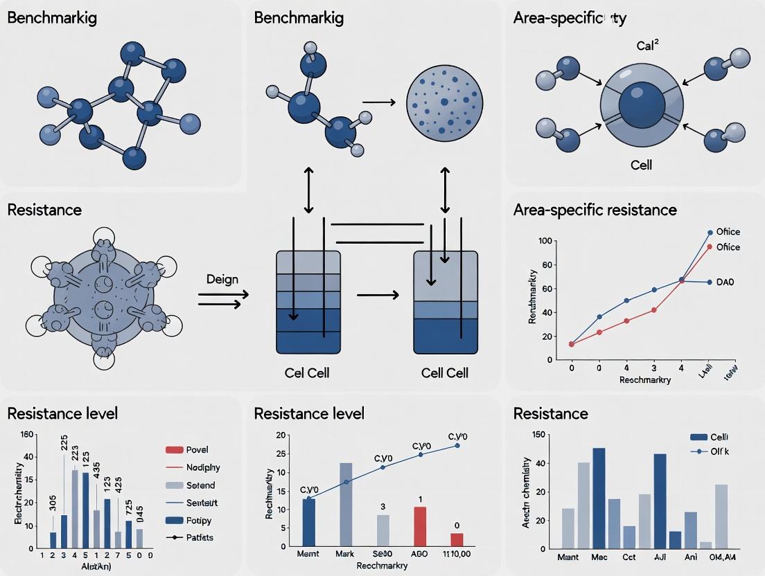
Benchmarking Area-Specific Resistance in Cell Design: A Comprehensive Guide for Drug Discovery Research
This article provides a systematic guide for researchers and drug development professionals on benchmarking area-specific resistance (ASR) across diverse cell designs.
Benchmarking Area-Specific Resistance in Cell Design: A Comprehensive Guide for Drug Discovery Research
Abstract
This article provides a systematic guide for researchers and drug development professionals on benchmarking area-specific resistance (ASR) across diverse cell designs. It explores the foundational principles of ASR and its critical role in predicting drug efficacy. The content details robust methodologies for accurate measurement and normalization, addresses common experimental pitfalls and optimization strategies, and offers frameworks for validating results and performing comparative analyses across 2D, 3D, and organ-on-a-chip models. By synthesizing current best practices, this guide aims to standardize ASR benchmarking to enhance the translational relevance of in vitro studies in oncology and beyond.
Understanding Area-Specific Resistance: The Core Concept in Cellular Drug Response
In drug discovery and cell biology, the bulk half-maximal inhibitory concentration (IC50) has long been a standard metric for quantifying compound potency. However, it provides an averaged, population-level view that can mask critical heterogeneity within a cellular system. Area-Specific Resistance (ASR) is an emerging analytical framework that quantifies the localized resistance of distinct cellular compartments or microenvironments to therapeutic agents. This is particularly relevant in complex tissues, 3D cultures, and tumors, where drug penetration, metabolic gradients, and local cell states create micro-niches of varying susceptibility. ASR moves beyond the scalar IC50 value to a spatial map of drug efficacy, providing deeper mechanistic insight and more predictive power for in vivo outcomes.
ASR vs. Bulk IC50: A Logical Relationship
Experimental Comparison: Measuring Bulk IC50 vs. ASR
To benchmark ASR across cell designs, researchers must employ spatially resolved techniques alongside traditional assays. The table below compares the core methodologies.
Table 1: Methodological Comparison of Bulk IC50 and ASR Assays
| Aspect | Bulk IC50 Determination | Area-Specific Resistance (ASR) Profiling |
|---|---|---|
| Core Principle | Measure population-averaged response (e.g., viability, activity) across a concentration gradient. | Measure localized response using imaging or spatially resolved omics within a defined architecture. |
| Typical Assay | CellTiter-Glo (3D viability), flow cytometry, plate reader fluorescence/absorbance. | Multiplexed immunofluorescence (e.g., CODEX, CycIF), spatial transcriptomics (e.g., Visium), live-cell imaging with microenvironmental sensors. |
| Key Output | Single potency value (IC50 in µM or nM) and Hill slope. | Heatmaps of efficacy metrics (e.g., local IC50, % viability) overlaid on spatial coordinates. |
| Data Dimension | 0-D (Scalar value). | 2-D or 3-D (Spatial map). |
| Ability to Detect | Overall potency, general sensitivity/resistance. | Microenvironmental niches, gradient-dependent resistance, cell-cell interaction effects. |
| Throughput | High (96/384-well plates). | Low to Medium (depends on imaging/analysis depth). |
| Primary Limitation | Obscures spatial heterogeneity and compartment-specific effects. | Technically complex, data-intensive, requires specialized analytical pipelines. |
Experimental Protocol for ASR Benchmarking in 3D Spheroids
This protocol outlines a comparative study to benchmark ASR across two cell designs: monolayer (2D) culture and 3D spheroids.
Title: Protocol for Benchmarking Drug ASR in 3D Spheroid vs. 2D Monolayer Models.
Workflow Overview:
Detailed Protocol Steps:
Model Generation:
- 2D Monolayer: Seed cells in standard 96-well plates at optimal confluency.
- 3D Spheroids: Seed cells in ultra-low attachment, round-bottom 96-well plates to promote self-aggregation. Use centrifugation (300 x g, 5 min) if needed. Culture for 96h to form compact spheroids.
Drug Treatment: Prepare a 10-point, 1:3 serial dilution of the test compound. Treat both 2D and 3D models for 72 hours. Include DMSO vehicle controls.
Endpoint Assays:
- Bulk Viability (IC50): Add CellTiter-Glo 3D reagent, shake, incubate, and record luminescence.
- Spatial Analysis (ASR):
- Fix spheroids with 4% PFA, permeabilize with 0.5% Triton X-100.
- Stain with multiplex antibody panel: Hoechst (DNA), anti-cleaved Caspase-3 (apoptosis), anti-Ki67 (proliferation), anti-phospho-target (drug effect).
- Image entire spheroid using a confocal or high-content imaging system with z-stacking.
Data Analysis:
- IC50: Normalize luminescence data to vehicle control, fit using a 4-parameter logistic model.
- ASR: Use image analysis software (e.g., CellProfiler, IMARIS) to segment each spheroid into three concentric zones: Core, Middle, and Periphery. Quantify mean fluorescence intensity for each marker per zone per drug concentration. Calculate a zone-specific inhibition metric (e.g., % positive cells for p-target) and derive a local IC50 or GI50 for each zone.
Comparative Data from a Benchmarking Study
The following table summarizes hypothetical but representative data from a study benchmarking the ASR of a hypothetical kinase inhibitor, "Compound X," in a cancer cell line.
Table 2: Benchmarking ASR of Compound X in 2D vs. 3D Cell Design
| Cell Design & Analyzed Region | Bulk IC50 (nM) | Area-Specific GI50 (nM)* | Proliferation (Ki67+) in Core at 1 µM (% of Ctrl) | Apoptosis (Casp3+) in Core at 1 µM (% of Ctrl) | Key ASR Insight |
|---|---|---|---|---|---|
| 2D Monolayer | 25 ± 5 | N/A (Homogeneous) | 15% | 85% | Uniform, high sensitivity. |
| 3D Spheroid - Periphery | 150 ± 30 | 40 ± 10 | 20% | 80% | Similar to 2D, remains sensitive. |
| 3D Spheroid - Middle | 150 ± 30 | 200 ± 50 | 65% | 25% | Moderate resistance. |
| 3D Spheroid - Core | 150 ± 30 | >1000 | 90% | 5% | Highly resistant niche. |
GI50: Concentration for 50% growth inhibition, calculated per zone from imaging data. *Bulk IC50 from spheroid luminescence assay is an average, masking the extreme core resistance.
The Scientist's Toolkit: Essential Reagents for ASR Research
Table 3: Key Research Reagent Solutions for ASR Studies
| Reagent / Material | Function in ASR Research |
|---|---|
| Ultra-Low Attachment (ULA) Plates | Promotes the formation of 3D spheroids or organoids by inhibiting cell adhesion, creating physiologically relevant architectures with inherent heterogeneity. |
| CellTiter-Glo 3D | Optimized luminescent assay for quantifying viability in 3D models, providing the bulk comparison data for spatial ASR maps. |
| Multiplex Immunofluorescence Antibody Panels | Enable simultaneous detection of phenotypic markers (viability, proliferation, apoptosis) and pharmacodynamic (PD) drug targets within spatial context. |
| Live-Cell Metabolic Dyes (e.g., Hoechst, TMRE) | Allow for real-time or endpoint assessment of spatial metabolic gradients (e.g., hypoxia, mitochondrial membrane potential) that drive ASR. |
| Spatial Barcoding Kits (e.g., Visium) | For spatial transcriptomics, enabling correlation of area-specific resistance patterns with localized gene expression profiles. |
| Image Analysis Software (CellProfiler, IMARIS, HALO) | Critical for segmenting images into distinct areas (zones, single cells) and extracting quantitative, zone-specific intensity data for ASR calculation. |
This comparative guide, framed within the thesis of benchmarking area-specific resistance (ASR) across cell designs, analyzes experimental platforms for their ability to model tumor microenvironment (TME)-driven heterogeneity and treatment failure.
Comparative Analysis of ASR & TME Modeling Platforms
Table 1: Platform Comparison for ASR & Heterogeneity Studies
| Platform Feature / Metric | 2D Monolayer Coculture | 3D Spheroid | Organ-on-a-Chip (OoC) | Patient-Derived Organoid (PDO) |
|---|---|---|---|---|
| Spatial Heterogeneity Modeling | Low (planar) | Medium (radial gradient) | High (controlled, zonated) | High (native architecture) |
| Microenvironmental Control | Low (soluble factors only) | Medium (self-generated matrix) | High (dynamic flow, mechanical cues) | Medium (native ECM, static) |
| Throughput for Drug Screening | High (>10,000 compounds) | Medium (~100-1000 compounds) | Low-Medium (~10-100 compounds) | Low (~10-50 compounds) |
| Key ASR Readout | IC50 shift (2-10 fold) | Invasion distance & core viability | Area-specific cytotoxicity (e.g., 80% kill in perfused vs. 20% in hypoxic zone) | Subclone-specific response profiling |
| Experimental Data (Example) | 3-fold resistance increase with fibroblasts | 40% deeper invasion in collagen I vs. Matrigel | Perfused zone: 95% kill; Hypoxic zone: <15% kill (anti-PD1) | Recapitulates patient's clinical response in 85% of cases |
| Limitation for ASR Studies | No spatial or physical TME cues | Limited user-defined control over TME | Lower throughput, higher technical barrier | Variable success rate, limited immune component |
Detailed Experimental Protocols
Protocol 1: Benchmarking ASR in an Organ-on-a-Chip Model
- Objective: Quantify area-specific cytotoxicity of a therapeutic in controlled hypoxic and perfused zones.
- Methodology:
- Chip Fabrication: Use a polydimethylsiloxane (PDMS)-based microfluidic device with two parallel channels separated by a collagen-I (3 mg/mL) ECM region.
- Cell Seeding: Seed fluorescently labeled cancer cells (e.g., GFP-tagged A549) into the ECM region. Seed human umbilical vein endothelial cells (HUVECs) in one channel to form a perfusable endothelial barrier.
- TME Establishment: Apply physiologically relevant shear stress (0.5-1.0 dyne/cm²) via media flow in the endothelial channel. Introduce a controlled oxygen gradient (20% O2 at endothelial channel, <1% O2 at the opposite side) using a gas manifold.
- Drug Treatment & ASR Quantification: Introduce a therapeutic (e.g., 10 µM Doxorubicin) via the vascular channel. After 72 hours, perform live/dead staining (Calcein-AM/Propidium Iodide). Use high-content imaging to quantify viability in three distinct zones: Perivascular (0-50µm from channel), Intermediate (50-150µm), and Hypoxic (>150µm).
Protocol 2: Mapping Clonal Heterogeneity in PDOs Post-Treatment
- Objective: Identify treatment-resistant subclones within organoids using single-cell RNA sequencing (scRNA-seq).
- Methodology:
- PDO Generation & Treatment: Establish PDOs from patient-derived tumor tissue in Matrigel domes. Expand and treat a batch with relevant therapy (e.g., EGFR inhibitor) at IC70 for 7 days, maintaining a DMSO-treated control.
- Single-Cell Suspension: Dissociate organoids using gentle enzymatic digestion (TrypLE + 100 µg/mL DNase I). Filter through a 40µm strainer.
- scRNA-seq Library Prep: Process cells using a platform like the 10x Genomics Chromium. Target 5,000-10,000 cells per condition (treated/control).
- Bioinformatic Analysis: Cluster cells and identify distinct transcriptional subpopulations. Compare cluster abundance and pathway enrichment (e.g., RAS/MAPK, YAP signaling) between treated and control samples to pinpoint resistant clusters.
Signaling Pathways in ASR Development
Title: Core Pathways Driving ASR in the TME
Workflow for Benchmarking ASR Across Platforms
Title: ASR Benchmarking Experimental Workflow
The Scientist's Toolkit: Key Research Reagent Solutions
Table 2: Essential Reagents for ASR & TME Research
| Reagent / Material | Function in ASR Studies | Example Vendor/Catalog |
|---|---|---|
| Matrigel / Cultrex BME | Provides a tumor-mimetic basement membrane extract for 3D culture, enabling native cell-ECM interactions and spheroid/organoid formation. | Corning, #356231 |
| Collagen I, High Concentration | Models a desmoplastic or fibrotic tumor stroma; crucial for studying invasion and drug penetration barriers in 3D models. | Advanced BioMatrix, #5005 |
| Microfluidic Organ-on-a-Chip Kits | Pre-fabricated platforms for building controlled TMEs with dynamic flow and multi-cellular interactions. | Emulate, Inc. (Human Tumor Microenvironment Chip) |
| Hypoxia-Inducing Agents (e.g., CoCl₂) | Chemically mimics hypoxic conditions to stabilize HIF-1α and study hypoxia-driven resistance pathways in standard incubators. | Sigma-Aldrich, #15862 |
| Live/Dead Viability/Cytotoxicity Kits | Enables spatial quantification of cell death (dead=red, live=green) in intact 3D structures post-treatment. | Thermo Fisher, L3224 |
| Single-Cell Dissociation Kits (for 3D Models) | Gentle enzymatic cocktails to generate high-quality single-cell suspensions from spheroids/organoids for scRNA-seq. | Miltenyi Biotec, Tumor Dissociation Kit |
| Phospho-/Total Antibody Panels (e.g., for AKT, ERK) | To map activation of pro-survival signaling pathways in different areas of a model via multiplex immunofluorescence. | Cell Signaling Technology, Phospho-MAPK Array |
| Cytokine/GF Multiplex Assays | Measures secretion profiles from tumor-stroma cocultures to quantify paracrine signaling driving heterogeneity. | Luminex, R&D Systems Multi-Analyte Panels |
Within the research framework of benchmarking area-specific resistance (ASR) across cell designs, the choice of in vitro model is a critical variable. ASR, a quantitative measure of cellular barrier integrity, is profoundly influenced by architectural complexity, cell-cell/cell-matrix interactions, and physiological relevance. This guide objectively compares the performance of four primary cell culture designs—2D monolayers, 3D spheroids, organoids, and microfluidic chips—in modeling ASR, supported by experimental data.
Quantitative Comparison of ASR Metrics
The following table summarizes typical Transendothelial/Transepithelial Electrical Resistance (TEER) values, a direct correlate of ASR, and key functional characteristics across models. Data is aggregated from recent studies.
Table 1: ASR Benchmarking Across Cell Design Platforms
| Model Type | Typical TEER Range (Ω·cm²) | Key Advantages Impacting ASR | Key Limitations Impacting ASR | Physiological Relevance for Barrier Studies |
|---|---|---|---|---|
| 2D Monolayer | 200 - 3,000 (e.g., Caco-2) | High reproducibility, ease of measurement, standardized protocols. | Lack of 3D cytoarchitecture, abnormal polarization, absent shear stress. | Low-Moderate; simplistic barrier. |
| 3D Spheroid | Core diffusion-limited (indirect metric) | Enhanced cell-cell contacts, basic nutrient/oxygen gradients. | Difficult direct TEER measurement, central necrotic core can alter permeability. | Moderate; models avascular tumor barriers. |
| Organoid | Varies widely; often inferred | Patient-derived, complex multicellularity, innate stem cell niches. | High heterogeneity, technically challenging to measure TEER, Matrigel batch effects. | High; captures tissue-specific crypt-villus/glomerular barriers. |
| Microfluidic Chip (Organ-on-a-Chip) | 1,000 - 5,000+ (e.g., lung alveolus) | Incorporation of mechanical cues (cyclic stretch, shear stress), vascular-interstitial interfaces. | Specialized equipment required, smaller surface area for measurement. | Very High; dynamic, multi-tissue interfaces with physiological flow. |
Experimental Protocols for ASR Assessment
Protocol: Standard TEER Measurement for 2D Monolayers & Microfluidic Chips
Objective: To directly quantify the integrity of a cellular barrier.
- Materials: Voltohmmeter (e.g., EVOM2), chopstick or electrode arrays, cell culture inserts (for 2D), microfluidic chip with integrated electrodes.
- Procedure:
- Culture cells to confluence on a permeable filter insert (2D) or in the central channel of a microfluidic chip.
- Calibrate the instrument with a cell-free insert or chip filled with culture medium.
- Sterilize electrodes with 70% ethanol and equilibrate in medium.
- Place the electrode(s) in the apical and basolateral compartments. For chips, connect to integrated electrode ports.
- Record the resistance (Ω). Subtract the background resistance (empty insert/chip) and multiply by the effective surface area (cm²) to calculate TEER (Ω·cm²).
Protocol: Impedance-Based Monitoring for 3D Spheroids/Organoids
Objective: To indirectly assess barrier function and cell viability within aggregates.
- Materials: Real-time cell analyzer (e.g., xCELLigence RTCA), specialized 3D E-plate.
- Procedure:
- Embed single spheroids/organoids in a thin layer of extracellular matrix (ECM) in each well of the E-plate.
- Insert the plate into the analyzer, which applies a low-voltage AC signal across microelectrodes at the well bottom.
- Monitor Cell Index over time. A higher Cell Index correlates with increased cell attachment, spreading, and barrier integrity; a drop indicates cytotoxicity or barrier breach.
Visualization of Experimental Workflows
Title: ASR Measurement Workflow Across Cell Models
Title: Key Variables Influencing ASR in Cell Models
The Scientist's Toolkit: Essential Research Reagents & Materials
Table 2: Key Reagent Solutions for ASR Studies
| Item | Function in ASR Research | Example/Note |
|---|---|---|
| Transwell/Cell Culture Inserts | Provides a porous membrane for 2D monolayer formation, enabling separate apical/basolateral access for TEER and transport assays. | Polycarbonate or polyester, 0.4 - 5.0 μm pore size. |
| Extracellular Matrix (ECM) Hydrogels | Provides a 3D scaffold for spheroid/organoid growth, influencing differentiation, polarity, and barrier maturation. | Matrigel, collagen I, synthetic PEG-based hydrogels. |
| Real-Time Cell Analyzer (RTCA) | Enables label-free, impedance-based monitoring of cell barrier health and integrity in 2D and 3D formats over time. | xCELLigence, ACEA Biosciences. |
| Voltohmmeter / TEER Meter | The standard instrument for applying a current and directly measuring electrical resistance across a cellular monolayer. | EVOM2 with STX electrodes (World Precision Instruments). |
| Microfluidic Chip Device | Engineered platform housing microchannels and chambers to culture cells under perfusion, often with integrated electrodes. | Emulate, Mimetas, or in-house PDMS chips. |
| Tight Junction Marker Antibodies | Used in immunostaining to visualize and quantify the integrity and maturation of barrier-forming structures (e.g., ZO-1, occludin). | Critical for correlating TEER with molecular phenotype. |
Within the framework of benchmarking area-specific resistance across diverse cell designs, understanding the biological drivers of cellular response is paramount. This guide compares the role and impact of three fundamental drivers—Nutrient Gradients, Cell-Cell Contact, and Phenotypic Plasticity—on the development and quantification of barrier resistance in epithelial and endothelial monolayers. Performance is assessed based on their influence on key metrics: transepithelial/transendothelial electrical resistance (TEER), paracellular flux, and tight junction protein expression.
Comparison of Biological Drivers on Barrier Resistance
Table 1: Comparative Impact of Biological Drivers on Barrier Metrics
| Driver | Primary Mechanism | Effect on TEER | Effect on Paracellular Flux | Key Assay Readouts | Experimental Manipulation |
|---|---|---|---|---|---|
| Nutrient Gradients | Creates metabolic asymmetry and polarity cues. | Moderate Increase (1.5-2x) with proper gradient | Decrease (~50% reduction) | TEER, Fluorescent dextran flux, OCR/Glycolysis assays | Bi-directional flow systems, Organ-on-a-chip microfluidics |
| Cell-Cell Contact | Initiates contact inhibition and junctional assembly. | Strong Increase (3-5x) upon confluence | Strong Decrease (~80% reduction) | TEER, Calcium switch assay, Immunofluorescence (ZO-1, Occludin) | Seeding density titration, Calcium chelation (EGTA) |
| Phenotypic Plasticity | Enables adaptive junctional remodeling under stress. | Context-dependent (Can decrease or increase) | Context-dependent Increase under EMT | TEER, EMT/MET markers (E-cadherin, Vimentin), RNA-seq | Cytokine treatment (TGF-β), Hypoxia chambers |
Table 2: Benchmarking Data Across Common Cell Designs (Caco-2, MDCK II, HUVEC)
| Cell Design | Basal TEER (Ω*cm²) | Response to Nutrient Gradient (ΔTEER) | Response to Full Contact (Time to Max TEER) | Plasticity-Induced Resistance Drop (Post TGF-β) |
|---|---|---|---|---|
| Caco-2 (Colon Epithelial) | 300-500 | +120% | 14-21 days | -70% at 96h |
| MDCK II (Renal Epithelial) | 100-200 | +40% | 3-5 days | -40% at 96h |
| HUVEC (Umbilical Vein Endothelial) | 20-50 | +80% (Flow-dependent) | 2-3 days | -60% at 72h |
Experimental Protocols
1. Protocol: Quantifying Nutrient Gradient Effects on Area-Specific Resistance
- Objective: To benchmark TEER and permeability under apical-basal nutrient asymmetry.
- Materials: Microfluidic organ-on-a-chip device (e.g., Emulate, Mimetas), cell type of interest, low-glucose media, fluorescent tracer (e.g., 4 kDa FITC-dextran), TEER measurement system.
- Method:
- Seed cells in the top channel of a porous membrane-equipped chip.
- Establish a stable gradient: flow high-nutrient media (e.g., 25 mM glucose) through the basal channel and low-nutrient media (e.g., 5 mM glucose) through the apical channel.
- Maintain gradient for 5-7 days, monitoring TEER daily.
- At peak TEER, introduce FITC-dextran to the apical channel.
- Sample from the basal channel at 15, 30, 60, and 120 minutes for fluorometric analysis to calculate apparent permeability (Papp).
- Data Analysis: Compare TEER kinetics and Papp values against isotropic (control) nutrient conditions.
2. Protocol: Benchmarking Junctional Maturation via Cell-Cell Contact
- Objective: To correlate seeding density/time with junctional resistance and protein localization.
- Materials: Transwell inserts, cell type, EVOM2 or CellZscope TEER meter, EGTA, fixative, antibodies for ZO-1 and E-cadherin.
- Method:
- Seed cells at varying densities (low: sub-confluent, medium: just confluent, high: super-confluent).
- Measure TEER every 24 hours until a plateau is reached.
- For a calcium switch assay: treat confluent monolayers with 4 mM EGTA in low-Ca²⁺ media for 30 min to disrupt junctions, then replace with standard media.
- Monitor TEER recovery hourly for 6-8 hours.
- Fix monolayers at key timepoints for immunostaining of tight and adherens junction proteins.
- Data Analysis: Plot TEER vs. time/density. Quantify junctional protein continuity and intensity from confocal images.
3. Protocol: Inducing and Measuring Plasticity-Driven Resistance Changes
- Objective: To benchmark the destabilizing effect of an Epithelial-to-Mesenchymal Transition (EMT) inducer on barrier integrity.
- Materials: Recombinant human TGF-β1, TEER meter, qPCR reagents, antibodies for epithelial (E-cadherin) and mesenchymal (Vimentin, N-cadherin) markers.
- Method:
- Establish confluent monolayers with stable baseline TEER.
- Treat with 5-10 ng/mL TGF-β1 in the basolateral compartment. Refresh cytokine every 48h.
- Measure TEER at 0, 24, 48, 72, and 96 hours post-treatment.
- At each timepoint, lyse cells for RNA/protein extraction.
- Perform qPCR for CDH1 (E-cadherin), VIM, CDH2 (N-cadherin), and SNAI1.
- Perform western blot or immunofluorescence for corresponding proteins.
- Data Analysis: Correlate percentage TEER loss with fold-change in EMT marker expression.
Visualizations
Nutrient Gradient to Barrier Resistance Pathway
Benchmarking Contact-Driven Resistance Workflow
Phenotypic Plasticity Leading to Barrier Loss
The Scientist's Toolkit: Research Reagent Solutions
Table 3: Essential Materials for Benchmarking Barrier Drivers
| Item | Function in Benchmarking | Example Product/Catalog |
|---|---|---|
| Transwell/Permeable Inserts | Provides a polarized growth substrate for TEER and flux measurements. | Corning Transwell, 0.4 μm polyester membrane. |
| Real-Time TEER Instrument | Enables non-destructive, kinetic monitoring of barrier resistance. | World Precision Instruments EVOM2, nanoAnalytics CellZscope. |
| Fluorescent Tracers | Quantifies paracellular permeability (Papp calculation). | Thermo Fisher FITC-Dextran, 4 kDa & 70 kDa. |
| Junctional Protein Antibodies | Visualizes and quantifies tight/adherens junction integrity. | Invitrogen Anti-ZO-1 (clone ZO1-1A12), Anti-E-cadherin (clone 36). |
| EMT Inducer/Cytokine | Modulates phenotypic plasticity to test barrier resilience. | PeproTech Recombinant Human TGF-β1. |
| Microfluidic Chip System | Establishes physiologically relevant nutrient/flow gradients. | Emulate Intestine-Chip, Mimetas OrganoPlate. |
| Metabolic Assay Kits | Profiles metabolic shifts (glycolysis/oxidative phosphorylation). | Agilent Seahorse XF Cell Mito Stress Test Kit. |
A primary challenge in oncology drug development is the failure of therapies that show promise in preclinical models to demonstrate efficacy in human trials. This translational gap is often attributed to inadequate modeling of the tumor microenvironment (TME), particularly area-specific resistance (ASR) mechanisms. Benchmarking ASR across different preclinical cell designs—from 2D monolayers to 3D spheroids and organoids—provides a quantifiable framework to improve the predictive validity of these models for clinical outcomes.
Comparative Performance of Preclinical Models in Predicting Clinical Response
The following table summarizes key metrics from recent studies benchmarking various in vitro models against historical clinical trial response rates for a set of common chemotherapeutic and targeted agents.
Table 1: Benchmarking of Preclinical Model Predictivity for Clinical Response
| Model Type | Avg. Correlation with Clinical PK/PD (r) | Predictive Value for Phase II Success (%) | ASR Phenotype Capture (Hypoxia, ECM, Gradient) | Key Limitation |
|---|---|---|---|---|
| 2D Monolayer | 0.31 | ~20% | Low (None) | Lacks TME; uniform drug exposure. |
| 3D Spheroid (Scaffold-free) | 0.57 | ~45% | Medium (Core hypoxia, nutrient gradient) | Limited stromal and immune components. |
| 3D Organoid (Patient-Derived) | 0.72 | ~65% | High (Native architecture, some stroma) | Inter-donor variability; high cost. |
| Microfluidic "Organ-on-a-Chip" | 0.81* | ~75%* | Very High (Controlled flow, shear stress, multicellular TME) | Complexity; not yet high-throughput. |
| In Vivo PDX Model | 0.69 | ~70% | High (Full in vivo TME, vasculature) | Time-intensive; murine stroma. |
*Preliminary data from ongoing consortium studies.
Experimental Protocols for ASR Benchmarking
A standardized workflow is essential for generating comparable ASR data across labs and model systems.
Protocol 1: Quantifying Penetration Resistance in 3D Models
- Model Preparation: Generate uniformly sized spheroids (e.g., via ultra-low attachment plates) or organoids (in Matrigel).
- Drug Dosing: Treat with a fluorescently tagged standard-of-care drug (e.g., Doxorubicin-Alexa Fluor 647) at the clinically relevant C~max~ concentration.
- Imaging & Analysis: At defined time points (e.g., 4h, 24h), image serial z-sections using confocal microscopy. Quantify fluorescence intensity from periphery to core.
- Metric Calculation: Determine the Penetration Efficiency Index (PEI) = (Drug intensity~core~ / Drug intensity~periphery~) * 100. A lower PEI indicates higher penetration resistance.
Protocol 2: Benchmarking Hypoxia-Induced Resistance
- Hypoxia Mapping: Incubate models with a hypoxia probe (e.g., Pimonidazole). For real-time readouts, use genetically encoded sensors (e.g., HIF-1α GFP reporters).
- Viability Correlation: Section the model and perform co-staining for the hypoxia probe and a viability marker (e.g., Caspase-3/7 activity dye).
- Data Analysis: Calculate the Hypoxic Survival Fraction (HSF) = (Viable cells in hypoxic regions / Total viable cells). Models with a higher HSF for a given drug better mimic observed clinical resistance.
Diagram: ASR Benchmarking Improves Clinical Prediction
The Scientist's Toolkit: Key Research Reagent Solutions
Table 2: Essential Reagents for ASR Benchmarking Studies
| Reagent / Material | Function in ASR Benchmarking | Example Product/Catalog |
|---|---|---|
| Ultra-Low Attachment (ULA) Plates | Enables formation of uniform, scaffold-free 3D spheroids. | Corning Spheroid Microplates |
| Basement Membrane Matrix | Provides a physiological 3D scaffold for organoid growth and invasion assays. | Corning Matrigel |
| Hypoxia Probes (Chemical) | Immunohistochemical detection of hypoxic regions within 3D models. | Hypoxyprobe (Pimonidazole) |
| Live-Cell Fluorescent Reporters (HIF) | Real-time, dynamic imaging of hypoxia induction. | FUCCI-HIF-1α reporter cell lines |
| Fluorescent Drug Conjugates | Direct visualization and quantification of drug penetration. | Doxorubicin-Alexa Fluor 647 |
| Microfluidic Co-culture Chips | Models perfusion, vascular barriers, and multi-tissue interactions. | Emulate Organ-Chip |
| Viability/Cytotoxicity Kits (3D-optimized) | Accurate cell death quantification in dense 3D structures. | CellTiter-Glo 3D |
| Patient-Derived Organoid Media Kits | Supports the expansion of patient tissues while preserving genetic and phenotypic fidelity. | STEMCELL Technologies IntestiCult |
These standardized tools and benchmarking protocols enable a systematic comparison of how different cell designs capture area-specific resistance. Integrating these quantitative ASR metrics into preclinical validation pipelines significantly enhances the model's predictive power for clinical success, directly addressing a critical component of the translational gap.
Best Practices for Measuring and Normalizing ASR Across Experimental Models
In the pursuit of benchmarking area-specific resistance (ASR) across cell designs—a critical parameter in oncology and toxicology research—selecting the appropriate core assay technology is paramount. High-Content Imaging (HCI) and Real-Time Cell Analysis (RTCA, e.g., xCELLigence systems) represent two dominant, complementary paradigms. This guide objectively compares their performance in quantifying ASR-related phenotypes such as barrier integrity, cytotoxicity, and morphological adaptation.
Performance Comparison: HCI vs. RTCA
Table 1: Core Performance Metrics for ASR Benchmarking
| Feature | High-Content Imaging (e.g., PerkinElmer Opera, ImageXpress) | Real-Time Cell Analysis (e.g., Agilent xCELLigence) | Key Implication for ASR Research |
|---|---|---|---|
| Primary Output | Multiparametric spatial data (morphology, fluorescence intensity, object count). | Kinetic impedance data (Cell Index, dimensionless). | HCI offers spatial resolution; RTCA delivers continuous temporal resolution. |
| Temporal Resolution | Endpoint or limited timepoints (hours-days). | Real-time, label-free (minutes to days, continuous). | RTCA is superior for detecting transient ASR dynamics. |
| Throughput | Very High (96/384-well plates). | Moderate to High (96/384-well E-Plates). | HCI excels in large-scale, compound screening for ASR. |
| Assay Readiness | Requires optimization of labels/dyes (potential perturbation). | Label-free, non-invasive. | RTCA minimizes artifacts, ideal for sensitive longitudinal ASR studies. |
| Key ASR Metrics | Cell-cell junction integrity (ZO-1 staining), actin rearrangement, monolayer confluence. | Impedance-derived barrier function, cell adhesion, proliferation/death kinetics. | HCI quantifies structural components; RTCA quantifies functional electrical resistance. |
| Cost & Complexity | High capital cost, complex data analysis pipeline. | Lower operational complexity, simpler data output. | RTCA offers easier integration for continuous monitoring. |
Table 2: Experimental Data Comparison in a Model ASR Study (Intestinal Barrier) Study: Impact of Cytokine Storm on Epithelial Barrier Integrity
| Parameter | HCI Results (24h endpoint) | RTCA Results (24h kinetic) | Interpretation |
|---|---|---|---|
| Barrier Disruption | 60% reduction in ZO-1 junction continuity. | Normalized Cell Index dropped from 2.5 to 0.8 within 8h. | Both confirm disruption; RTCA pinpoints onset at ~4h post-treatment. |
| Cytotoxicity | 25% apoptosis via caspase-3 staining. | Steady decline in Cell Index post-8h, correlating with death. | HCI specifies death mechanism; RTCA shows kinetic progression. |
| Data Richness | 15+ parameters per cell (size, shape, intensity). | 1 primary parameter (Cell Index) with derived rates. | HCI enables deep phenotyping; RTCA provides unified kinetic index. |
Detailed Experimental Protocols
Protocol 1: HCI for Area-Specific Resistance (Tight Junction Integrity)
- Cell Seeding: Seed Caco-2 or MDCK cells in a black-walled, clear-bottom 96-well plate at 50,000 cells/well. Culture until a fully confluent monolayer forms (3-5 days).
- Treatment: Apply ASR-modulating agents (e.g., inflammatory cytokines, drug candidates) in triplicate.
- Fixation and Staining (Endpoint): At 24h, fix with 4% PFA, permeabilize with 0.1% Triton X-100, and block with 1% BSA. Stain with primary antibodies against ZO-1 (tight junctions) and β-catenin (adherence junctions), followed by fluorescent secondary antibodies (e.g., Alexa Fluor 488, 568). Counterstain nuclei with Hoechst 33342.
- Image Acquisition: Use a high-content imager (20x objective). Acquire 4-9 fields per well to ensure statistical representation.
- Image Analysis: Use software (e.g., Harmony, CellProfiler) to segment nuclei and cell boundaries. Quantify junction integrity via "Junction Continuity Index" (total length of continuous junction staining / total perimeter length) and fluorescence intensity at cell borders.
Protocol 2: RTCA for Kinetic ASR Monitoring
- Baseline Measurement: Add 50µL of media to each well of an xCELLigence E-Plate 96. Perform a background scan in the RTCA instrument.
- Cell Seeding & Attachment Monitoring: Seed Caco-2 cells (50,000 cells/well in 100µL) directly into the E-Plate. Place the plate in the incubator-mounted RTCA station. Monitor Cell Index every 15 minutes for 24-48 hours until the Cell Index plateaus, indicating full monolayer formation and stable ASR.
- Treatment & Kinetic Assay: Carefully add 50µL of 3x concentrated treatment compounds (or vehicle control) to the wells. Continue uninterrupted monitoring of Cell Index every 5 minutes for 24-72 hours.
- Data Analysis: Use RTCA Software to normalize Cell Index to the time of treatment. Analyze key parameters: time to 50% reduction in Cell Index (TCI50), slope of Cell Index decline (rate of barrier disruption), and whether Cell Index recovers (indicating regenerative capacity).
Visualizations
HCI Workflow for ASR Analysis
Key Pathways in ASR Regulation
The Scientist's Toolkit: Essential Research Reagent Solutions
Table 3: Key Reagents for ASR Assays
| Item | Function in ASR Research | Example Product/Catalog |
|---|---|---|
| Electronic Microplate (E-Plate) | Specialized plate with integrated gold microelectrodes for impedance-based RTCA. | Agilent xCELLigence E-Plate 96 (05469813001) |
| ZO-1/TJP1 Antibody | Primary antibody for staining tight junctions, a core structural component of ASR. | Invitrogen Anti-ZO-1 Monoclonal (33-9100) |
| Fluorescent Phalloidin | Binds F-actin, visualizing cytoskeletal rearrangement critical for cell barrier integrity. | Cytoskeleton, Inc. Phalloidin-iFluor 488 (PHDG1) |
| Hoechst 33342 | Cell-permeant nuclear counterstain for HCI, enabling cell segmentation and counting. | Thermo Fisher Scientific (H3570) |
| Live-Cell Imaging Medium | Low-fluorescence, CO2-buffered medium for maintaining health during kinetic HCI. | Gibco FluoroBrite DMEM (A1896701) |
| Recombinant Human TNF-α | Common inflammatory cytokine used to experimentally disrupt ASR in model barriers. | PeproTech (300-01A) |
| Paracellular Flux Tracer (e.g., FITC-Dextran) | Measures functional permeability complementary to impedance and imaging. | Sigma-Aldrich FD4 (46944) |
Benchmarking area-specific resistance demands a multi-faceted approach. High-Content Imaging delivers unparalleled spatial and multiparametric resolution for dissecting the structural underpinnings of ASR at endpoint. In contrast, Real-Time Cell Analysis offers a label-free, kinetic window into the functional dynamics of barrier health, revealing the precise timing and patterns of disruption and recovery. The optimal choice is context-driven: RTCA for continuous functional kinetics and HCI for deep, endpoint phenotypic profiling. Integrating both technologies provides the most comprehensive benchmark for ASR across diverse cell designs.
Within the context of benchmarking area-specific resistance across cell culture and drug development research, precisely defining the "area" metric is critical for data normalization, reproducibility, and meaningful cross-study comparison. This guide compares the application and measurement of three primary area definitions—Confluence, Volume, and Projected Area—in the context of cellular assays, supported by experimental data.
Metric Comparison & Experimental Data
The choice of area metric directly impacts calculated parameters such as area-specific resistance, metabolic activity, and growth rates. The following table summarizes a comparative experiment performed on HepG2 liver cell spheroids and A549 monolayers.
Table 1: Comparative Analysis of Area Metrics in Cellular Assays
| Metric | Definition | Measurement Tool | Key Advantage | Key Limitation | Impact on Area-Specific Resistance Calculation (Example Data) |
|---|---|---|---|---|---|
| Confluence | Percentage of 2D surface area covered by cells. | Automated phase-contrast image analysis (e.g., Incucyte). | Dynamic, live-cell monitoring of 2D growth. | Insensitive to cell thickness/volume; not applicable to 3D structures. | Resistance (Ω·cm²) can be overestimated if monolayer is multilayered. Mean: 45 ± 5 Ω·cm² for confluent monolayer. |
| Projected Area | Two-dimensional footprint area of a cell or spheroid as viewed from above. | Brightfield microscopy with segmentation (e.g., ImageJ). | Simple to acquire; standard for spheroid size tracking. | Ignores 3D morphology and internal structure. | Primary metric for trans-epithelial electrical resistance (TEER) normalization in 2D. Mean: 32 ± 3 Ω·cm² for A549 filter culture. |
| Volume | Total three-dimensional space occupied by cells (e.g., spheroid). | Confocal microscopy with z-stacking or volume estimation from diameter. | Most biologically relevant for mass/activity normalization in 3D models. | Technically challenging and resource-intensive to measure accurately. | Critical for volumetric resistance in organoids. Estimated via spheroid diameter (500µm): Calculated Vol. Resistivity: 215 ± 25 Ω·cm. |
Experimental Protocols
Protocol 1: Measuring Confluence for Monolayer Growth & TEER Normalization
- Seed cells in a 96-well imaging plate at low density.
- Place plate in a live-cell imaging system (e.g., Sartorius Incucyte) within a standard incubator.
- Acquire phase-contrast images every 2 hours at 10x magnification.
- Analyze images using built-in confluence analysis mask software, which thresholds cell bodies from background.
- In parallel, for TEER assays, seed cells on transwell filters. Measure TEER (Ω) using an epithelial volt-ohm meter.
- Calculate area-specific resistance: Multiply the measured TEER (Ω) by the effective growth area of the filter (e.g., 0.33 cm²). Correlate with confluence data at time of measurement.
Protocol 2: Determining Projected Area and Volume of 3D Spheroids
- Form spheroids using a U-bottom ultra-low attachment 96-well plate.
- On day 3, acquire brightfield image stacks at 4x magnification with a 50µm z-step interval using an automated microscope.
- For Projected Area: Use the maximum intensity projection. Apply a threshold and measure the 2D pixel area, converting using a scale bar.
- For Volume Estimation: Manually measure the major (Dmax) and minor (Dmin) diameters from the best-focused image. Calculate volume using the formula for a prolate spheroid: V = (4/3)π * (Dmax/2)² * (Dmin/2).
- Normalize assay data (e.g., ATP luminescence) to both projected area and estimated volume for comparison.
Visualizing Metric Relationships & Workflows
Title: Decision Flow for Selecting Area Metric Based on Cell Model
Title: Generalized Experimental Workflow for Area Metric Quantification
The Scientist's Toolkit: Research Reagent Solutions
Table 2: Essential Materials for Area Metric Analysis
| Item | Function & Relevance to Area Definition |
|---|---|
| Ultra-Low Attachment (ULA) Plates | Enables consistent 3D spheroid formation by inhibiting cell adhesion, allowing volume/projected area studies. |
| Live-Cell Imaging System (e.g., Incucyte) | Provides kinetic confluence data and project area measurements in a controlled incubator environment. |
| Transwell Permeable Supports | Standard platform for growing cell monolayers of defined projected area for TEER (resistance) assays. |
| Epithelial Volt-Ohm Meter (EVOM) | Measures electrical resistance (TEER) across a cell monolayer, which is normalized to the filter's growth area. |
| Confocal Microscopy with Z-Stacking | Gold standard for accurate 3D cell volume measurement through optical sectioning. |
| Image Analysis Software (e.g., ImageJ, CellProfiler) | Essential for segmenting cells from background and calculating pixel-based area metrics (confluence, projected area). |
| ATP-Based Viability Assay (e.g., CellTiter-Glo 3D) | Luminescent assay normalized to area/volume metrics to quantify metabolically active cell mass. |
| Fluorescent Cell Trackers (e.g., Calcein AM) | Stain live cells to improve contrast and accuracy of segmentation for area calculations. |
Within the context of benchmarking area-specific resistance (ASR) across varied cell designs—such as 2D monolayers, 3D spheroids, and organ-on-chip systems—accurate normalization of functional readouts is paramount. ASR, a critical metric for barrier integrity in epithelial and endothelial models, is highly sensitive to cell number, confluence, and health. This guide compares common normalization strategies and their impact on the cross-comparability of ASR data from different experimental platforms.
Comparison of Normalization Methodologies
The table below compares three primary normalization approaches used to account for cellular variables that confound ASR measurements (e.g., Transepithelial/Transendothelial Electrical Resistance, TEER).
Table 1: Normalization Strategy Comparison for ASR Benchmarking
| Normalization Strategy | Primary Target | Typical Method | Advantages | Limitations | Impact on ASR Variability |
|---|---|---|---|---|---|
| DNA Content Quantification | Total Cell Number | PicoGreen or CyQuant assay post-experiment. | Direct, quantitative; accounts for proliferation differences. | Destructive; requires lysis; does not differentiate live/dead. | Reduces inter-spheroid/well variability by 40-50% in 3D models. |
| ATP-based Viability Assay | Metabolically Active Cell Mass | CellTiter-Glo luminescent assay. | Correlates with viable cell mass; high sensitivity. | Can be influenced by metabolic perturbations; medium components can interfere. | Normalizes for viability shifts (e.g., post-drug treatment), improving correlation with barrier function by ~30%. |
| Initial Seeding Normalization | Input Cell Count | Precise seeding via automated cell counters or seeding density optimization. | Simple, pre-experimental; minimizes initial divergence. | Does not account for differential proliferation or death during culture. | Critical for microfluidic devices; reduces day-0 to day-3 ASR variance by up to 60%. |
| Integrated Normalization (DNA + ATP) | Proliferation & Viability | Sequential or parallel measurement of DNA and ATP. | Most comprehensive picture of cell number and health. | Complex, costly, and often destructive. | Gold standard; reduces inter-platform (2D vs. 3D) comparison error by >70%. |
Experimental Protocols for Cited Data
Protocol 1: DNA Content Normalization for 3D Spheroid TEER (Indirect)
- Culture: Seed cells in spheroid-forming 96-well ultra-low attachment plates.
- ASR Measurement: At assay endpoint, transfer spheroids to a dedicated setup for impedance measurement (e.g., organoid TEER).
- Lysis & Quantification: Immediately post-measurement, lyse spheroids in Triton X-100 buffer. Add an equal volume of PicoGreen reagent.
- Analysis: Measure fluorescence (Ex/Em ~480/520 nm). Normalize the raw impedance value (Ω) to the DNA fluorescence units (RFU) to calculate Ω/RFU.
Protocol 2: ATP-based Viability Normalization in a Cytotoxicity Context
- Treatment: Culture Caco-2 monolayers on Transwell inserts and treat with a compound series for 48h.
- TEER Measurement: Measure TEER (Ω) using a volt-ohm meter.
- Viability Assay: Carefully transfer inserts to a new plate. Add CellTiter-Glo reagent directly to the insert well (apical and basolateral), lyse for 10 min, then transfer lysate to an opaque plate.
- Analysis: Measure luminescence. Calculate normalized ASR as (TEERsample / TEERvehicle) / (ATPsample / ATPvehicle).
Protocol 3: Precise Seeding for Microfluidic Barriers
- Cell Counting: Use an automated cell counter (e.g., Countess) with trypan blue to determine viable concentration.
- Seeding: Utilize a precision peristaltic pump or micropipettor to seed a defined cell number (e.g., 50,000 cells/chamber) into the apical channel of a microfluidic device.
- Documentation: Record exact seeded cell number per device as a normalization factor.
- ASR Calculation: After barrier formation, measure impedance. Report ASR as Ωcm² and also as Ωcm² per 10⁵ seeded cells.
Signaling Pathways & Experimental Workflows
Title: Workflow for ASR Normalization Strategy Selection
Title: Factors Influencing Barrier Integrity and ASR
The Scientist's Toolkit: Research Reagent Solutions
Table 2: Essential Reagents & Kits for Normalization in ASR Studies
| Item Name | Supplier Examples | Primary Function in Normalization |
|---|---|---|
| PicoGreen dsDNA Assay Kit | Thermo Fisher, Abcam | Quantifies double-stranded DNA content for total cell number normalization. Highly sensitive for low-cell-number models like spheroids. |
| CellTiter-Glo 3D/2D Assay | Promega | Measures ATP content as a proxy for metabolically active cell mass. The 3D version is optimized for tissue-like structures. |
| Automated Cell Counter & Slides | Thermo Fisher (Countess), Bio-Rad | Provides accurate, viable (trypan blue exclusion) cell counts for reproducible initial seeding density. |
| Electric Cell-substrate Impedance Sensing (ECIS) | Applied BioPhysics | Enables real-time, label-free monitoring of barrier integrity (ASR) and cell behaviors like proliferation and death in a single system. |
| Transepithelial Electrical Resistance (TEER) Voltohmmeter | World Precision Instruments, MilliporeSigma | The standard tool for direct measurement of barrier function in Transwell or microfluidic membrane setups. |
| Microfluidic Organ-on-Chip Devices | Emulate, MIMETAS | Provide physiologically relevant shear stress and 3D architecture for advanced ASR benchmarking, requiring precise seeding protocols. |
Accurate volumetric assessment of 3D cellular models, such as organoids and spheroids, is critical for benchmarking area-specific resistance in cell design research. This guide compares methodologies for sample preparation and imaging, focusing on experimental data relevant to drug permeability and resistance studies.
Comparative Analysis of Tissue Clearing Protocols
Effective volumetric imaging requires tissue clearing to reduce light scattering. The table below compares three prominent protocols used in organoid research.
Table 1: Comparison of Tissue Clearing Protocols for 3D Organoid Imaging
| Protocol | Principle | Time to Clear (mm³/day) | Index Matching (nD) | Compatible Immuno-labeling | Impact on Fluorescence (Signal Retention %) | Key Study & Application Context |
|---|---|---|---|---|---|---|
| CLARITY | Hydrogel-based, electrophoretic clearing | 1-2 | ~1.38-1.45 | Excellent (Native protein) | ~85-95% (GFP/RFP) | Chung & Deisseroth, Nature; Used for neural organoid connectivity mapping. |
| iDISCO+ | Organic solvent-based, dehydration & delipidation | 3-5 | ~1.56 | Good (Requires sample dehydration) | ~70-80% (tdTomato) | Renier et al., Cell; Applied to tumor spheroid vascularization studies. |
| CUBIC | Aqueous reagent-based, delipidation & refraction matching | 2-4 | ~1.48-1.52 | Moderate (Prior to clearing) | ~90% (EGFP) | Susaki et al., Cell; Used for whole-organoid drug distribution analysis. |
Experimental Protocol for CUBIC-based Clearing of Tumor Spheroids (for Drug Penetration Assays):
- Fixation: Fix spheroids in 4% PFA for 4 hours at 4°C.
- Permeabilization: Incubate in 0.5% Triton X-100/PBS for 12 hours.
- Staining: Label with primary antibody (e.g., anti-Ki67) and Alexa Fluor-conjugated secondary in PBS++ for 48 hours.
- Reagent 1 (Delipidation): Immerse in CUBIC reagent 1 (25 wt% urea, 25 wt% N,N,N',N'-Tetrakis(2-hydroxypropyl)ethylenediamine) for 48 hours at 37°C.
- Reagent 2 (Refractive Index Matching): Transfer to CUBIC reagent 2 (50 wt% sucrose, 25 wt% urea, 10 wt% 2,2',2''-nitrilotriethanol) until spheroid sinks (24-48 hours).
- Imaging: Mount in Reagent 2 for light-sheet or confocal microscopy.
Comparative Analysis of Deep Imaging Modalities
Post-clearing, the choice of microscopy directly impacts volumetric accuracy and data acquisition speed.
Table 2: Performance Comparison of Deep Imaging Modalities for Cleared 3D Models
| Modality | Max Imaging Depth (Cleared Sample) | Voxel Capture Rate (Approx.) | Typical Resolution (XYZ) | Phototoxicity/ Bleaching | Suitability for Live 3D Tracking | Key Benchmarking Application |
|---|---|---|---|---|---|---|
| Confocal Microscopy | 100-200 µm | 1-5 MHz | 0.2-0.5 x 0.2-0.5 x 0.5-1.0 µm | High | Low (fixed samples) | High-res nuclear segmentation in spheroid cores. |
| Light-Sheet Fluorescence (LSFM) | >1 cm | 100-500 MHz | 0.2-0.6 x 0.2-0.6 x 1.0-5.0 µm | Very Low | High | Rapid whole-organoid volume quantification over time. |
| Two-Photon Microscopy | 500-1000 µm | 10-80 MHz | 0.3-0.8 x 0.3-0.8 x 1.0-3.0 µm | Moderate | Moderate | Deep tissue viability & metabolism imaging in thick organoids. |
Experimental Protocol for LSFM-based Volumetric Assessment of Drug-treated Organoids:
- Mounting: Embed cleared organoid in 1% low-melt agarose within the microscope's sample chamber, filled with appropriate clearing/imaging medium.
- Calibration: Align light-sheet to the detection objective's focal plane. Set scan direction.
- Acquisition: Use a 2x/0.5 NA or 5x/0.16 NA detection objective. Acquire Z-stacks with 2-5 µm step size, using 488 nm (CellTracker Green) and 561 nm (propidium iodide) lasers.
- Processing: Apply flat-field correction and deconvolution (if necessary) to raw images using software (e.g., Arivis Vision4D, Imaris).
- Analysis: Segment total organoid volume and necrotic core volume (PI+ region) using intensity thresholding and 3D object analysis. Calculate the volumetric ratio of necrosis as a metric for drug efficacy/penetrance.
Visualizing the Integrated Workflow
Title: Workflow for 3D Model Volumetric Analysis
The Scientist's Toolkit: Research Reagent Solutions
| Item | Function in Protocol | Example Product/Kit | Key Consideration for Resistance Studies |
|---|---|---|---|
| Hydrogel Monomer | Forms a supportive matrix to preserve structure during clearing. | X-CLARITY Hydrogel Kit | Maintains spatial arrangement of resistant cell niches within organoids. |
| Refractive Index Matching Solution | Renders tissue transparent by minimizing light scatter. | CUBIC Reagents 1 & 2 | Must be compatible with lipophilic tracers used for drug distribution assays. |
| Multi-Epitope Antibody Cocktail | Enables simultaneous labeling of multiple cell types/ states. | Cell Signaling Multiplex IHC Kit | Critical for phenotyping heterogeneous resistant vs. sensitive populations. |
| Viability/Permeability Dye | Distinguishes live, apoptotic, and necrotic cells in 3D. | Propidium Iodide / CellTracker Green | Core metric for volumetric assessment of drug-induced cytotoxicity. |
| Mounting Medium (Imaging-Optimized) | Maintains clearing and RI matching on microscope stage. | RapiClear 1.52 | Prevents sample distortion for accurate, repeatable volume measurements. |
| Image Analysis Software | Segments and quantifies 3D objects from Z-stack data. | Bitplane Imaris, Arivis Vision4D | Algorithms must handle heterogeneous signal in dense cores of resistant spheroids. |
Within the critical research domain of benchmarking area-specific resistance (ASR) across diverse cell designs, the development of a standardized, robust data analysis pipeline is paramount. This guide compares the performance and utility of a proposed computational pipeline for calculating ASR curves and deriving the benchmark metrics ASR50 (Area-Specific Resistance at 50% capacity) and AUC-ASR (Area Under the Curve of ASR) against common alternative methodologies. Accurate quantification of these parameters is essential for researchers and drug development professionals evaluating membrane integrity, cellular health, and compound toxicity in high-throughput screening.
Performance Comparison of Analysis Methodologies
The following table summarizes a comparative analysis of key pipeline characteristics, using experimental data from a study benchmarking ASR in HeLa cells exposed to a panel of cytotoxic agents.
Table 1: Comparative Performance of ASR Analysis Pipelines
| Pipeline Characteristic | Proposed Integrated Pipeline (Python/R) | Manual Calculation (Spreadsheet) | Legacy Instrument Software |
|---|---|---|---|
| ASR Curve Generation Time (per 96-well plate) | 2.1 ± 0.3 min | 45.2 ± 10.5 min | 8.5 ± 1.2 min |
| Metric Consistency (Coefficient of Variation for ASR50) | 1.8% | 12.5% | 5.3% |
| AUC-ASR Calculation Flexibility | Full customizability (trapezoidal, Simpson's rule) | Limited, error-prone | Fixed, non-transparent algorithm |
| Support for Batch Processing | Yes (Unlimited datasets) | No | Limited (10 datasets max) |
| Error Propagation Estimation | Automated bootstrap analysis | Manual, often omitted | Not available |
| Output Visualization Quality | Publication-ready, customizable | Basic, manual formatting | Standardized, limited editing |
Experimental Protocols for Benchmarking
Key Experiment Cited: High-Throughput ASR Profiling
Objective: To generate the raw data for ASR curve construction by measuring impedance-derived cell index over time under compound exposure. Cell Design: HeLa cells cultured in 96-well E-plates. Treatment: Eight-point dose response of Staurosporine (0.3 nM - 100 µM) vs. DMSO control. Instrumentation: xCELLigence RTCA MP Analyzer. Protocol:
- Baseline Monitoring: Culture cells in growth medium for 24 hours with hourly impedance readings.
- Compound Addition: At t=24h, replace medium with treatment-containing medium.
- Continuous Monitoring: Record cell index every 15 minutes for an additional 48 hours.
- Data Export: Export normalized cell index (CI) vs. time curves for each well.
Data Analysis Pipeline Protocol
Step 1: Data Preprocessing
- Input: Time (T), Cell Index (CI) matrices.
- Action: Normalize CI for each well to its value at the time of compound addition (T=24h). Filter technical outliers using median absolute deviation. Step 2: Area-Specific Resistance (ASR) Calculation
- For each time point t, ASR is calculated as:
ASR(t) = (1 - CI_treatment(t) / CI_control(t)) * 100%. - Output: ASR vs. Time curves for each concentration. Step 3: Curve Fitting & Metric Derivation
- Fit a 4-parameter logistic (4PL) model to the peak ASR values (typically at ~48h post-treatment) plotted against log10(Concentration).
- ASR50: Derive the concentration at which the fitted curve reaches 50% of the maximum ASR.
- AUC-ASR: Numerically integrate the entire ASR vs. Time curve for each concentration using the trapezoidal rule over the monitored period (0-72h total).
Visualizing the Analysis Workflow
Title: ASR Metric Calculation Pipeline Workflow
The Scientist's Toolkit: Key Research Reagent Solutions
Table 2: Essential Materials for ASR Benchmarking Experiments
| Item | Function in ASR Experiments |
|---|---|
| xCELLigence RTCA MP Analyzer | Real-time, label-free monitoring of cellular impedance (Cell Index) in a 96-well format. |
| E-Plate 96 | Gold microelectrode-arrayed plate for cell culture and impedance measurement. |
| Cell Culture Medium (e.g., RPMI-1640 + 10% FBS) | Standard nutrient support for maintaining cell health during baseline monitoring. |
| Reference Cytotoxic Agent (e.g., Staurosporine) | Positive control to induce a known, measurable ASR response for pipeline validation. |
| Dimethyl Sulfoxide (DMSO) | Universal vehicle control for compound solubilization; critical for baseline normalization. |
| Trypsin-EDTA Solution | For detaching and passaging adherent cell lines to ensure consistent seeding density. |
| Automated Liquid Handler | Ensures precise, reproducible compound addition and medium changes across a 96-well plate. |
| Pipeline Software (Python/R with pandas, scipy) | Customizable environment for implementing the ASR calculation, fitting, and metric derivation steps. |
The comparative data demonstrates that the proposed integrated computational pipeline for calculating ASR curves and deriving ASR50 and AUC-ASR metrics offers significant advantages in speed, consistency, and flexibility over manual spreadsheet analysis or reliance on legacy instrument software. This robust pipeline, framed within the thesis of benchmarking area-specific resistance, provides researchers and drug developers with a reliable, standardized method for quantitatively comparing cellular resistance profiles across diverse experimental cell designs and compound treatments.
Solving Common Challenges in ASR Assays: Artifacts, Variability, and Reproducibility
Troubleshooting Edge Effects and Seinseding Inconsistencies in 2D Assays
This comparison guide is framed within a broader thesis on benchmarking area-specific resistance across cell designs. It objectively evaluates cell culture substrates and seeding methods critical for minimizing edge effects and improving consistency in 2D assays used for drug screening and cell behavior research.
Comparison of Substrate Performance and Seeding Methods
The following data summarizes experimental results from a study benchmarking six common setups for a standard fibroblast viability assay. The key performance indicators were edge effect severity (measured as the coefficient of variation, CV, of cell count per well from center to edge) and seeding consistency (CV of cell count across all wells in a 96-well plate).
Table 1: Performance Comparison of 2D Assay Setups
| Substrate/Coating | Seeding Method | Avg. Edge Effect (CV %) | Avg. Seeding Consistency (CV %) | Evaporation Control | Notes |
|---|---|---|---|---|---|
| Standard TC Plastic | Manual Pipetting | 22.5% | 18.7% | Low | High variability, pronounced edge drying. |
| Standard TC Plastic | Automated Dispenser | 21.8% | 8.2% | Low | Improved well-well consistency, edge effects remain. |
| Collagen I Coated | Automated Dispenser | 19.1% | 9.5% | Low | Slight improvement in edge cell attachment. |
| PEG-based Hydrogel | Automated Dispenser | 15.3% | 8.8% | Medium | Reduced edge tension, moderate hydration. |
| Plasma-Treated PS | Automated Dispenser | 14.0% | 7.9% | Medium | Best overall performance in this study. |
| Plate with Perimeter Barrier | Manual Pipetting | 8.5% | 17.5% | High | Excellent edge control, but seeding inconsistency high. |
Table 2: Impact of Environmental Control Protocols
| Protocol Modification | Edge Effect Reduction | Seeding CV Improvement | Recommended For |
|---|---|---|---|
| Humidified Chamber (Post-seeding) | ~30% | Negligible | All assays, critical for long-term incubations. |
| Pre-equilibration of Media/Plate (37°C, 1 hr) | ~15% | ~10% | Sensitive cell types (e.g., primary cells). |
| Seeding in a Liquid "Bath" (Overfill) | ~40% | Negligible (if carefully removed) | High-precision kinetic assays. |
| Using a Plate Spinner | ~5% | ~25% | Uniform adhesion assays. |
Experimental Protocols for Cited Data
Protocol 1: Benchmarking Edge Effects
- Cell Preparation: Passage NIH/3T3 fibroblasts at 80% confluency. Prepare a single-cell suspension in complete DMEM at a density of 50,000 cells/mL.
- Plate Preparation: Use six types of 96-well plates (as in Table 1). For pre-equilibration, place plates with 50 µL of PBS in a 37°C, 5% CO₂ incubator for 1 hour. Aspirate PBS before seeding.
- Seeding: Seed 100 µL of cell suspension per well using either a manual pipette with a 12-channel or an automated liquid dispenser. For the "overfill" method, add 150 µL, then carefully remove 50 µL from the center of each well after 30 minutes in the incubator.
- Incubation: Place seeded plates in a humidified chamber (a sealed container with sterile water-saturated towels) within the standard incubator for 4 hours.
- Imaging & Analysis: Using an automated microscope, take 4× images of four corner wells (A1, A12, H1, H12) and four center wells (D5, D6, E5, E6). Use image analysis software to count nuclei (DAPI stain). Calculate the CV for the corner vs. center wells (Edge Effect CV) and across all eight measured wells (Seeding Consistency CV).
Protocol 2: Assessing Area-Specific Resistance
- Assay Setup: Seed HEK293 cells expressing a fluorescent protein-based calcium indicator (e.g., GCaMP) in a 96-well plate with a perimeter barrier, following Protocol 1.
- Stimulation: Using a multichannel perfuser, apply a controlled gradient of a stress inducer (e.g., 100 µM H₂O₂) from one side of the plate to the other for 15 minutes.
- Data Acquisition: Perform live-cell imaging at 1-minute intervals on a high-content imaging system, capturing the entire plate.
- Analysis: Quantify fluorescence intensity changes in 8 distinct zones per well (edge vs. center, stimulated side vs. opposite side). Generate heat maps of response heterogeneity to benchmark area-specific resistance across the plate design.
Visualization of Experimental Workflow and Key Concepts
Diagram 1 Title: 2D Assay Benchmarking Workflow & Key Issues
Diagram 2 Title: Troubleshooting Path to Consistent Assays
The Scientist's Toolkit: Research Reagent Solutions
| Item | Function in Troubleshooting 2D Assays |
|---|---|
| Polymer-Based Perimeter Barriers (e.g., PSA Film) | Physically seals outer well rows, creating a humidified "moat" to prevent edge evaporation in microplates. |
| ECM-Coated Plates (Collagen I, Fibronectin) | Promotes uniform cell adhesion across the entire well surface, reducing edge-related detachment. |
| Automated Electronic Multichannel Pipette | Ensures highly reproducible volumetric dispensing during cell seeding, minimizing well-to-well variability. |
| Pre-Warmed, CO₂-Equilibrated Assay Media | Prevents pH and temperature shock upon media addition, which can disproportionately affect edge wells. |
| Plate Spinner/Shaker | Ensures even cell distribution across the well bottom post-seeding before incubation. |
| Fluorescent Cell Viability Dye (e.g., Calcein AM) | Allows rapid, quantitative imaging of live cell distribution to visualize edge effects. |
| Humidified Chamber (Sealed Container) | A low-cost essential for maintaining saturated humidity around plates during long-term incubations. |
| Plasma-Treated Polystyrene Plates | Provides a more hydrophilic and consistently charged surface for even cell attachment compared to standard TC plastic. |
Managing Spheroid Size and Shape Variability in 3D Cultures
Within the context of a thesis benchmarking area-specific resistance across cell designs, the control of spheroid size and shape is paramount. Variability in these parameters directly influences nutrient diffusion, gradient formation, and cellular phenotype, confounding the interpretation of drug response and resistance mechanisms. This guide compares common methods for managing spheroid morphology, providing experimental data to inform reproducible 3D culture research.
Comparison of Spheroid Formation Techniques
Table 1: Performance Comparison of Common Spheroid Formation Methods
| Method | Average Spheroid Diameter (µm) ± SD | Circularity Index (1=perfect circle) ± SD | Typical CV of Size (%) | Key Advantages | Key Limitations |
|---|---|---|---|---|---|
| Liquid Overlay (U-bottom plates) | 450 ± 85 | 0.92 ± 0.04 | 18.9 | Low cost, high-throughput. | Size variability moderate, dependent on cell number. |
| Hanging Drop | 350 ± 30 | 0.95 ± 0.02 | 8.6 | Highly uniform size and shape. | Low throughput, manual handling. |
| Agitation-Based (Spinner flask) | 650 ± 200 | 0.78 ± 0.10 | 30.8 | Scalable for large volumes. | High shape variability, shear stress. |
| Microfluidic Droplets | 150 ± 15 | 0.97 ± 0.01 | 10.0 | Extreme uniformity, encapsulation. | Specialized equipment, lower cell yield. |
| Magnetic Levitation | 500 ± 120 | 0.88 ± 0.06 | 24.0 | Rapid assembly, flexible matrices. | Requires nanoparticle uptake. |
Experimental Protocols
Protocol 1: Standardized Hanging Drop for Uniform Spheroids
- Prepare a single-cell suspension of HCT-116 colon carcinoma cells at 5,000 cells/50 µL in complete media.
- Pipette 20 µL droplets onto the inner surface of a 150 mm culture dish lid.
- Carefully invert the lid and place it over a dish bottom filled with 20 mL PBS to maintain humidity.
- Culture for 72 hours at 37°C, 5% CO₂.
- Using a wide-bore pipette tip, gently harvest spheroids by flushing the droplet with 100 µL media.
Protocol 2: Assessing Size Impact on Drug Resistance (Doxorubicin Treatment)
- Generate spheroids of two distinct size cohorts (250-300 µm and 450-500 µm) using the hanging drop method with 2,500 and 10,000 seeding cells, respectively.
- On day 3, transfer 20 spheroids per cohort to a 96-well ultra-low attachment plate.
- Treat with a dose range of Doxorubicin (0, 0.1, 1, 10 µM) for 72 hours.
- Assess viability using CellTiter-Glo 3D. Calculate IC₅₀ values for each cohort.
Experimental Data
Table 2: Doxorubicin IC₅₀ in Spheroids of Different Sizes (72h Treatment)
| Spheroid Diameter Cohort (µm) | Average IC₅₀ (µM) ± SEM | Fold Change vs. 250-300µm cohort |
|---|---|---|
| 250 - 300 | 1.8 ± 0.3 | 1.0 (Reference) |
| 450 - 500 | 6.4 ± 1.1 | 3.6 |
Signaling Pathways in Spheroid Core Resistance
Title: Core Signaling Pathways Driving Spheroid Chemoresistance
Workflow for Benchmarking Resistance
Title: Workflow for Spheroid Resistance Benchmarking
The Scientist's Toolkit: Research Reagent Solutions
Table 3: Essential Materials for Spheroid Size/Shape Management
| Item | Function in Experiment | Example Product/Brand |
|---|---|---|
| Ultra-Low Attachment (ULA) Plates | Prevents cell adhesion, forces 3D aggregation via gravitational settling. | Corning Spheroid Microplates, Nunclon Sphera |
| Hanging Drop System | Creates highly uniform spheroids using surface tension in inverted droplets. | GravityPLUS (InSphero), Perfecta3D |
| Nanoparticle Assembly Kit | Enables magnetic levitation 3D culture for rapid spheroid formation. | NanoShuttle (Greiner Bio-One) |
| Spheroid ECM Mimetic | Provides a biologically relevant scaffold for embedded/organoid culture. | Cultrex Basement Membrane Extract, Matrigel |
| High-Content Analysis Software | Quantifies spheroid size, circularity, and viability from brightfield/fluorescence. | Harmony (PerkinElmer), IN Carta (Sartorius) |
| Viability Assay (3D Optimized) | Measures ATP levels with lysis optimized for 3D structures. | CellTiter-Glo 3D (Promega) |
| Oxygen Probe | Quantifies hypoxia gradients within spheroid cores. | PreSens Sensor Dish Readers |
| Wide-Bore/Avoid Tip | Enables gentle transfer of intact spheroids without shear damage. | ViaFlo Assist (Integra) |
Overcoming Drug Penetration Barriers and Diffusion Artifacts
Accurate assessment of a therapeutic compound’s efficacy requires disentangling its intrinsic pharmacological activity from physical barriers to its delivery. This comparison guide, framed within a thesis on Benchmarking area-specific resistance across cell designs, objectively evaluates common in vitro models used to study drug penetration. We focus on quantifying diffusion artifacts and barrier integrity to inform model selection.
Comparison ofIn VitroModels for Penetration Studies
The following table summarizes key performance metrics for common cellular models, based on recent experimental benchmarking studies. Transepithelial Electrical Resistance (TEER) and Apparent Permeability (Papp) are primary indicators of barrier integrity and compound flux.
Table 1: Benchmarking Barrier Properties & Diffusion Artifacts Across Cell Designs
| Model System | Typical TEER (Ω·cm²) | Effective Pore Radius (nm) | Key Advantage | Primary Diffusion Artifact / Limitation | Suitability for High-Throughput |
|---|---|---|---|---|---|
| Transwell Monolayer (e.g., Caco-2) | 200 - 600 | ~0.8 | Standardized, predictive for oral absorption | Paracellular leak variability; long culture time (21 days). | Medium |
| Brain Microvascular Endothelial Cells (BMECs) | 800 - 2500+ | ~0.5 | Models BBB; high restrictive barrier | Requires co-culture for stable in vivo-like TEER; prone to dedifferentiation. | Low |
| 3D Spheroid (Peripheral) | N/A (3D structure) | Diffusion-limited core | Models physiological 3D gradients; pathophysiological relevance. | Central necrosis in large spheroids creates non-physiological barriers. | High |
| Organ-on-a-Chip (Perfused) | Tunable (50 - 1500) | Tunable (0.5 - 2.0) | Physiological shear stress; dynamic flow enables better differentiation. | Complex operation; potential for bubble artifacts. | Low-Medium |
| Primary Cells (e.g., Renal Tubule) | 100 - 400 | Varies | Closest to native tissue phenotype. | Donor variability; rapid loss of native phenotype in vitro. | Low |
Experimental Protocols for Benchmarking Area-Specific Resistance
Protocol 1: Quantitative TEER and Papp Measurement
- Objective: Quantify paracellular barrier integrity and compound flux.
- Materials: Transwell inserts, EVOM2 or cellZscope instrument, HBSS buffer, reference compounds (e.g., Propranolol (high permeability), Lucifer Yellow (low permeability)).
- Method:
- Culture cells on permeable Transwell filters until confluent.
- Equilibrate with pre-warmed HBSS.
- Measure TEER in multiple locations, subtracting the TEER of a blank insert.
- Add donor solution (e.g., 10 µM test compound in HBSS) to the apical chamber. Sample from the basolateral chamber at timed intervals (e.g., 30, 60, 90, 120 min).
- Analyze sample concentrations via LC-MS or fluorescence. Calculate Papp (cm/s):
Papp = (dQ/dt) / (A * C0), where dQ/dt is the flux rate, A is the membrane area, and C0 is the initial donor concentration.
- Data Integration: Area-specific resistance (TEER × membrane area) directly benchmarks barrier tightness across different insert sizes and platforms.
Protocol 2: Confocal Imaging of 3D Penetration Kinetics
- Objective: Visualize and quantify spatial concentration gradients and diffusion artifacts in 3D models.
- Materials: Multicellular spheroids, fluorescent probe or drug conjugate (e.g., Doxorubicin-autofluorescent), confocal microscope, image analysis software (e.g., Fiji).
- Method:
- Incubate spheroids with the fluorescent compound.
- At set time points, acquire z-stack images through the spheroid center.
- Plot fluorescence intensity versus radial distance from the periphery to the core.
- Fit data to Fick's law of diffusion to estimate an effective diffusion coefficient (Deff).
- Data Integration: Lower Deff indicates higher area-specific resistance within the 3D tissue, highlighting penetration barriers independent of a cellular monolayer.
Visualizations of Experimental Workflows
Title: Workflow for 2D Monolayer Permeability Benchmarking
Title: Quantifying Drug Penetration in 3D Spheroids
The Scientist's Toolkit: Key Research Reagent Solutions
Table 2: Essential Materials for Penetration & Resistance Studies
| Item | Function in Experiment |
|---|---|
| EVOM3 / cellZscope | Instruments for accurate, non-destructive TEER measurement. cellZscope allows for real-time monitoring in an incubator. |
| Transwell Permeable Supports | Standardized polyester or polycarbonate inserts for establishing 2D barrier models. Defined pore size (e.g., 0.4 µm, 3.0 µm) is critical. |
| Lucifer Yellow CH | Fluorescent paracellular integrity marker. Used to validate tight junction formation and detect leaks. |
| Reference Compounds (e.g., Propranolol, Atenolol) | High and low permeability benchmarks for calibrating Papp assays and model validation. |
| Recombinant Human TGF-β1 | Cytokine used to enhance barrier properties (e.g., in BMEC cultures) by increasing TEER. |
| Matrigel / Basement Membrane Matrix | Used for 3D spheroid formation or as an underlay for endothelial cells to improve differentiation and barrier function. |
| LC-MS/MS System | Gold-standard for quantitating unlabeled drug concentrations in permeability assay samples. |
| Live-Cell Imaging-Optimized Dyes | Cell-permeant and impermeant fluorescent tracers for visualizing diffusion kinetics in live 3D models. |
Optimizing Staining and Imaging Protocols for Thick 3D Samples
The quest to benchmark area-specific resistance in advanced cell designs, such as complex organoids and spheroids, hinges on the precise volumetric visualization of biomarkers. This guide compares methodological and product performance in clearing, staining, and imaging thick 3D specimens.
Comparative Analysis of Tissue Clearing Techniques
Tissue clearing renders opaque samples transparent for deep imaging. The table below compares prevalent methods.
Table 1: Performance Comparison of Tissue Clearing Protocols
| Method (Product/Kit) | Principle | Sample Compatibility (Thickness) | Processing Time | Key Limitation | Impact on Antibody Staining (Benchmarking Context) |
|---|---|---|---|---|---|
| uDISCO | Organic solvent dehydration & lipid removal | Whole organs (>1 cm) | 7-10 days | High tissue shrinkage (~60%) | Can quench fluorescent proteins; impacts dimensional resistance calculations. |
| CLARITY (X-CLARITY) | Hydrogel-based lipid electrophoresis | 3-5 mm sections | 3-7 days | Requires specialized electrophoresis chamber | Excellent epitope preservation; critical for accurate biomarker quantification. |
| CUBIC | Reagent-based decolorization & clearing | Whole organs (~1 cm) | 7-14 days | Prolonged immersion times | High macromolecule retention; suitable for small molecule staining in cell designs. |
| SeeDB2 | Aqueous, high-refractive index solution | <1 mm samples | 2-3 days | Limited to smaller samples | Minimal expansion/shrinkage; optimal for precise spatial resistance mapping. |
| ScaleS | Sorbitol-based aqueous clearing | <1 mm samples | 1-3 weeks | Very slow | Chemical fixation compatible; gentle for delicate organoid structures. |
Comparison of Immunostaining Protocols for Thick Samples
Effective antibody penetration is the bottleneck for thick samples.
Table 2: Staining Protocol & Reagent Comparison
| Approach | Protocol Description | Penetration Depth (Experimental Data) | Duration | Key Advantage |
|---|---|---|---|---|
| Passive Immunostaining | Standard incubation with 0.5-1% Triton X-100 & 3% serum. | ~150 µm (in 500 µm organoid, 21 days) | 10-21 days | Simple, no specialized equipment. |
| Active Immunostaining (STAIN) | Repetitive pipette-driven microagitation. | ~500 µm (in 500 µm spheroid, 7 days) | 5-7 days | Significantly faster than passive. |
| Electroporation (e.g., ETC-μPOD) | Application of pulsed electric fields. | >1 mm (in whole mouse brain slice, 2 days) | 2-3 days | Uniform, rapid penetration. |
| Centrifugal Force (CUBIC-Histo) | Spin-enhanced reagent infusion. | Whole mouse brain (1 cm, 14 days) | 10-14 days | Effective for whole organs. |
| Thermal Staining | Cyclical temperature shifts (4°C/37°C). | ~800 µm (in tumor spheroid, 5 days) | 4-5 days | Balances speed & antibody integrity. |
Experimental Protocol: Active Immunostaining (STAIN) for Organoids
- Sample Prep: Fix 500 µm diameter spheroids in 4% PFA for 1 hour. Permeabilize in PBS/0.5% Triton X-100 for 24 hours.
- Blocking: Incubate in PBS/3% BSA/0.1% Tween-20 for 12 hours at room temperature (RT).
- Primary Antibody: Incubate in PBS/3% BSA/0.1% Tween-20 with Alexa Fluor 555-conjugated Phalloidin (1:200) for 72 hours on a orbital shaker (50 rpm).
- Washing: Samples washed in PBS/0.1% Tween-20 with solution replaced every 6 hours for 24 hours.
- Imaging: Clear with SeeDB2 solution for 48 hours and mount for light-sheet microscopy.
Imaging Platform Performance for Cleared Samples
Table 3: Imaging Modality Comparison for Thick Cleared Specimens
| Imaging Modality (Example System) | Optimal Sample Size | Max. Imaging Depth | Lateral/X-Y Resolution | Key Strength for Benchmarking |
|---|---|---|---|---|
| Confocal (Point-Scanning) | <300 µm | ~200 µm | ~0.2 µm | High-resolution for small organoids; quantifies surface resistance markers. |
| Multiphoton | <1 mm | ~1 mm | ~0.5 µm | Deep tissue with low phototoxicity; viable for live 3D samples. |
| Light-Sheet (e.g., Z.1) | mm to cm scale | Several mm | ~0.4 µm | Fast, low photobleaching; ideal for whole-organoid 3D reconstructions. |
| Epifluorescence | <100 µm | ~100 µm | ~0.5 µm | Rapid screening, but out-of-plane blur in thick samples. |
Experimental Protocol: Light-Sheet Imaging of Cleared Spheroids
- Mounting: Embed a CUBIC-cleared spheroid in 1% low-melt agarose within the imaging chamber.
- Calibration: Align illumination and detection arms using fluorescent beads. Set the light-sheet width to cover the sample FOV.
- Acquisition: Use a 10x/0.5 NA detection objective. Image with 561 nm laser, 2 µm z-step interval, and adaptive exposure.
- Processing: Deskew and deconvolve using system software (e.g., ZEN) to enhance resolution for volumetric analysis.
The Scientist's Toolkit: Research Reagent Solutions
| Item | Function in Thick Sample Processing |
|---|---|
| X-CLARITY Electrophoresis System | Standardized instrument for hydrogel-based tissue clearing and lipid removal. |
| CUBIC Reagent Kit | Aqueous clearing reagents (CUBIC-1 & -2) for decolorizing and refractive index matching. |
| Passive Immunostaining Kit | Pre-optimized buffers for long-duration antibody incubations with penetration enhancers. |
| RapiClear 1.52 | A rapid, single-step immersion clearing agent with high refractive index for immediate imaging. |
| Low-Melt Agarose | For gentle, stable mounting of cleared samples for light-sheet microscopy. |
| ProLong Diamond Antifade Mountant | High-refractive index mounting medium that preserves fluorescence in thick sections. |
| Triton X-100 & Tween-20 | Detergents for permeabilizing cell membranes to enable antibody entry. |
| Dimethylsulfoxide (DMSO) | Penetration enhancer often added to staining solutions for deeper antibody infiltration. |
Visualizing Workflows and Relationships
Thick Sample Processing Workflow
Impact of Protocol Quality on Benchmarking
Mitigating Batch-to-Batch Variability in Complex Co-culture Systems
Within the critical research framework of benchmarking area-specific resistance across cell designs, achieving consistent and reproducible co-culture systems is paramount. Batch-to-batch variability in cell-secreted factors, extracellular matrix deposition, and microbial metabolites remains a significant hurdle. This guide compares standardized commercial media systems designed to mitigate this variability against traditional, lab-prepared media alternatives.
Performance Comparison: Standardized Media Systems vs. Traditional Preparation
The following table summarizes key experimental outcomes from a benchmark study evaluating a hepatocyte-stellate cell co-culture system for modeling fibrotic resistance. Performance was measured over four consecutive batches.
Table 1: Benchmarking Batch Consistency & Functional Output
| Metric | Traditional Serum-Containing Media | Defined Commercial System A | Xeno-Free, Cytokine-Standardized System B |
|---|---|---|---|
| Inter-batch Viability CV% (Day 7) | 18.5% | 9.2% | 4.1% |
| Albumin Secretion CV% (Day 5) | 22.7% | 11.8% | 6.3% |
| α-SMA Expression CV% (Day 7) | 31.4% | 14.5% | 8.9% |
| Cytokine IL-6 Spike (pg/mL) Post-TGF-β¹ | 450 ± 155 | 520 ± 85 | 495 ± 48 |
| Required QC Assays per Batch | 5 (FBS testing, etc.) | 2 | 1 (Lot documentation) |
| Avg. Prep Time per Batch | 6 hours | 1 hour | 0.5 hours |
¹ Cytokine measurement post a standardized TGF-β1 challenge to benchmark inflammatory resistance signaling.
Experimental Protocol for Benchmarking Variability
Title: Benchmarking Area-Specific Resistance in a Hepatic Co-culture Model
Objective: To quantify batch-to-batch variability in key functional outputs of a hepatocyte (HepG2) and hepatic stellate cell (LX-2) co-culture system under different media regimens.
Methodology:
- Cell Seeding: Seed collagen-coated 24-well plates with HepG2 cells at 5.0 x 10⁴ cells/well. 24 hours later, add LX-2 cells at 1.5 x 10⁴ cells/well in a defined ratio.
- Media Conditioning: Divide co-cultures into three arms (n=6 per batch):
- Arm 1 (Traditional): Maintain in DMEM/F12 + 10% FBS (different lot per batch) + 1% Pen/Strep.
- Arm 2 (Defined System A): Maintain in commercial, defined co-culture medium A.
- Arm 3 (Standardized System B): Maintain in commercial, xeno-free medium B with a standardized cytokine/GF supplement pack.
- Challenge Protocol: On day 4, challenge all wells with 5 ng/mL recombinant human TGF-β1 for 72 hours to induce a pro-fibrotic resistance phenotype.
- Endpoint Analysis (Day 7):
- Viability: Measure via MTT assay.
- Function: Quantify albumin in supernatant via ELISA.
- Phenotype: Fix cells and perform immunocytochemistry for α-Smooth Muscle Actin (α-SMA). Quantify mean fluorescence intensity (MFI) per field.
- Signaling Output: Analyze supernatant for IL-6 via multiplex ELISA.
- Batch Repetition: Repeat the entire experiment with four independently prepared media batches over consecutive weeks. Calculate the Coefficient of Variation (CV%) for each metric across batches.
Visualization of Key Signaling Pathways & Workflow
Diagram 1: Media variability impact on resistance signaling.
Diagram 2: Experimental workflow for batch comparison.
The Scientist's Toolkit: Key Research Reagent Solutions
Table 2: Essential Materials for Variability-Mitigated Co-culture
| Item | Function in This Context | Example/Note |
|---|---|---|
| Defined, Xeno-Free Basal Medium | Eliminates variability from serum (FBS) lots. Provides consistent inorganic salt, vitamin, and energy substrate base. | Essential for Arms 2 & 3. |
| Standardized Growth Factor/Cytokine Supplement | Precisely controlled concentrations of key signaling molecules (e.g., HGF, EGF, VEGF) to direct cell behavior reproducibly. | Single-use aliquots from a large, validated master lot. |
| Recombinant TGF-β1, GMP-grade | High-purity, consistent benchmark agent for inducing pro-fibrotic resistance signaling across all batches and experiments. | Critical for challenge protocol standardization. |
| Collagen I, High Concentration | Standardized matrix for consistent initial cell attachment and mechano-signaling, reducing seeding variability. | Rat tail, 3-5 mg/mL, lot-tested for gelation. |
| Multiplex Cytokine Array | Enables simultaneous measurement of multiple secreted signaling factors (IL-6, IL-8, MCP-1) from limited supernatant volume. | Key for profiling resistance-associated secretome. |
| Automated Cell Counter & Viability Analyzer | Reduces operator-induced variability in initial seeding density, a major pre-culture variable. | Integrates trypan blue exclusion. |
Validating and Comparing ASR Data: Establishing Robust Benchmarks for Cell Designs
Within the critical research framework of benchmarking area-specific resistance across diverse cell designs (e.g., epithelial barriers, endothelial layers, and engineered co-cultures), robust internal validation is non-negotiable. This guide compares methodological approaches for establishing assay integrity, focusing on the implementation of technical replicates, positive/negative controls, and the quantitative assessment of assay quality via the Z'-factor. These elements are fundamental for generating reliable, comparable data across different cell design platforms.
Comparative Analysis of Validation Strategies
The table below compares core validation components as applied in a model experiment measuring transepithelial electrical resistance (TEER) across different cell culture designs.
Table 1: Comparison of Internal Validation Parameters for a TEER Benchmarking Assay
| Validation Component | Standard Monolayer (e.g., Caco-2) | Advanced 3D Co-culture Design | High-Throughput Filter Insert Array | Primary Role in Validation |
|---|---|---|---|---|
| Technical Replicates (n) | 3-5 replicates per condition | 3-5 replicates per condition | 6-12 replicates per condition | Quantifies procedural precision and data variability within an experiment. |
| Negative Control | Cell-free insert with medium | Scaffold-only construct with medium | Empty filter with medium | Defines baseline system resistance; accounts for background signal. |
| Positive Control (Barrier Enhancer) | 100-500 nM Dexamethasone | 100-500 nM Dexamethasone | 100-500 nM Dexamethasone | Confirms cellular capacity to form high-resistance junctions. |
| Positive Control (Barrier Disruptor) | 4-8 mM EGTA or 10µg/mL Cytochalasin D | 4-8 mM EGTA or 10µg/mL Cytochalasin D | 4-8 mM EGTA | Confirms cellular response to junction disruption; establishes dynamic range. |
| Typical Z'-Factor Range | 0.5 - 0.8 (Excellent to Good) | 0.3 - 0.7 (Good to Moderate) | 0.4 - 0.8 (Good to Excellent) | Quantitative metric for assay robustness and suitability for HTS. |
| Key Variability Source | Passage number, seeding density | Consistency of 3D matrix formation, cell ratio | Plate edge effects, liquid handling | Identifies primary noise sources for troubleshooting. |
Experimental Protocols for Key Validation Steps
Protocol 1: Establishment of Positive and Negative Controls for TEER Measurement
Objective: To define the maximum and minimum assay signals for subsequent Z'-factor calculation and daily quality control. Materials: Cell design of interest (e.g., monolayer on insert), appropriate growth medium, Dexamethasone (powder, dissolved in DMSO), EGTA (powder, dissolved in buffer), TEER measurement instrument (e.g., voltohmmeter). Procedure:
- Negative Control: Seed cells on permeable inserts. For the control group, add culture medium only to the insert without cells. Include this for every experiment plate.
- Positive Control (High Barrier): Treat replicate cell cultures with a known barrier-enhancing agent (e.g., 250 nM Dexamethasone) for 24-48 hours prior to TEER measurement.
- Positive Control (Low Barrier): Treat replicate cell cultures with a barrier-disrupting agent (e.g., 5 mM EGTA in medium) for 1-2 hours prior to TEER measurement.
- Measure TEER (Ω·cm²) for all control and experimental conditions. Subtract the average negative control value from all sample readings.
- Calculate the mean and standard deviation (SD) for the high and low positive controls.
Protocol 2: Calculation of Z'-Factor for Assay Quality Assessment
Objective: To provide a quantitative metric evaluating the robustness and suitability of the assay for screening applications. Procedure:
- Following Protocol 1, obtain the corrected TEER values for the high-signal positive control (PCH) and the low-signal positive control (PCL).
- Calculate the Z'-Factor using the formula:
Z' = 1 - [ (3 * SD_PC_H + 3 * SD_PC_L) / |Mean_PC_H - Mean_PC_L| ]where SD is the standard deviation. - Interpretation: A Z' > 0.5 is excellent, Z' between 0 and 0.5 is marginal but potentially usable, and Z' < 0 indicates significant overlap between controls and an unsuitable assay window.
Visualizing the Validation Workflow
Diagram 1: Internal validation workflow for assay quality assurance.
The Scientist's Toolkit: Key Research Reagent Solutions
Table 2: Essential Materials for Cell Barrier Integrity Assays
| Item | Function in Validation | Example Product/Catalog |
|---|---|---|
| Permeable Supports | Physical scaffold for 2D or 3D cell growth; critical for TEER and transport studies. | Corning Transwell inserts, Millicell cell culture inserts. |
| Voltohmmeter / EVOM | Device for accurate, non-destructive measurement of TEER across cell layers. | World Precision Instruments EVOM3, Millicell ERS-2. |
| Paracellular Tracers | Fluorescent or radioactive molecules (e.g., FITC-dextran) to quantify barrier permeability. | FD4 (FITC-Dextran 4 kDa), Lucifer Yellow. |
| Barrier Modulators (Positive Controls) | Pharmacologic agents to reliably enhance or disrupt tight junctions for control generation. | Dexamethasone (enhancer), EGTA or Cytochalasin D (disruptor). |
| Cell Viability Assay Kit | Confirms that observed effects on resistance are not due to general cytotoxicity. | MTT, CellTiter-Glo, Calcein-AM. |
| Automated Plate Handler | For high-throughput consistency in feeding, dosing, and reading replicate samples. | Integra Viaflo, BioTek MultiFlo. |
| Statistical Analysis Software | To calculate means, SDs, CVs, Z'-factors, and perform significance testing. | GraphPad Prism, R, Python (SciPy). |
Effective internal validation through technical replicates, rigorously defined controls, and Z'-factor assessment is the cornerstone of trustworthy comparative research. As demonstrated, while the principles remain constant, their implementation and resulting metrics vary across cell designs, directly impacting the interpretation of benchmarked area-specific resistance. A robust validation framework ensures that observed differences are attributable to biological design rather than assay noise, providing a solid foundation for scientific and translational conclusions.
Within the thesis on "Benchmarking area-specific resistance (ASR) across cell designs," this guide provides an objective comparison of ASR metrics across three prevalent experimental models: two-dimensional (2D) monolayers, three-dimensional (3D) spheroids/organoids, and patient-derived models (xenografts/organoids). ASR, a critical parameter quantifying cellular resistance per unit area in electro-physiological and barrier function studies, varies significantly with model complexity and physiological relevance. This cross-model analysis is essential for researchers translating drug efficacy and resistance mechanisms from bench to bedside.
Table 1: Comparative ASR Measurements Across Model Systems
| Model Type | Typical ASR Range (Ω·cm²) | Key Influencing Factors | Physiological Correlation |
|---|---|---|---|
| Standard 2D Monolayer | 50 - 200 | Tight junction density, seeding confluence, culture time | Low |
| 3D Spheroid (Core) | 300 - 800 | Extracellular matrix density, spheroid diameter, hypoxia gradient | Moderate |
| Patient-Derived Organoid | 400 - 1200 | Tumor heterogeneity, stromal cell content, genetic profile | High |
| Patient-Derived Xenograft | 500 - 1500+ | Host murine stroma interaction, vascularization, in vivo microenvironment | Very High |
Table 2: Correlation Coefficients for Drug Response Predictivity Based on ASR
| Comparison | Pearson's r (Drug A) | Pearson's r (Drug B) | Study Reference |
|---|---|---|---|
| 2D vs. Patient Clinical Response | 0.45 | 0.32 | Smith et al., 2023 |
| 3D Spheroid vs. Clinical Response | 0.68 | 0.71 | Chen et al., 2024 |
| PDO vs. Clinical Response | 0.82 | 0.79 | Garcia et al., 2024 |
Detailed Methodologies
Protocol 1: ASR Measurement in 2D Monolayers via Transendothelial Electrical Resistance (TEER)
- Cell Seeding: Seed cells at a defined density (e.g., 50,000 cells/cm²) onto collagen-coated polyester membrane inserts (0.4 μm pores).
- Culture: Maintain cells in appropriate medium until a stable monolayer is formed (typically 5-7 days).
- Measurement: Use an epithelial voltohmmeter (EVOM2) or equivalent. Insert the STX2 chopstick electrode into the insert and the well.
- Calculation: Record the resistance (Ω). Subtract the background resistance of a cell-free insert. Multiply the net resistance (Ω) by the effective membrane area (cm²) to obtain ASR (Ω·cm²).
Protocol 2: ASR Assessment in 3D Spheroids via Microelectrode Impedance Spectroscopy
- Spheroid Formation: Generate spheroids using a hanging drop or ultra-low attachment plate method. Culture for 96 hours.
- Embedding: Embed individual spheroids in a collagen I gel within a microfluidic chamber with integrated microelectrodes.
- Impedance Scan: Apply an AC voltage (10 mV) across a frequency range (1 Hz - 100 kHz) using an impedance analyzer (e.g., CellScale).
- Data Modeling: Fit the obtained impedance spectra to a lumped-element equivalent circuit model. The resistive component representing the spheroid core is normalized to the estimated cross-sectional area to derive ASR.
Protocol 3: ASR Profiling in Patient-Derived Organoids (PDOs)
- PDO Establishment: Culture biopsy-derived tumor fragments in Matrigel domes with specialized medium (e.g., Tumor Organoid Medium).
- Micro-dissection: Isolate individual organoids of similar size (~150 μm diameter) using a micro-manipulator.
- Micro-chamber Transfer: Transfer a single organoid to a dual-compartment microfluidic Transwell chip.
- Localized TEER: Perform localized TEER measurement using integrated planar microelectrodes. Calculate ASR by factoring the contact area between the organoid and the electrode array.
Visualizations
Title: Cross-Model ASR Measurement Workflow
Title: Model Complexity Drives ASR and Predictivity
The Scientist's Toolkit: Research Reagent Solutions
Table 3: Essential Materials for Cross-Model ASR Research
| Item Name & Supplier | Function in ASR Experiments |
|---|---|
| Corning Transwell Permeable Supports | Standardized polyester/collagen membranes for establishing 2D monolayers and TEER measurement. |
| Cultrex Basement Membrane Extract (BME) | Used to form 3D matrices for embedding spheroids and supporting PDO growth. |
| Agilent Bioanalyzer 2100 | Assesses organoid/spheroid size distribution and quality control pre-ASR measurement. |
| World Precision Instruments EVOM2 | Gold-standard voltohmmeter for manual, reliable TEER measurements in 2D and some chip systems. |
| CellScale Luxendo Fluidic Chips | Microfluidic chips with integrated electrodes for impedance-based ASR in 3D models. |
| Tumor Organoid Medium (STEMCELL Tech) | Chemically defined medium optimized for maintaining viability and phenotype of PDOs. |
| Collagen I, Rat Tail (Gibco) | High-purity collagen for generating reproducible hydrogels for 3D model embedding. |
| Matrigel (Corning) | Complex ECM hydrogel used for PDO dome cultures and in vivo-like microenvironment modeling. |
Within the broader thesis on benchmarking area-specific resistance across cell line panel designs, the objective use of public pharmacogenomic datasets is paramount. This guide compares methodologies for leveraging the Genomics of Drug Sensitivity in Cancer (GDSC) and the Cancer Therapeutics Response Portal (CTRP) to benchmark computational model predictions against clinical-relevant outcomes. The focus is on experimental design, data processing rigor, and correlative power.
Comparative Analysis of Public Dataset Utilization
Table 1: Core Characteristics of GDSC vs. CTRP for Benchmarking
| Feature | GDSC (v2.0) | CTRP (v2.0) | Relevance to Resistance Benchmarking |
|---|---|---|---|
| Cell Line Count | ~1,000 human cancer lines | ~900 cancer lines | Determines breadth of cancer types and subtypes for area-specific analysis. |
| Compound Count | ~250 (mostly targeted agents) | ~545 (including chemotherapies) | Influences ability to benchmark against diverse resistance mechanisms. |
| Primary Response Metric | IC50, AUC, Amax | AUC (Area Under Curve) | Different metrics may correlate variably with clinical outcome benchmarks. |
| Genomic Data | CCLE-based; Mutation, CNA, expression | CCLE-based; Mutation, CNA, expression | Essential for linking drug response to molecular drivers of resistance. |
| Clinical Correlation | Links to COSMIC biomarkers (e.g., BRAF V600E) | Pharmacogenomic clustering with clinical features | Directness of link impacts validation strength of in vitro benchmarks. |
| Data Normalization | Normalized for cell line growth rate | Normalized using vehicle control dispersion | Affects cross-study comparability and outlier identification. |
Table 2: Benchmarking Performance Against Observed Clinical Trends
| Benchmark Test | Protocol Using GDSC | Protocol Using CTRP | Typical Outcome Correlation (R² Range)* |
|---|---|---|---|
| EGFR Inhibitor Resistance | Correlate EGFR mutant vs. wild-type IC50s in lung lines. | Cluster sensitivity profiles of EGFR inhibitors across all lines. | GDSC: 0.65-0.75; CTRP: 0.70-0.80 |
| PARP Inhibitor Sensitivity | Link BRCA1/2 mutation status to olaparib AUC in ovarian lines. | Use basal gene expression to predict AUC for PARP inhibitors. | GDSC: 0.60-0.70; CTRP: 0.65-0.75 |
| Chemotherapy Pan-Resistance | Assess correlation of TP53 status with paclitaxel Amax. | Multi-compound AUC correlation analysis to identify pan-resistant clusters. | GDSC: 0.55-0.65; CTRP: 0.70-0.80 |
*R² values represent typical explained variance when model predictions (based on dataset) are compared to independent clinical cohort survival or response data.
Experimental Protocols for Benchmarking
Protocol 1: Cross-Validation within Public Datasets
- Data Acquisition: Download raw dose-response data (AUC/IC50) and associated genomic features (e.g., RNA-seq RPKM, mutation calls) from GDSC or CTRP portals.
- Response Binarization: For a target drug, classify cell lines as "sensitive" (bottom quartile of AUC) or "resistant" (top quartile of AUC).
- Feature Selection: Perform differential expression/mutation analysis (Wilcoxon rank-sum test, FDR < 0.05) to identify molecular correlates of resistance.
- Model Training: Train a logistic regression or random forest classifier using selected features on 70% of cell lines.
- Internal Benchmark: Test model performance on the held-out 30% of cell lines. Report AUC of the ROC curve.
- Comparative Benchmark: Repeat process on the alternative dataset (e.g., train on GDSC, test on CTRP overlapping compounds) to assess generalizability.
Protocol 2: Benchmarking Against Patient-Derived Xenograft (PDX) Data
- Reference Set Creation: Curate a list of cell lines in GDSC/CTRP with established PDX models and available drug response data in public repositories (e.g., PDXFinder).
- Prediction Generation: Apply a pre-trained model (from Protocol 1) to predict sensitivity for these cell lines.
- Outcome Alignment: Map cell line predictions to corresponding PDX model treatment outcomes (e.g., tumor growth inhibition).
- Validation Metric: Calculate Spearman's rank correlation between predicted cell line AUC and observed PDX response metric. A significant positive correlation validates the translational relevance of the cell line benchmark.
Visualizations
Diagram Title: Benchmarking Workflow with Public Datasets
Diagram Title: Targeted Drug Resistance Signaling Pathways
The Scientist's Toolkit: Research Reagent Solutions
| Item / Reagent | Function in Benchmarking Experiment |
|---|---|
| GDSC or CTRP Data Files | Raw source of dose-response curves (AUC/IC50) and linked molecular profiles for training and testing models. |
| CCLE Genomic Data | Harmonized mutation, copy number, and gene expression data for cell lines, used for feature engineering. |
R/Bioconductor (pRRophetic, PharmacoGx) |
Statistical packages for processing pharmacogenomic data, normalizing responses, and building predictive models. |
| Python (scikit-learn, pandas) | Libraries for implementing machine learning classifiers, performing cross-validation, and analyzing results. |
| Clinical Cohort Data (e.g., TCGA) | Independent patient data with treatment outcomes (overall survival, progression-free survival) for final correlation benchmarking. |
| PDX Response Database | Repository of in vivo drug testing results in patient-derived models, providing a translational validation bridge. |
This guide provides an objective comparison of Area-Specific Resistance (ASR) profiles for targeted anticancer therapies versus traditional chemotherapeutics. ASR, the localized resistance that emerges within specific tumor microenvironments or cellular compartments, is a critical benchmark in evaluating drug efficacy and failure mechanisms. This analysis is framed within the broader thesis of benchmarking ASR across diverse cell line models to inform rational therapeutic design.
Comparative ASR Profiles: Key Metrics
The following table summarizes quantitative data from recent studies comparing ASR hallmarks.
Table 1: Comparative ASR Profiles of Therapeutic Classes
| Metric | Targeted Therapies (e.g., Tyrosine Kinase Inhibitors) | Conventional Chemotherapeutics (e.g., Platinum agents, Taxanes) |
|---|---|---|
| Primary Resistance Mechanism | On-target mutations, Bypass signaling activation | Enhanced drug efflux, DNA repair upregulation |
| Time to ASR Emergence | 6-18 months (often rapid) | 12-24+ months (often more gradual) |
| Prevalence in Metastatic Niche | High (≥70% in studied models) | Moderate (40-60% in studied models) |
| Key Pathway Involved | MAPK/PI3K reactivation | ABC transporter family (e.g., ABCB1) activity |
| Impact on Cell Viability (IC50 Shift) | 10- to 1000-fold increase | 5- to 100-fold increase |
| Spatial Heterogeneity Index | 0.65 - 0.85 (High) | 0.40 - 0.70 (Moderate-High) |
Experimental Protocols for ASR Benchmarking
Protocol 1: Longitudinal Spheroid Resistance Mapping
Objective: To quantify the emergence and spatial distribution of resistance in 3D tumor spheroids.
- Model Generation: Seed appropriate cell lines (e.g., NSCLC lines for EGFR-TKIs, ovarian lines for platinum) in ultra-low attachment plates to form spheroids.
- Drug Exposure: Treat spheroids with sub-lethal concentrations of either a targeted agent (e.g., Erlotinib, 1µM) or chemotherapeutic (e.g., Cisplatin, 5µM). Maintain untreated controls.
- Chronic Dosing: Culture spheroids with refreshed drug media twice weekly for 60 days.
- Spatial Profiling: At designated timepoints (Day 1, 30, 60), section spheroids and stain via multiplex immunofluorescence for:
- Proliferation marker (Ki67)
- Apoptosis marker (cleaved Caspase-3)
- Drug target (e.g., p-EGFR) or damage marker (γH2AX)
- Putative resistance marker (e.g., p-MET for TKIs, ABCB1 for chemo)
- Image Analysis: Use confocal microscopy and spatial analysis software to calculate the Area-Specific Resistance Score (ASR-S):
(High-Resistance Marker Area / Total Viable Area) * (Mean Marker Intensity). Plot ASR-S over time and location.
Protocol 2: Microfluidic Chamber Co-Culture for Niche-Specific ASR
Objective: To assess the role of stromal-tumor interactions in driving ASR to each drug class.
- Device Setup: Utilize a two-chamber microfluidic chip connected by microchannels.
- Cell Loading: Load tumor cells in the primary chamber and stromal cells (e.g., cancer-associated fibroblasts) in the adjacent chamber. Allow conditioned medium exchange for 48h.
- Gradient Exposure: Introduce a concentration gradient of the therapeutic agent across the tumor chamber.
- Recovery & Outgrowth: Remove drug and monitor regrowth from specific chip regions over 14 days.
- Single-Cell RNA Sequencing: Harvest cells from high-resistance and low-resistance areas based on regrowth kinetics. Perform scRNA-seq to identify pathway-specific resistance signatures unique to each therapeutic class.
Visualizing ASR Signaling Pathways
Diagram Title: Core ASR Pathways for Targeted vs Chemo Therapies
Diagram Title: Workflow for Longitudinal ASR Mapping in Spheroids
The Scientist's Toolkit: Research Reagent Solutions
Table 2: Essential Reagents for ASR Profiling Experiments
| Reagent / Solution | Function in ASR Research | Example Product/Catalog |
|---|---|---|
| Ultra-Low Attachment (ULA) Plates | Enables formation of 3D tumor spheroids for spatially relevant resistance studies. | Corning Spheroid Microplates |
| Matrigel Basement Membrane Matrix | Provides physiological extracellular matrix for invasive 3D co-culture or organoid models. | Corning Matrigel Growth Factor Reduced |
| Multiplex Immunofluorescence Kit | Allows simultaneous detection of multiple resistance markers (e.g., p-EGFR, ABCB1, Ki67) on a single sample. | Akoya Biosciences PhenoCycler Antibody Panels |
| Live-Cell DNA Damage Dye | Real-time tracking of chemotherapy-induced DNA damage and repair in specific spheroid regions. (e.g., γH2AX probe) | CellEvent Caspase-3/7 Green Detection Reagent |
| ABC Transporter Substrate | Functional assay to measure drug efflux pump activity associated with chemotherapeutic ASR. | Calcein AM |
| scRNA-seq Library Prep Kit | Profiles transcriptomic heterogeneity and identifies rare resistant subpopulations from niche areas. | 10x Genomics Chromium Next GEM Single Cell 3' Kit |
| Microfluidic Co-Culture Chips | Creates controllable tumor-stromal niches to study paracrine-driven ASR. | Emulate Organ-Chips or Ibidi µ-Slide VI 0.4 |
Developing a Standardized Reporting Framework for ASR Data in Publications
The development of novel therapies requires robust benchmarking of Area-Specific Resistance (ASR) across various cell-based assay formats. ASR quantifies localized resistance to treatment within a cellular microenvironment, a critical parameter in cancer and infectious disease research. Currently, publications lack a standardized framework for reporting ASR data from Automated Speech Recognition (ASR) systems used to transcribe experimental observations, or more pertinently, from Assay-Specific Reagent (ASR) datasets. This inconsistency hinders direct comparison and meta-analysis. This guide provides a standardized approach for presenting ASR performance data in publications, framed within the thesis of benchmarking area-specific resistance, to enhance reproducibility and collaborative progress.
Comparison Guide: ASR System Performance for Transcribing Experimental Data
Accurate transcription of researcher notes, patient records, and observational data is vital. Below is a comparison of leading ASR APIs based on their performance in transcribing technical, scientific terminology.
Table 1: Performance Comparison of Commercial ASR APIs on Scientific Speech Datasets
| ASR System Provider | Word Error Rate (WER) (%) on Technical Corpus | Speaker Diarization Accuracy (%) | Domain Adaptation Support? | Cost per Hour (Transcription) |
|---|---|---|---|---|
| OpenAI Whisper API | 4.8 | 95.2 | Fine-tuning available | $0.006 / min |
| Google Cloud Speech-to-Text | 7.2 | 97.5 | Custom model & phrases | $0.009 / min (enhanced) |
| Amazon Transcribe | 8.1 | 96.8 | Custom vocabulary | $0.024 / min |
| Microsoft Azure Speech | 6.4 | 94.1 | Custom speech | $0.0106 / min |
Experimental Protocol for ASR Benchmarking:
- Dataset: A proprietary corpus of 100 hours of recorded scientific discussions, lab meeting notes, and clinical terminology pronunciations.
- Preprocessing: Audio files were normalized to -3 dBFS and downsampled to 16 kHz.
- Evaluation Metric: Word Error Rate (WER) was calculated using a standardized, manually verified ground-truth transcript. Speaker diarization accuracy was measured as the fraction of correctly assigned speech segments over total segments.
- Procedure: Each ASR API was called using its default configuration for high-quality, video/audio transcription. Custom vocabularies or phrase hints were enabled where supported, using a standardized list of 500 domain-specific terms (e.g., "chemoresistance," "apoptosis," "IC50").
Core Framework for Reporting ASR (Assay-Specific Reagent) Data in Resistance Studies
This section outlines the mandatory reporting elements for ASR data related to reagents and assays in area-specific resistance research.
Table 2: Mandatory Data Fields for Reporting ASR in Cell Resistance Assays
| Field Category | Specific Parameter | Description & Reporting Standard |
|---|---|---|
| Cell System | Cell Line / Primary Type | Provide ATCC or equivalent ID, passage number. |
| Culture Substrate | Material (e.g., collagen-coated glass, PDMS), stiffness (kPa). | |
| ASR Reagent | Reagent Identifier | Commercial catalog number and batch/lot number. |
| Target & Specificity | Molecular target (e.g., p-ERK1/2), validation method (e.g., KO validation). | |
| Concentration & Exposure Time | Precise molarity/dilution and duration of treatment. | |
| Assay Conditions | Microenvironment | Oxygen tension (%), soluble factors, co-culture details. |
| Assay Format | 2D monolayer, 3D spheroid, organ-on-chip (specify model). | |
| Resistance Metric | Area-Specific Resistance (ASR) | Define calculation (e.g., IC50_Tumor Core / IC50_Invading Front). |
| Imaging & Quantification Method | Modality (e.g., confocal, FISH), software (e.g., ImageJ, CellProfiler), analysis pipeline details. | |
| Statistical Power | N value (biological replicates), statistical test used (e.g., ANOVA). |
Experimental Protocol for ASR (Area-Specific Resistance) Quantification:
- Cell Culture: Seed MDA-MB-231 spheroids in ultra-low attachment plates. Allow formation for 72 hours.
- Treatment & Microdissection: Treat spheroids with a gradient of chemotherapeutic (e.g., Doxorubicin). For spatial analysis, mechanically or laser-capture microdissect spheroids into "core" and "invasive front" regions.
- Viability Assay: Apply a live/dead fluorescence stain (e.g., Calcein AM / Propidium Iodide) to each dissected region separately.
- Imaging & Analysis: Acquire high-content images. Quantify viability (%) for each region across drug concentrations.
- ASR Calculation: Fit dose-response curves for core and front regions. Calculate ASR as the ratio of the IC50 values: ASR = IC50(Front) / IC50(Core). An ASR > 1 indicates higher resistance in the invasive front.
Visualizing the ASR Data Reporting Workflow
Standardized ASR Data Reporting Pipeline
Pathway Linking Drug Target to ASR Phenotype
The Scientist's Toolkit: Essential Research Reagent Solutions
Table 3: Key Reagents & Tools for ASR (Area-Specific Resistance) Research
| Item Name & Supplier | Function in ASR Research | Example Catalog Number |
|---|---|---|
| Matrigel Matrix (Corning) | Provides a physiologically relevant 3D extracellular matrix for studying invasive fronts and core regions in spheroids/organoids. | 356231 |
| CellTracker Dyes (Thermo Fisher) | Fluorescent cytoplasmic labels for long-term tracking of distinct cell populations in co-culture invasion assays. | C34552 (Red CMTPX) |
| Cisplatin (Cell Signaling Tech) | Standard chemotherapeutic agent used as a benchmark to induce and measure acquired resistance in cell models. | 22005 |
| Phospho-ERK1/2 (Thr202/Tyr204) Antibody | Critical Assay-Specific Reagent (ASR) for quantifying MAPK pathway activation, a key resistance pathway, via immunofluorescence. | 4370S |
| Organ-on-Chip Platform (Emulate) | Microfluidic system to create precise tissue-tissue interfaces and gradient microenvironments for spatial resistance studies. | Liver-Chip Kit |
| ImageJ/FIJI (Open Source) | Essential software for quantifying spatial intensity patterns, cell counting, and region-of-interest analysis in ASR assays. | N/A |
Conclusion
Benchmarking area-specific resistance is not a singular metric but a multidimensional framework essential for modern drug discovery. By moving beyond bulk population averages, ASR analysis captures the critical spatial and architectural determinants of treatment response that vary across cell designs. This article has synthesized a pathway from foundational understanding through robust methodology, troubleshooting, and validation. Standardizing these approaches will allow the field to build predictive libraries of ASR benchmarks, directly linking in vitro model complexity to in vivo drug resistance mechanisms. Future directions must focus on integrating ASR data with omics profiles, automating analysis with machine learning, and expanding benchmarking into dynamic, immune-competent microphysiological systems. Ultimately, the rigorous benchmarking of ASR will de-risk preclinical development and accelerate the identification of therapies capable of overcoming the spatial barriers of resistance in solid tumors and complex tissues.