BioMEMS: The Invisible Revolution in Medicine
How microscopic machines are transforming healthcare and biological research
Explore the TechnologyIntroduction: BioMEMS - The Invisible Revolution in Medicine
Imagine a world where tiny devices, smaller than a grain of dust, can navigate through your bloodstream to diagnose diseases, deliver drugs precisely to cancerous cells, or even repair damaged tissue from within.
This isn't science fiction—it's the reality being created right now in laboratories around the world through BioMEMS technology. These microscopic marvels represent one of the most exciting intersections of engineering and biology, offering revolutionary approaches to age-old medical challenges.
The journey of BioMEMS from scientific curiosity to transformative technology spans decades, with origins that might surprise you. In this article, we'll explore the fascinating history, fundamental concepts, and groundbreaking applications of BioMEMS that are reshaping medicine as we know it.

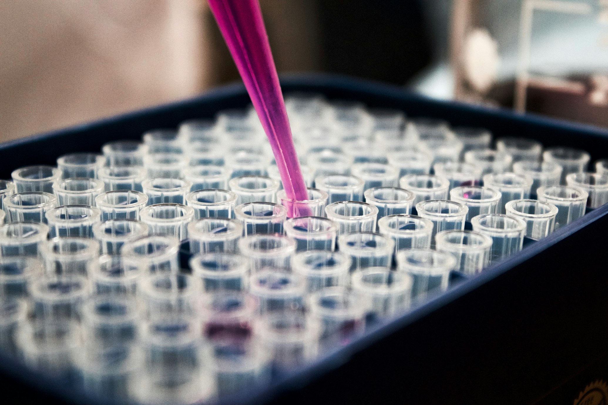
BioMEMS devices operate at the same scale as biological cells, enabling precise interactions with living systems.
What Are BioMEMS? The Meeting of Biology and Microtechnology
Bio-microelectromechanical systems (BioMEMS) are defined as micro or nano-scale devices or systems used for processing, delivering, manipulating, or analyzing biological and chemical entities for biological or biomedical applications 1 .
Essentially, they are miniaturized machines that interact with biological systems at a scale that matches the body's natural structures—from individual cells down to proteins and DNA molecules.
What makes BioMEMS special is their interdisciplinary nature, combining principles from engineering, materials science, biology, chemistry, and medicine. They can be fabricated from various materials including silicon, glass, polymers, and even biological elements themselves 2 .
These devices typically range in size from micrometers (millionths of a meter) to millimeters, allowing them to operate in spaces that would be impossible for conventional medical devices to access.
Scale Comparison
- Human hair 50-100 μm
- Red blood cell 8 μm
- Typical BioMEMS device 1-1000 μm
- Bacteria 1-5 μm
Historical Milestones: From Ancient Concepts to Modern Technology
Early Foundations
First glucose enzyme electrode (1962), early neural electrodes, and cochlear implants (1977) established foundation for bio-interfacing 1 .
Sensor Development
Ion-sensitive field-effect transistor (ISFET) developed (1972), early drug delivery devices emerged, demonstrating medical applications 1 .
MEMS Revolution
Rapid advancement of MEMS technology, first silicon-based microelectrodes for neuron studies (1988) 1 .
BioMEMS Emergence
Term "BioMEMS" coined, microfluidic devices, integrated DNA analysis, microfabricated microneedles, and PDMS-based soft lithography developed 1 .
Commercial Expansion
Exponential growth in research and commercialization, diagnostic chips, advanced drug delivery systems, and point-of-care devices 5 .
Nanoscale Applications
Nanoscale BioMEMS, organ-on-chip technology, advanced biosensors with unprecedented sensitivity and complexity 6 .
| Time Period | Key Developments | Significance |
|---|---|---|
| 1960s | First enzyme electrode, early neural electrodes | Established foundation for bio-interfacing |
| 1970s | ISFET sensors, early drug delivery devices | Demonstrated medical applications |
| 1980s | Silicon-based neural interfaces, micromachined sensors | Adapted semiconductor technology for bio applications |
| 1990s | Microfluidics, lab-on-a-chip, soft lithography | Created distinct BioMEMS field with specialized techniques |
| 2000s | Personalized diagnostics, point-of-care devices | Expanded commercial applications |
| 2010s-2020s | Nanoscale BioMEMS, organ-on-chip, advanced biosensors | Achieved unprecedented sensitivity and complexity |
A Key Experiment: Ultrasensitive Detection of Digoxin With Optical BioMEMS
To understand how BioMEMS work in practice, let's examine a recent breakthrough experiment that demonstrates the capabilities of this technology.
Methodology Overview
In 2024, researchers developed a multi-purpose optical BioMEMS platform for ultrasensitive detection of biomolecules, using the heart medication digoxin as a case study 6 .
- Cantilever design with extreme sensitivity
- Surface functionalization with specific antibodies
- Optical system setup with interferometric detection
- Sample introduction and measurement procedure
- Image processing algorithm for quantification
Performance Metrics
| Parameter | Value | Significance |
|---|---|---|
| Detection Limit | 300 fM | Extremely low concentration detection |
| Sensitivity | 5.5 × 10¹² AU/M | High response to concentration changes |
| Response Time | < 8 minutes | Much faster than conventional lab tests |
| Spring Constant | 0.0032 N/m | Extreme mechanical sensitivity |
Results and Scientific Importance
The experiment demonstrated extraordinary sensitivity, achieving a detection limit of 300 fM (femtromolar, or 10⁻¹⁵ molar) and a maximum sensitivity of 5.5 × 10¹² AU/M 6 . This means the system could detect digoxin at concentrations that are incredibly low—equivalent to finding a single grain of sand in an Olympic-sized swimming pool!
The researchers also found that their system could complete measurements in less than eight minutes for all samples, significantly faster than conventional laboratory tests. The platform proved to be stable, reproducible, and capable of functioning with very small sample volumes.
Key Advantages Demonstrated:
- Ultra-sensitivity: Enables early disease diagnosis and therapeutic drug monitoring
- Rapid results: Facilitates point-of-care testing rather than centralized laboratory analysis
- Multi-purpose platform: Versatile and cost-effective with different bioreceptors
- Cost-effectiveness: Could reduce healthcare costs by enabling regular monitoring

Optical detection methods enable highly sensitive measurements of molecular interactions.
The Scientist's Toolkit: Essential Components for BioMEMS Research
BioMEMS research requires a diverse array of specialized materials and techniques. Here are some of the most important elements in the BioMEMS toolkit:
Fabrication Materials
- Silicon: Excellent mechanical properties 2
- Glass: Optical transparency and insulation
- Polymers: Especially PDMS for flexibility
- Biological Materials: Proteins, antibodies, DNA
Essential Research Reagent Solutions in BioMEMS
| Reagent/Material | Function | Application Examples |
|---|---|---|
| PDMS (Polydimethylsiloxane) | Flexible, transparent elastomer for microfluidics | Lab-on-chip devices, cell culture platforms |
| Photoresist | Light-sensitive material for pattern transfer | Creating microfabrication masks |
| SAMs (Self-Assembled Monolayers) | Molecular layers that form spontaneously on surfaces | Surface functionalization, biosensor interfaces |
| Biolinkers | Molecules that connect biological elements to surfaces | Antibody immobilization, cell patterning |
| Fluorescent dyes | Light-emitting markers for detection | Cell viability assays, target detection |
Applications: How BioMEMS Are Revolutionizing Healthcare
BioMEMS technology has found applications across virtually all areas of biomedical research and clinical practice:
Research Tools
Cell Manipulation
BioMEMS devices can sort, trap, and manipulate individual cells for research purposes 5 .
Tissue Engineering
Microfabricated scaffolds guide cell growth and organization, helping to create functional artificial tissues 3 .
High-Throughput Screening
Microarrays allow researchers to simultaneously test thousands of biological interactions 4 .
Future Directions: Where BioMEMS Technology Is Headed
The field of BioMEMS continues to evolve at a rapid pace. Future developments will likely focus on:

Emerging Trends
- Increased Integration: Combining multiple functions on single devices
- Nanoscale Applications: Interaction with subcellular components and individual molecules
- Smart Implants: Autonomous implantable devices for monitoring and therapy
- Artificial Intelligence Integration: AI algorithms for data analysis and decision support
- Personalized Medicine: Patient-specific devices and treatments
Transforming Healthcare
As BioMEMS technology continues to advance, it promises to transform healthcare from a largely reactive practice to one that is predictive, preventive, and personalized.
The invisible revolution of BioMEMS reminds us that sometimes the biggest advances come in the smallest packages—and that the future of medicine may be written not in medical charts, but in microfabrication labs where biology and technology meet at the smallest of scales.
The history of BioMEMS demonstrates how the convergence of different disciplines can create revolutionary technologies that benefit human health.